The document provides information about right hepatectomy surgery:
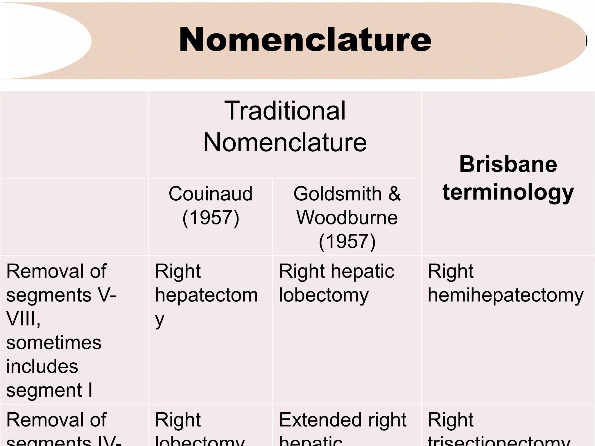
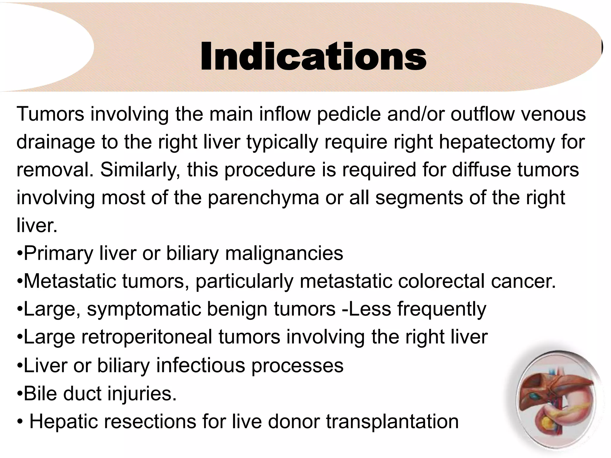
1. It discusses the traditional and Brisbane terminology used to describe right hepatectomy and the indications for the procedure such as primary liver tumors, metastatic tumors, and large benign tumors.
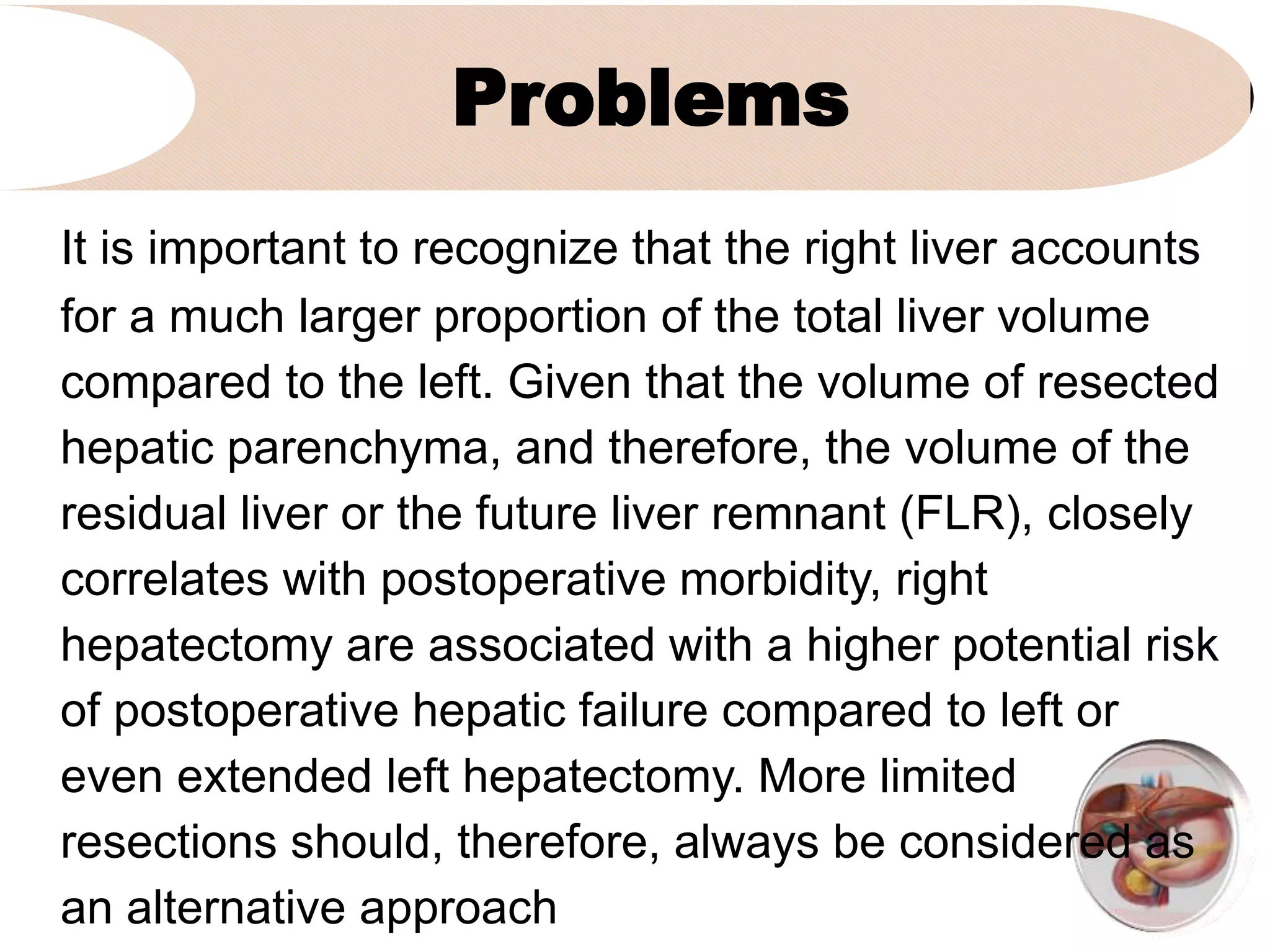
2. It notes some of the challenges with right hepatectomy due to the large volume of the right liver and higher risk of postoperative hepatic failure compared to other procedures.
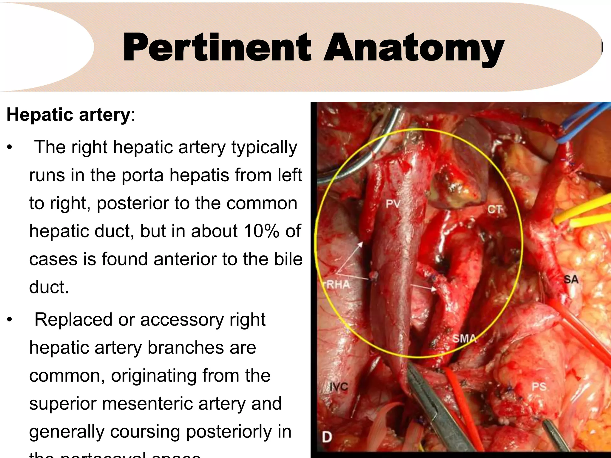
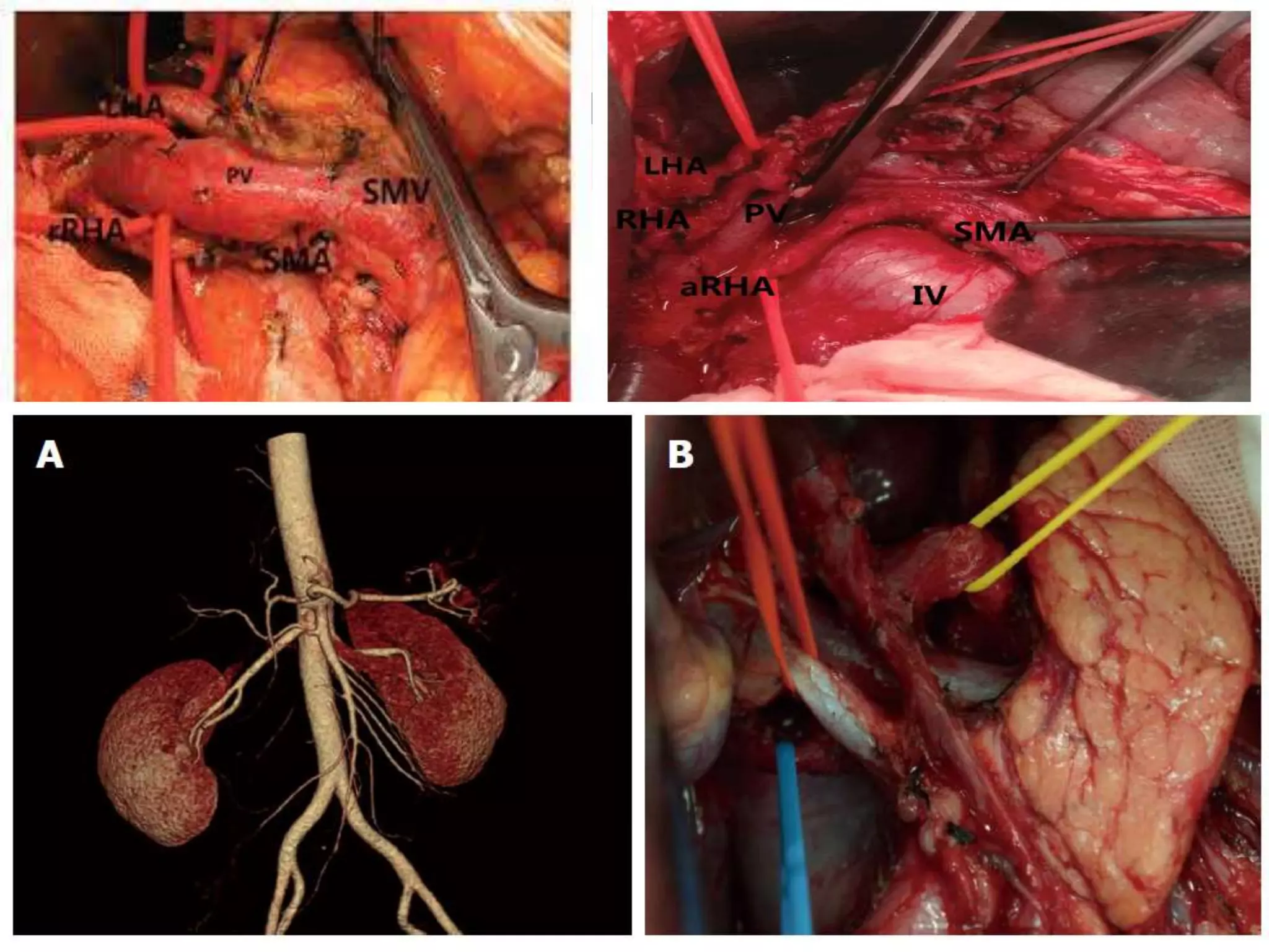
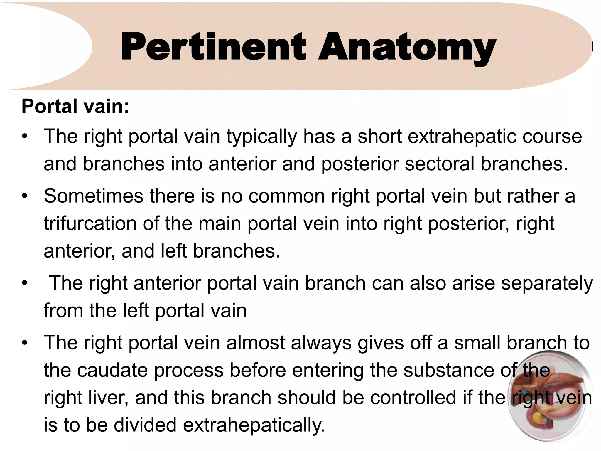
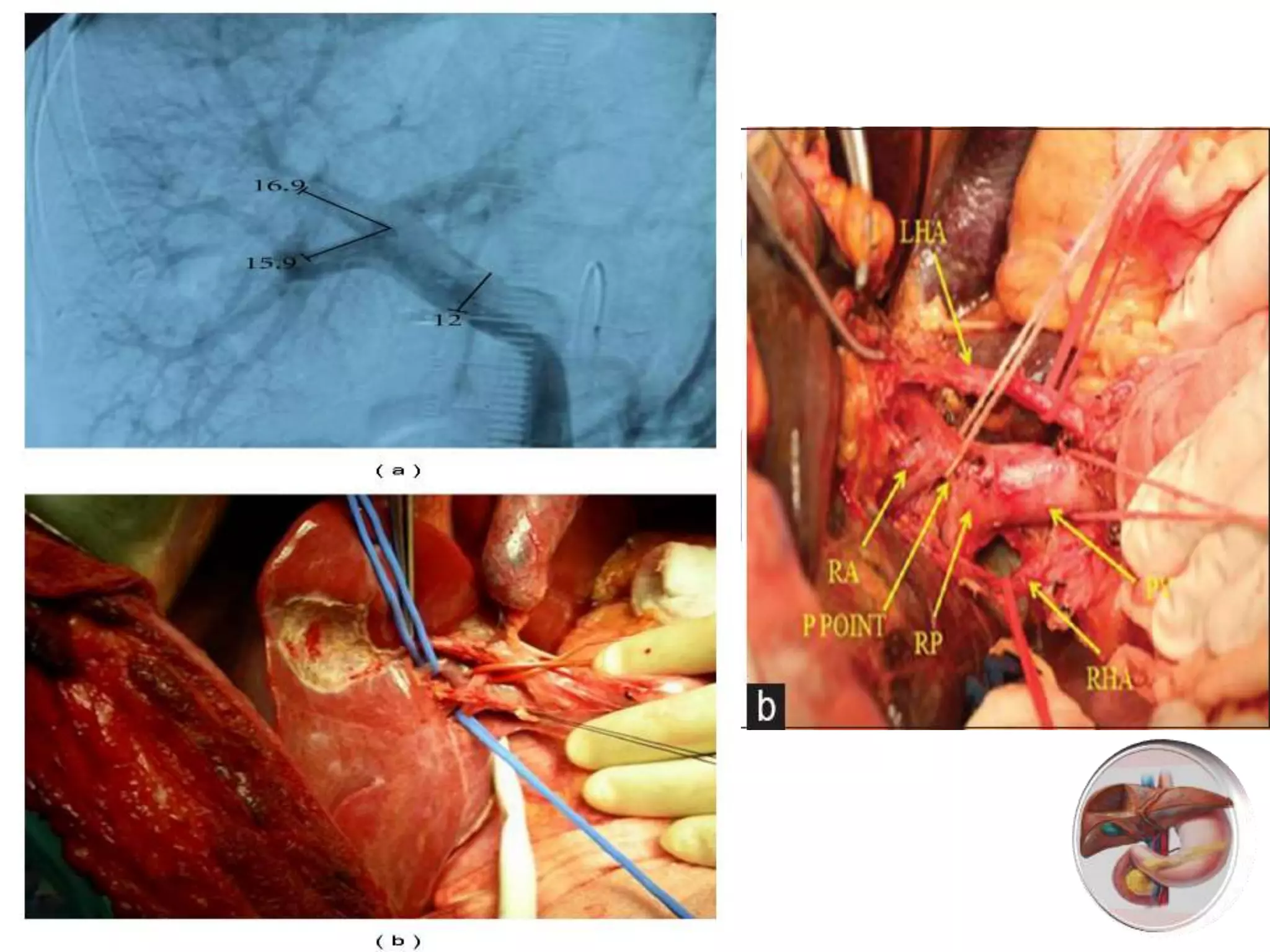
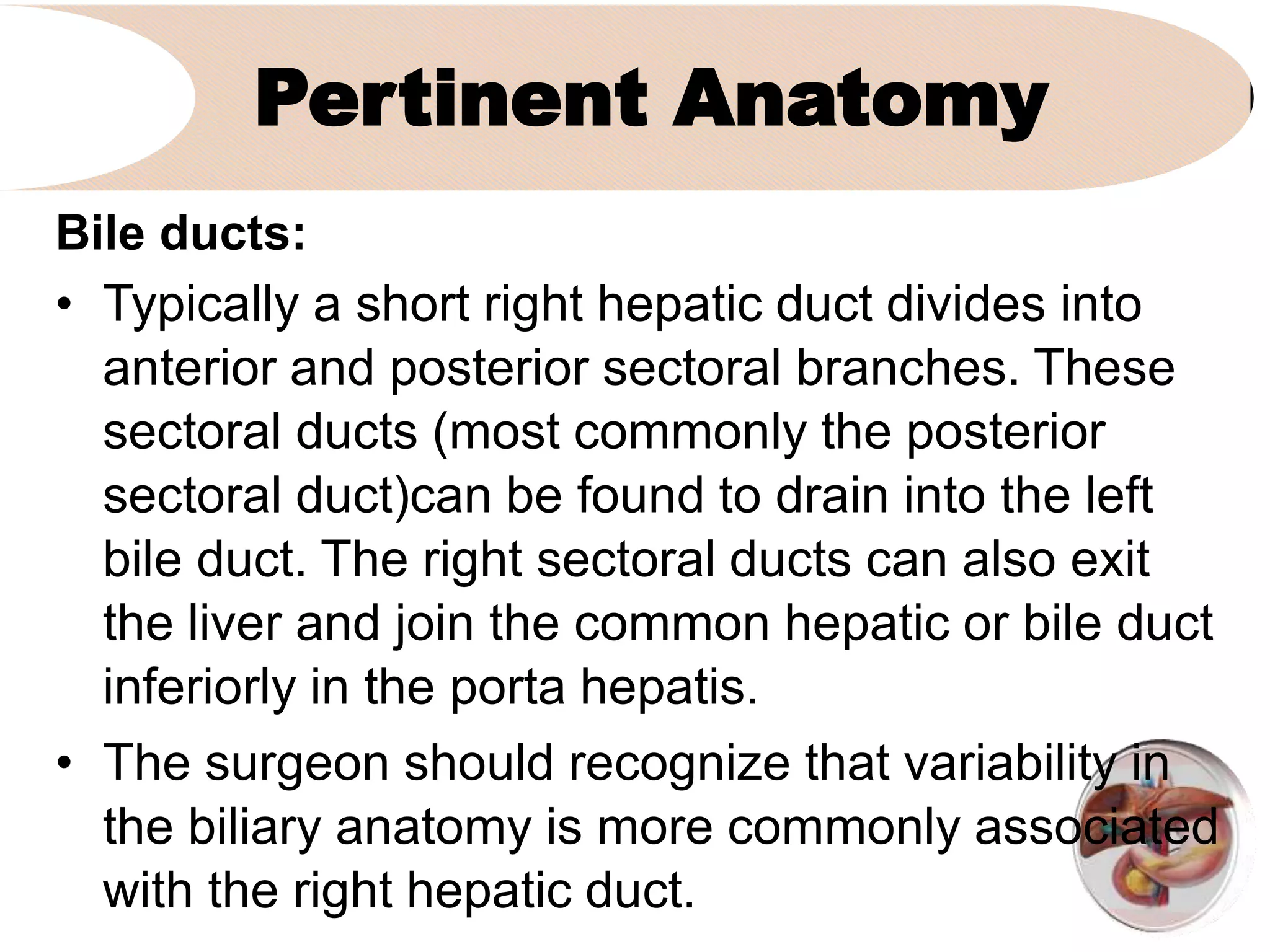
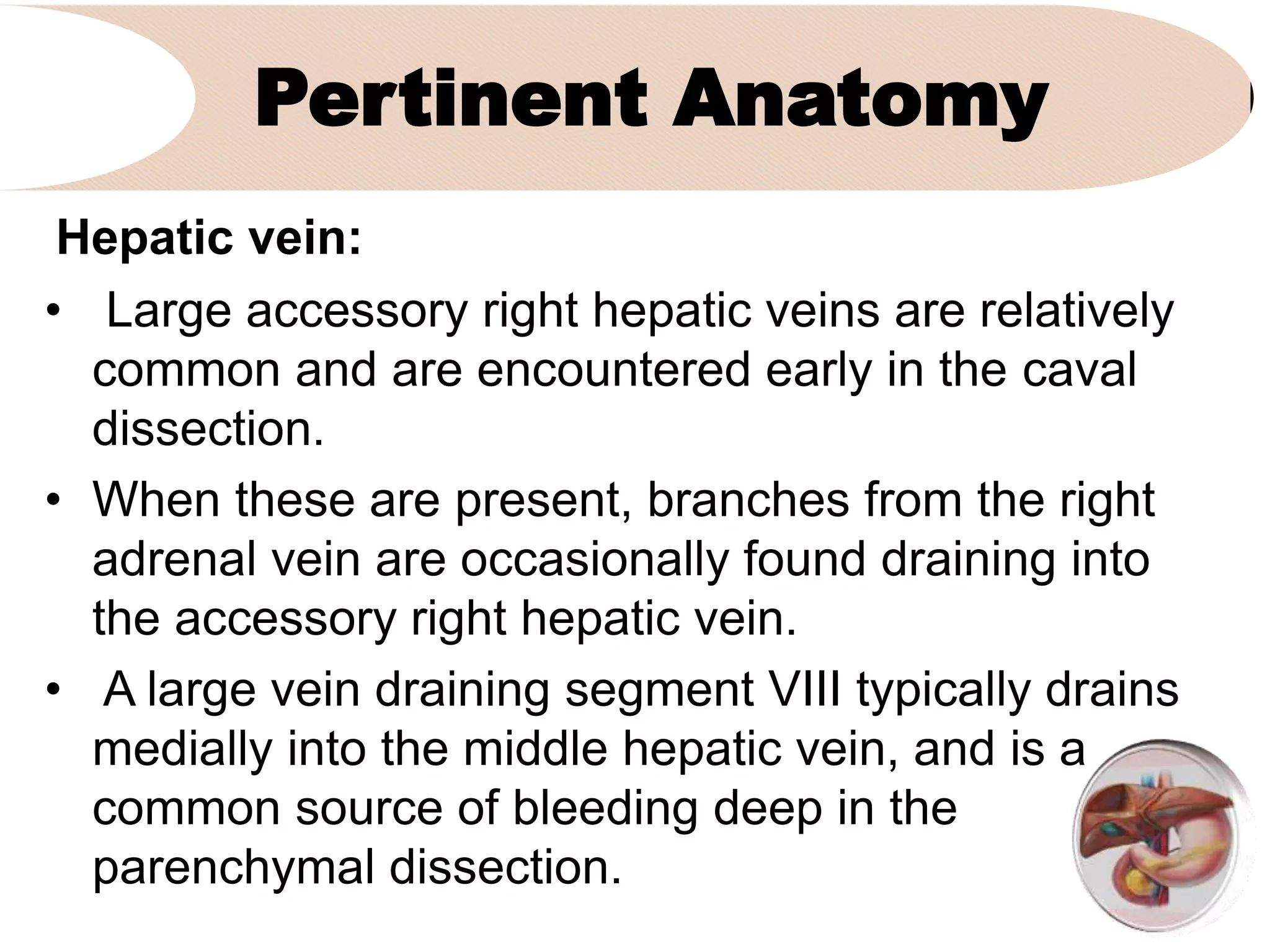
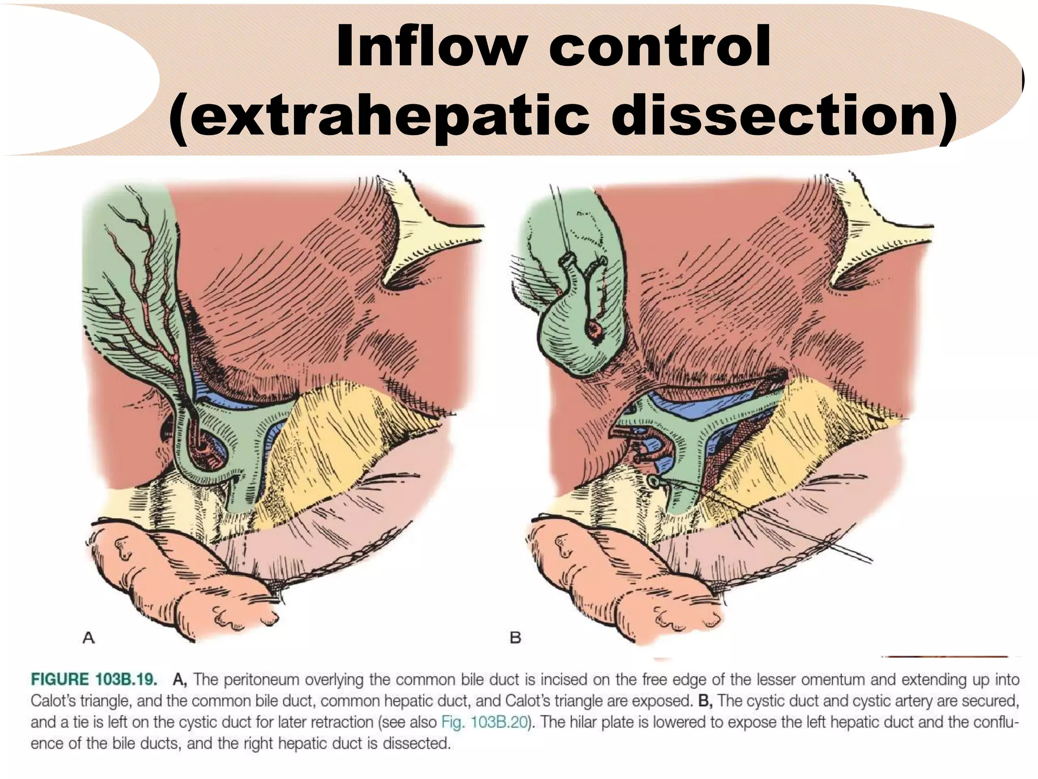
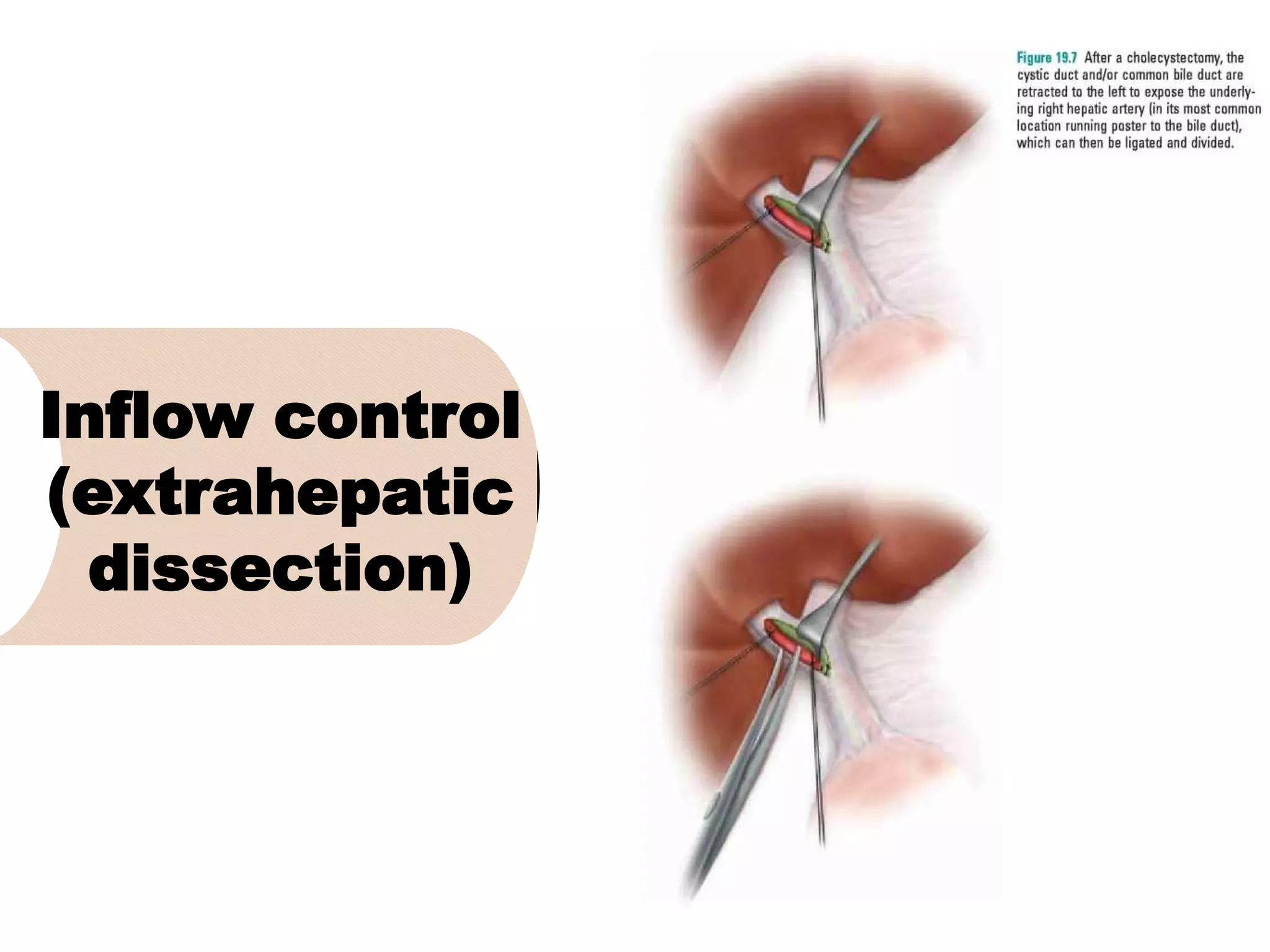
3. It describes the pertinent anatomy of the hepatic artery, portal vein, bile ducts, and hepatic vein that are important to understand for the surgery.
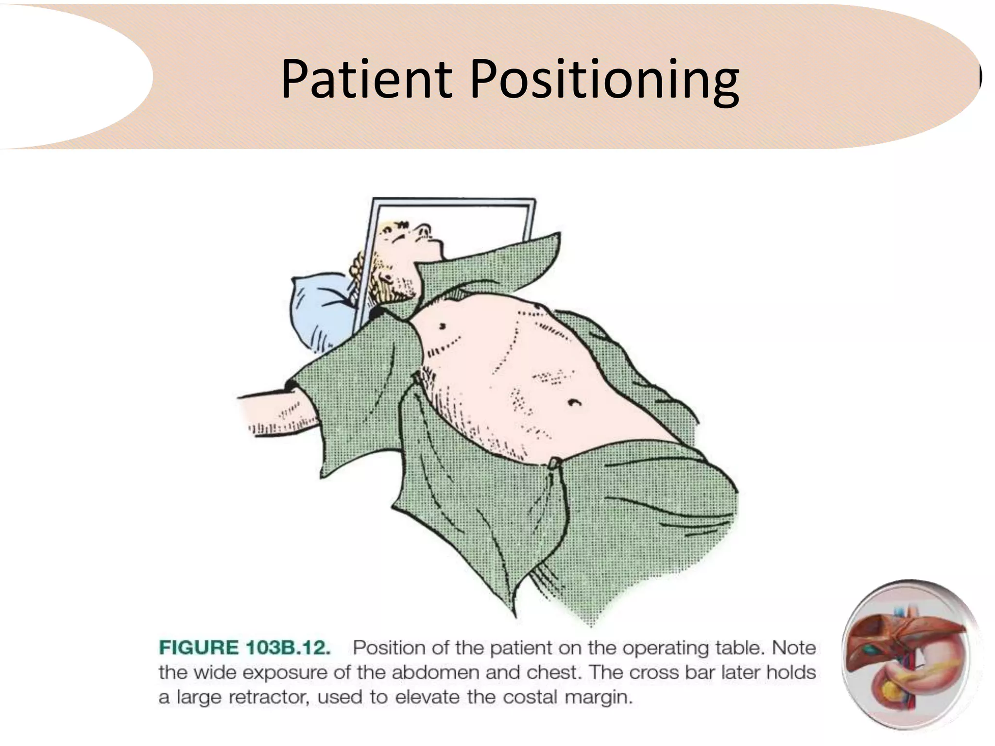
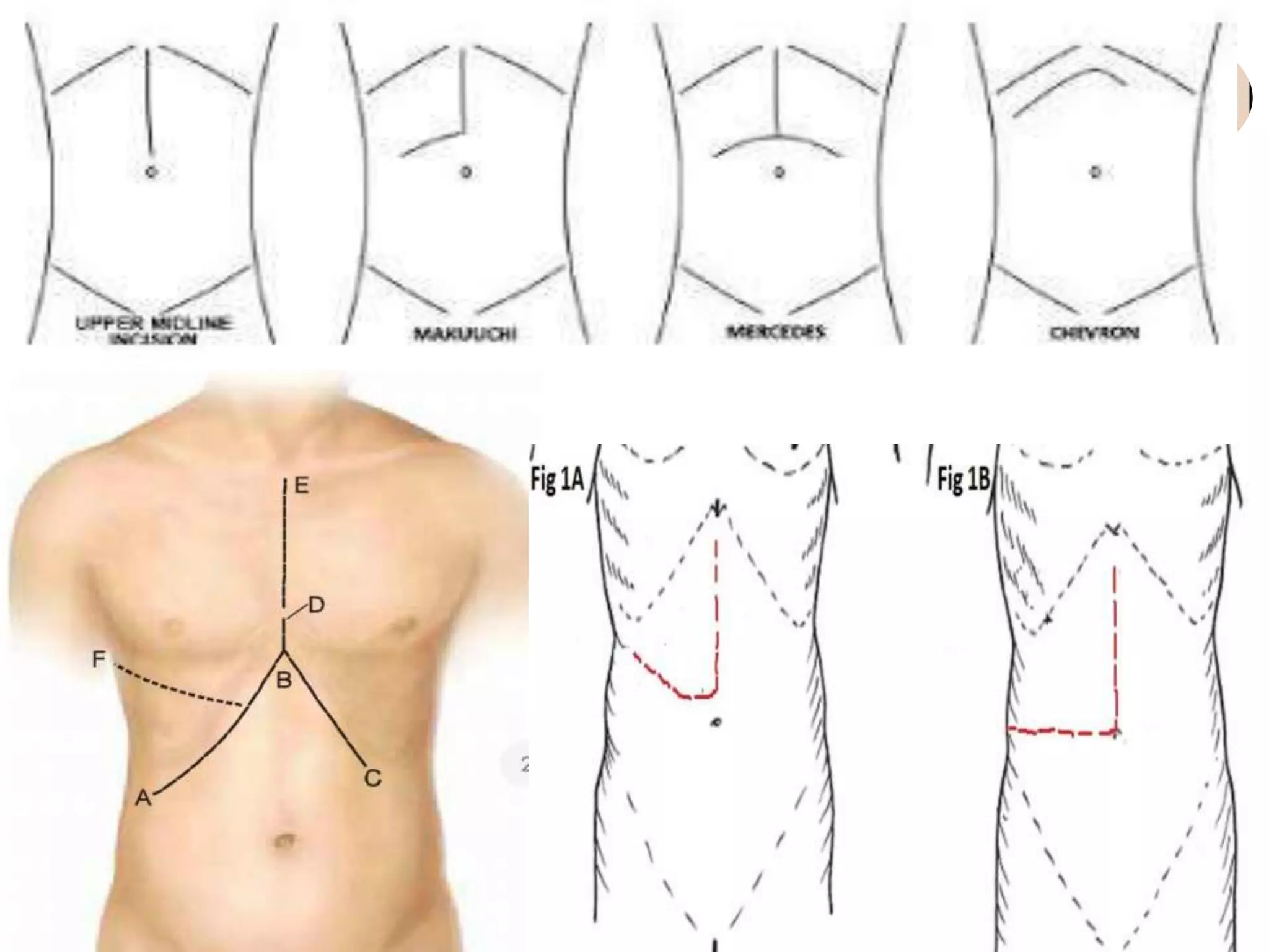
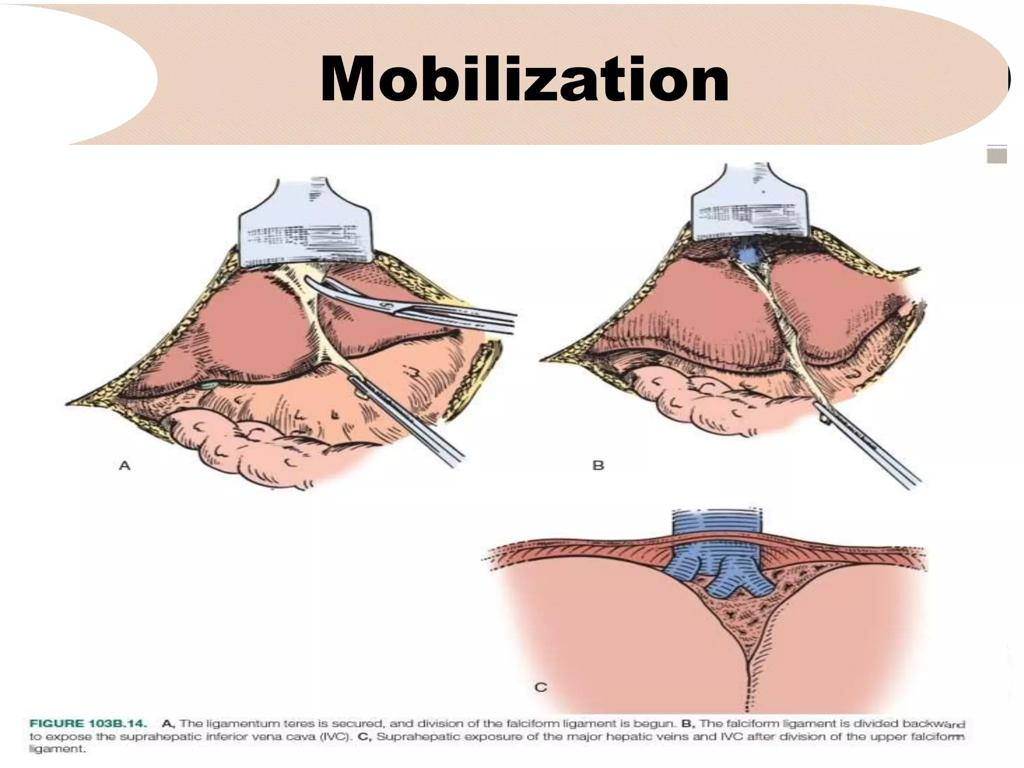
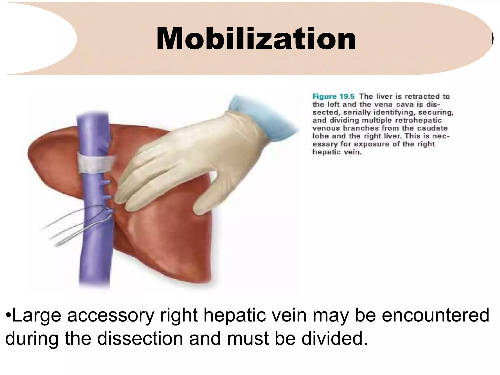
4. It outlines the key steps of the operative procedure including patient positioning, incisions, retraction, exploration, liver mobilization
3. Nomenclature
1. Click to add subtitle
Traditional
Nomenclature
Brisbane
terminology
Couinaud
(1957)
Goldsmith &
Woodburne
(1957)
Removal of
segments V-
VIII,
sometimes
includes
segment I
Right
hepatectom
y
Right hepatic
lobectomy
Right
hemihepatectomy
Removal of Right Extended right Right
4. Indications
1. Click to add subtitle
Tumors involving the main inflow pedicle and/or outflow venous
drainage to the right liver typically require right hepatectomy for
removal. Similarly, this procedure is required for diffuse tumors
involving most of the parenchyma or all segments of the right
liver.
•Primary liver or biliary malignancies
•Metastatic tumors, particularly metastatic colorectal cancer.
•Large, symptomatic benign tumors -Less frequently
•Large retroperitoneal tumors involving the right liver
•Liver or biliary infectious processes
•Bile duct injuries.
• Hepatic resections for live donor transplantation
5. Problems
It is important to recognize that the right liver accounts
for a much larger proportion of the total liver volume
compared to the left. Given that the volume of resected
hepatic parenchyma, and therefore, the volume of the
residual liver or the future liver remnant (FLR), closely
correlates with postoperative morbidity, right
hepatectomy are associated with a higher potential risk
of postoperative hepatic failure compared to left or
even extended left hepatectomy. More limited
resections should, therefore, always be considered as
an alternative approach
6. Pertinent Anatomy
Hepatic artery:
• The right hepatic artery typically
runs in the porta hepatis from left
to right, posterior to the common
hepatic duct, but in about 10% of
cases is found anterior to the bile
duct.
• Replaced or accessory right
hepatic artery branches are
common, originating from the
superior mesenteric artery and
generally coursing posteriorly in
8. Pertinent Anatomy
Portal vain:
• The right portal vain typically has a short extrahepatic course
and branches into anterior and posterior sectoral branches.
• Sometimes there is no common right portal vein but rather a
trifurcation of the main portal vein into right posterior, right
anterior, and left branches.
• The right anterior portal vain branch can also arise separately
from the left portal vain
• The right portal vein almost always gives off a small branch to
the caudate process before entering the substance of the
right liver, and this branch should be controlled if the right vein
is to be divided extrahepatically.
9.
10. Pertinent Anatomy
Bile ducts:
• Typically a short right hepatic duct divides into
anterior and posterior sectoral branches. These
sectoral ducts (most commonly the posterior
sectoral duct)can be found to drain into the left
bile duct. The right sectoral ducts can also exit
the liver and join the common hepatic or bile duct
inferiorly in the porta hepatis.
• The surgeon should recognize that variability in
the biliary anatomy is more commonly associated
with the right hepatic duct.
11. Pertinent Anatomy
Hepatic vein:
• Large accessory right hepatic veins are relatively
common and are encountered early in the caval
dissection.
• When these are present, branches from the right
adrenal vein are occasionally found draining into
the accessory right hepatic vein.
• A large vein draining segment VIII typically drains
medially into the middle hepatic vein, and is a
common source of bleeding deep in the
parenchymal dissection.
14. Incisions
• The incision depends on body habitus and tumor
location
• A midline incision is often most comfortable for the
patient.
• An extended right subcostal or a bilateral subcostal
with a vertical extension are other common incision
choices-"Mercedes"-type incision
• a midline incision from the xiphoid process to the
umbilicus, with right lateral extension toward a point
midway between the inferior costal margin and
anterior superior iliac spine
15. Incisions
• A right subcostal incision with midline vertical
extension to the xiphoid ("hockey stick"
incision).
• A limited upper midline incision may be
made first to allow preliminary manual and
visual exploration to exclude obviously
unresectable disease or the presence of
extrahepatic spread
• Thoraco-abdominal approach, but this is
rarely necessary
17. Retractors
• The key issue for exposure is cephalad retraction
of the costal margin at approximately a 45 degree
angle.
• Any number of retractor systems can provide this
retraction combined with lateral retraction of the
right chest wall and inferior retraction of the lower
abdominal viscera
18.
19. Anesthetic Considerations
• The most common source of bleeding from the hepatic
parenchyma is from the hepatic veins.
• Maintaining a low central venous pressure (CVP) during
resection is invaluable in limiting blood loss
• Mild trendelenburg position, limitation of IV fluids and
pharmacologic management with vasodialators are
helpful. Once resection is completed anaesthesiologist
should be informed to hydrate the patient
20. Exploration
• Diagnistic laparoscopy is used selectively on the risk
of occult unresectable diseases.
• Thorough open exploration for occult metastasis
disease should include bimenual palpation of the
liver as well as inspection/palpation of all peritoneal
surfaces and relevant nadal basins.
• Intraoperative US is used to identify occult hepatic
tumours and assess the details of the tumour and
vascular anatomy
22. Mobilization
• With a sharp dissection, the right hepatic vein is
and common trank of the middle and left hepatic
veins are axposed anteriorly. The groove
between the right and middle hepatic vein is
dissected down to the vena cava.
• The coronary ligament are divided, exposing the
right hepatic veins.
23. Mobilization
• The liver is turned to the left and the diaphragm is
retracted laterally and caphalad. This expose the
peritoneal reflexion of the right triangular ligament,
which is divided laterally and on the inferior
surface of the right liver.
• The dissection is continued lateral to medial,
separating the right adrenal gland from the liver
and dropping it posteriorly, exposing the lateral
wall of the vena cava.
26. Mobilization
• At this stage, inferior border of the right hepatic
vein is sharply dissected and a tunnel between
the right and middle hepatic vein is dissected.
• Once this tunnel is finished, the right hepatic
vein is encircled with vessel loop.
29. Inflow control
(extrahepatic dissection)
• On occasions, the artery will be present anterior to the
bile duct or in an accessory/replaced position in the
portocaval space, requiring dissection in these area.
• It is always prudent to check a pulse in the left hepatic
artery at the base of the umbilical fissure with the
presumed right hepatic artery clumped to confirm
anatomy.
• In this typical position, the proximal right hepatic artery
stump can be used as a sling to retract the biliary tree
superiorly and to the left, exposing underlying portal
vein
30. Inflow control
(extrahepatic dissection)
• The portal vein anatomy is then dissected
sharply. The main, right and left portal veins
should be dissected and visualized keeping the
variations in mind.
• The right posrtal vein is then dissected and
relatively constant branch to the caudate process
is exposed, encircled, tied and divided for
maximum exposure.
32. Inflow control
(extrahepatic dissection)
• Unless mandate by the tumour proximity, the right
bile duct should be divided intrahepatically within
glissonian sheath to minimise chance of left bile
duct injury.
33. Inflow control
(Intratrahepatic approach
• If tumor proximity does not mandate hilar
dissection, the inflow to the liver can be taken
intrahepatically as a pedicle within the invaginated
glissonian peritoneal sheath.
• This can be taken as a single main right pedicle or a
separate division of the right anterior and posterior
pedicles.
• The right inflow pedicles can be encircled via
anterior and posterior hepatotomies or by dividing
the parenchyma down to the pedicles.
• As in the extrahepatic approach, the hilar plate
should be lowered to protect the left bile duct
35. Inflow control
(Intratrahepatic approach
• In the hepatotomy approach, incisions are made
vertically along the gallbladder fossa and continued
transversely along the base of segment IV and
separately at the caudate process.
• The right main pedicle can then be encircled with the
thumb and forefinger or with a large right angle clamp.
Hepatic parenchyma can be cleared and the right
anterior and posterior padicles can also be separately
encircled.
36. Inflow control
(Intratrahepatic approach
• One must be wary of inadvertent injury to middle
hepatic vein branches.
• Division of the pedicles can then be carried out with
clamping and suturing or with vascular staplers.
• It is always prudent to clamp and check demarcation
and/or flow to the left liver prior to division. Similarly, if
there is any concern for compromise of the left hepatic
duct, a cholangiogram should be obtained
39. Inflow control
(Intratrahepatic approach
• The right pedicles can also be approached by
dividing the hepatic parenchyma in the plane of
the planned resection down to the anterior portion
of the right inflow pedicle.
• An incision in the caudate process is made and
the pedicles encircled, checked, and divided.
40. Outflow control
• The dissection of the right hepatic vain is done
during mobilization.
• After division of the right inflow, we typically
divide the vein with a vascular stapler, but
controlling the vein with vascular clamps
followed by division and suturing is reasonable.
• If the tumor is close or if there is any concern for
caval injury, control of the infra- and
suprahepatic vana cava is prudent.
42. Outflow control
• The middle hepatic vein runs in the main portal scissura (Cantlie's
line). Therefore, a right or extended right hepatectomy may involve
resection of a significant length of the middle vein.
• If the middle hepatic vein is to be divided somewhere in its central
portion, this requires intraparenchymal dissection and division
within the substance of the liver.
• If a division of the middle hepatic vein at its origin is required,
there are two approaches that can be used.
• An extrahepatic division can be accomplished by encircling the
middle hepatic vein with a clamp in its usually short extrahepatic
course.
• The middle vein can be encircled above the liver but is much
44. Outflow control
• Regardless of the approach, it is of critical importance to
protect the outflow of the left hepatic vein, since
compromise of this vein will result in venous ischemia of
the remnant left liver, as well as torrential hemorrhage as
the liver will be devoid of venous outflow.
• To ensure that the left hepatic vein is intact, the presumed
middle hepatic vein can be clamped with patent left-sided
inflow. If there is compromise of the left hepatic vein,
venous congestion will be seen grossly.
• In addition, the outflow can be tested with Doppler
ultrasound. In experienced hands, the left hepatic vein
45. Parenchymal Transection
• A right hepatectomy requires division of the hepatic
parenchyma in the main portal scissura in the plane of
the middle hepatic vein.
• If possible, the middle hepatic vein should be
preserved to optimize venous drainage of the remnant.
In this situation, the transection line is just to the right of
the middle hepatic vein.
• This requires dissection of the right-sided branches of
the middle hepatic vein, with careful identification and
ligation.
46. Parenchymal Transection
• There is typically a large branch off the
middle hepatic vein to segment VIII that
must be carefully controlled.
• A right hepatectomy can also be performed
in this central plane to the left of the middle
hepatic vein with preservation of segment
IV.
47. Parenchymal Transection
• There are numerous instruments and methods used
to transect hepatic parenchyma,none necessarily
better than the other.
• Among the many techniques, some prefer use of
instruments such as the Cavitron ultrasonic
aspirator (CUSA). others prefer pre-coagulation with
radiofrequency ablation devices and still others
prefer a simple clamp-crushing technique with a
Kelly clamp.
48. Parenchymal Transection
• The preferred technique is dependent on training,
comfort level, instrument availability and local
expertise.
• Intraparenchymal structures should be
anticipated, identified and divided safely with full
knowledge of one's location inside the liver.
50. Special Considerations
• For large tumors of the right liver, classical
mobilization as described above may not be possible
or safe. Sometimes an anterior approach is necessary.
In this technique, the inflow to the right liver is
controlled and the parenchymal transection is
performed without any mobilization or dissection of the
vena cava.
• The transection is carried out back to the vena cava,
and the right hepatic vein (and other retrohepatic
venous branches) is encircled and divided. Only then
is the liver dissected off of its diaphragmatic and
retroperitoneal attachments.
51. Special Considerations
• This technique has been associated with a survival
benefit in patients with large hepatocellular
carcinoma of the right liver in a randomized trial.
• The anterior approach can also be facilitated by a
"hanging maneuver" in which a large clamp is used to
bluntly dissect the plane between the vena cava and
the liver inferiorly up to the groove between the right
and middle hepatic vein. This clamp is then used to
pass a tape underneath the liver which can guide the
anterior approach parenchymal transection
54. Post resection
• An oozing raw surface is common after resection
and requires some degree of hemostasis. Use of
the argon beam coagulator is common. Significant
bleeding should be sutured.
• If there is any concern about placing deep sutures
and injury to underlying structures, topical
hemostatics can be used instead of sutures.
• The falciform ligament is re-attached to prevent
extreme rotation of the left liver.
55. Post resection
• Perfusion and venous drainage of the remnant liver
should be observed grossly and assessed with
Doppler ultrasound if necessary.
• Although it is common practice to place drains after a
hepatic resection, randomized trials have not shown
any benefit.
• Inspection of the cut surface of the liver and the porta
hopatis for bile leak which should be addressed with
fine absorbable suture.
• If an ongoing bile leak cannot be corrected, a drain
should be placed