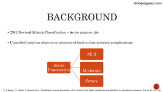
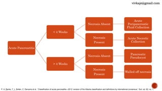
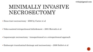
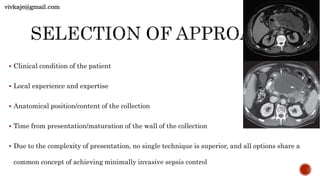
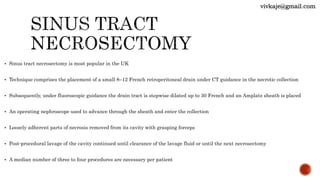
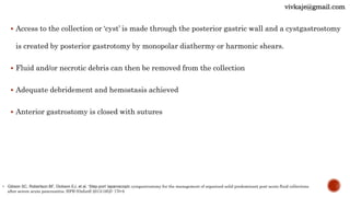
 2012 Revised Atlanta Classification – Acute pancreatitis
 Classified based on absence or presence of local and/or systemic complications
Acute
Pancreatits
Mild
Moderate
Severe
• P. A. Banks, T. L. Bollen, C. Dervenis et al., “Classification of acute pancreatitis—2012: revision of the Atlanta classification and definitions by international consensus,” Gut, vol. 62, no. 1, pp.
vivkaje@gmail.com
Acute Pancreatitis
< 4 Weeks
Necrosis Absent
Acute
Peripancreatic
Fluid Collection
Necrosis
Present
Acute Necrotic
Collection
> 4 Weeks
Necrosis Absent
Pancreatic
Pseudocyst
Necrosis
Present
Walled off necrosis
P. A. Banks, T. L. Bollen, C. Dervenis et al., “Classification of acute pancreatitis—2012: revision of the Atlanta classification and definitions by international consensus,” Gut, vol. 62, no. 1,
vivkaje@gmail.com
 Half of the deaths attributable to AP occur within the first 7 days of admission
 Patients with severe AP who survive this first phase of illness particularly those with
persistent
SIRS or organ failure are at risk of developing secondary infection of pancreatic necrosis
 Mortality in patients with infected necrosis and organ failure ~ 20–30%
• C. J. McKay, S. Evans, M. Sinclair,C.R.Carter, andC.W. Imrie, “High early mortality rate from acute pancreatitis in Scotland, 1984–1995,” The British Journal of Surgery, vol. 86, no. 10, pp. 1302–
1999.
vivkaje@gmail.com
 Step Up - Management of postacute peripancreatic collections
 PANTER randomized controlled trial - 35% of patients with established necrotic collections did
not require any further intervention over simple small diameter percutaneous catheter drainage
 Consensus that in those patients with persistent sepsis, a minimally invasive approach is
preferred over open surgical necrosectomy
• H. C. Van Santvoort, M. G. Besselink, O. J. Bakker et al., “A step-up approach or open necrosectomy for necrotizing pancreatitis,”The New England Journal ofMedicine, vol. 362, no. 16,
pp. 1491–1502, 2010.
vivkaje@gmail.com
 Sinus tract necrosectomy - 2000 by Carter et al
 Video-assisted retroperitoneal debridement – 2001 Horvath et al
 Laparoscopic necrosectomy - transperitoneal or a retroperitoneal approach
 Endoscopic transluminal drainage and necrosectomy – 2000 Seifert et al
vivkaje@gmail.com
 Clinical condition of the patient
 Local experience and expertise
 Anatomical position/content of the collection
 Time from presentation/maturation of the wall of the collection
 Due to the complexity of presentation, no single technique is superior, and all options share a
common concept of achieving minimally invasive sepsis control
vivkaje@gmail.com
 Sinus tract necrosectomy is most popular in the UK
 Technique comprises the placement of a small 8–12 French retroperitoneal drain under CT guidance in the necrotic collection
 Subsequently, under fluoroscopic guidance the drain tract is stepwise dilated up to 30 French and an Amplatz sheath is placed
 An operating nephroscope used to advance through the sheath and enter the collection
 Loosely adherent parts of necrosis removed from its cavity with grasping forceps
 Post-procedural lavage of the cavity continued until clearance of the lavage fluid or until the next necrosectomy
 A median number of three to four procedures are necessary per patient
vivkaje@gmail.com
 Conversion to open surgery for further necrosectomy or treatment of complications needed in 14– 26%
patients
 Fistula formation (4–22%) and bleeding (10–17%) are the most prominent procedure-related complications
 Mortality of patients treated with sinus tract necrosectomy was reported between 9 and 19%
• Carter CR, McKay CJ, Imrie CW. Percutaneous necrosectomy and sinus tract endoscopy in the management of infected pancreatic necrosis: an initial experience. Ann Surg
2000;232(2):175-80
• Connor S, Ghaneh P, Raraty M, et al. Minimally invasive retroperitoneal pancreatic necrosectomy. Dig Surg 2003; 20(4):270-7
• Connor S, Raraty MG, Howes N, et al. Surgery in the treatment of acute pancreatitis – minimal access pancreatic necrosectomy. Scand J Surg 2005;94(2): 135-42
vivkaje@gmail.com
vivkaje@gmail.com
 Under GA, patient is placed in a supine position with the left flank raised to 30–40°
 A midaxillary, subcostal incision of 5 cm is made close to the exit point of the drain
 Guided by the drain and the CT images, the surgeon enters the retroperitoneum and clears
purulent material by suction
 Grasping forceps are used to remove the first necrosis under irrigation and suction
 When the directly visible necrosis is removed, a videoscope is introduced and CO2 can be infused
through the initial drain to inflate the cavity and enhance vision
vivkaje@gmail.com
 With a laparoscopic forceps or long grasping clamp, more necrosis is removed
 By limiting the debridement to only the loosely adherent necrosis, the risk of bleedi g from
viable underlying tissue is brought to a minimum
 The initial drain is removed and careful irrigation of the cavity performed
 After placement of two large-bore single- lumen drains for postoperative lavage, the fascia
and skin are closed
 Lavage with normal saline (up to 10 l/24 h) is continued until the effluent is clear
• Horvath KD, Kao LS, Wherry KL, et al. A technique for aparoscopic-assisted percutaneous drainage of infected pancreatic necrosis and pancreatic abscess. Surg Endosc 2001;15(10):1221-5
• van Santvoort HC, Besselink MG, Horvath KD, et al. Videoscopic assisted retroperitoneal debridement in infected necrotizing pancreatitis. HPB (Oxford) 2007;9(2):156-9
vivkaje@gmail.com
 Laparoscopic direct necrosectomy was described in the 1990’s but failed to gain popularity due to
technical difficulty
 Laparoscopic cystogastrostomy is the most frequently used
 In this procedure, three or four ports are used for operating
 After insertion and inflation of the abdomen, an anterior gastrostomy is made and the pancreatic collection
located
• M. Gagner, “Laparoscopic treatment of acute necrotizing pancreatitis,” Seminars in Laparoscopic Surgery, vol. 3, no. 1, pp. 21– 28, 1996.
vivkaje@gmail.com
 Access to the collection or ‘cyst’ is made through the posterior gastric wall and a cystgastrostomy
is created by posterior gastrotomy by monopolar diathermy or harmonic shears.
 Fluid and/or necrotic debris can then be removed from the collection
 Adequate debridement and hemostasis achieved
 Anterior gastrostomy is closed with sutures
• Gibson SC, Robertson BF, Dickson EJ, et al. ‘Step-port’ laparoscopic cystgastrostomy for the management of organized solid predominant post-acute fluid collections
after severe acute pancreatitis. HPB (Oxford) 2013;16(2): 170-6
vivkaje@gmail.com
vivkaje@gmail.com
 Endoscopic transluminal drainage and necrosectomy can be considered if the infected collection
lies within few centimeters of the gastric or duodenal lumen
 Endoscopic ultrasound aids in
 Locating the collection
 Confirm its contents
 Measure the distance to the intestinal lumen
 Trace vascular structures in the intended puncture site
 Distinguish the collection from other structures
 Most endoscopists use a 19 G FNA needle to puncture the collection through the intestinal wall
vivkaje@gmail.com
 Content is aspirated or contrast injected, to confirm access into the collection
 Guide wire is advanced into the cavity
 A fistula tract between the intestinal lumen and the necrotic cavity is then created using
electrocautery
 Tract is dilated to approximately 10 mm using balloon dilators, hemostasis achieved
 Two double pigtails and a nasocystic flushing catheter are placed
 The nasocystic catheter is rinsed with 1 L NS/24 h to keep the cystgastrostomy open
 If necessary, multiple noncommunicating collections can be drained in one session
vivkaje@gmail.com
 In case patient does not improve clinically within the first 72 h after drainage, an endoscopic
necrosectomy is performed
 A forward viewing endoscope is used to balloon dilate the tract up to 15 – 20 mm and the
endoscope is advanced into the collection
 Necrosectomy can be performed using various instruments including snares, waterjets, nets,
baskets and grasping forceps to pull the necrotic tissue out of the collection and into the stomach
• Park DH, Lee SS, Moon SH, et al. Endoscopic ultrasound-guided versus conventional transmural drainage for pancreatic pseudocysts: a prospective randomized trial. Endoscopy 2009;41(10): 842-8
• Varadarajulu S, Christein JD, Tamhane A, et al. Prospective randomized trial comparing EUS and EGD for transmural drainage of pancreatic pseudocysts (with videos). Gastrointest Endosc
2008;68(6): 1102-11
• Rana SS, Bhasin DK, Rao C, et al. Non-fluoroscopic endoscopic ultrasound-guided transmural drainage of symptomatic non-bulging walled-off pancreatic necrosis. Dig Endosc 2013;25(1): 47-52
vivkaje@gmail.com
 Irrigating the cavity with hydrogen peroxide might loosen necrosis, but there is no convincing
evidence for this method
 After removing the loosely adherent necrosis, again multiple double pigtail stents and a
nasocystic catheter are placed to maintain the fistula tract
 Over time, the infected fluid and potentially necrotic tissue drain to the intestine and the cavity
collapses
• Park DH, Lee SS, Moon SH, et al. Endoscopic ultrasound-guided versus conventional transmural drainage for pancreatic pseudocysts: a prospective randomized trial. Endoscopy
2009;41(10): 842-8
• Varadarajulu S, Christein JD, Tamhane A, et al. Prospective randomized trial comparing EUS and EGD for transmural drainage of pancreatic pseudocysts (with videos). Gastrointest
Endosc 2008;68(6): 1102-11
• Rana SS, Bhasin DK, Rao C, et al. Non-fluoroscopic endoscopic ultrasound-guided transmural drainage of symptomatic non-bulging walled-off pancreatic necrosis. Dig Endosc
vivkaje@gmail.com
 Guidelines advocate catheter drainage as first step in the treatment of infected necrosis
 percutaneous or endoscopic transluminal
 Preferably delay the drainage at least 4 weeks after the initial presentation
 Intervention in sterile necrosis is best avoided to reduce secondary infection
 When patients experience ongoing symptoms, intervention can be considered in a late phase,
beyond 8 weeks after the initial presentation
Working Group IAP/APA Acute Pancreatitis Guidelines. IAP/ APA evidence-based guidelines for the management of acute pancreatitis. Pancreatology 2013;13(4 Suppl 2):e1-e15
vivkaje@gmail.com
 No definitive evidence exists as to what procedure should be the treatment of choice
 Many suggest that minimally invasive interventions are superior to open necrosectomy - not yet proven by well
designed trials
 The only RCT - the PANTER trial (compared a step-up approach with primary open necrosectomy)
 The obvious advantages of this step-up approach over open necrosectomy cannot blindly be attributed to the
minimally invasive surgery
 primary open necrosectomy was not preceded by drainage of the collection
 Laparoscopic (i.e. transperitoneal) necrosectomy may be superior to open necrosectomy when considering the
postoperative complications
 Laparoscopic necrosectomy should not be recommended as a standard approach to infected pancreatic necrosis
 lack of well-designed prospective and comparative studies
 Laparoscopic necrosectomy requires extensive surgical experience
• Freeman ML, Werner J, van Santvoort HC et al. Interventions for
necrotizing pancreatitis: summary of a multidisciplinary
consensus conference. Pancreas 2012;41(8): 1176-94
 Simple percutaneous drainage of the dominant collection is indicated as a first step
 Careful clinical observation with monitoring of biochemical and haematological indices will
determine whether enhanced drainage is required
 Minimally invasive necrosectomy establishing a postoperative continuous closed lavage system,
will improve sepsis control and optimise outcome
 The results of a number of randomised studies are awaited to inform the debate as to the
optimal choice of enhanced surgical or endoscopic intervention within a step up environment
vivkaje@gmail.com

Minimally invasive necrosectomy

  • 2.
     2012 RevisedAtlanta Classification – Acute pancreatitis  Classified based on absence or presence of local and/or systemic complications Acute Pancreatits Mild Moderate Severe • P. A. Banks, T. L. Bollen, C. Dervenis et al., “Classification of acute pancreatitis—2012: revision of the Atlanta classification and definitions by international consensus,” Gut, vol. 62, no. 1, pp. vivkaje@gmail.com
  • 3.
    Acute Pancreatitis < 4Weeks Necrosis Absent Acute Peripancreatic Fluid Collection Necrosis Present Acute Necrotic Collection > 4 Weeks Necrosis Absent Pancreatic Pseudocyst Necrosis Present Walled off necrosis P. A. Banks, T. L. Bollen, C. Dervenis et al., “Classification of acute pancreatitis—2012: revision of the Atlanta classification and definitions by international consensus,” Gut, vol. 62, no. 1, vivkaje@gmail.com
  • 4.
     Half ofthe deaths attributable to AP occur within the first 7 days of admission  Patients with severe AP who survive this first phase of illness particularly those with persistent SIRS or organ failure are at risk of developing secondary infection of pancreatic necrosis  Mortality in patients with infected necrosis and organ failure ~ 20–30% • C. J. McKay, S. Evans, M. Sinclair,C.R.Carter, andC.W. Imrie, “High early mortality rate from acute pancreatitis in Scotland, 1984–1995,” The British Journal of Surgery, vol. 86, no. 10, pp. 1302– 1999. vivkaje@gmail.com
  • 5.
     Step Up- Management of postacute peripancreatic collections  PANTER randomized controlled trial - 35% of patients with established necrotic collections did not require any further intervention over simple small diameter percutaneous catheter drainage  Consensus that in those patients with persistent sepsis, a minimally invasive approach is preferred over open surgical necrosectomy • H. C. Van Santvoort, M. G. Besselink, O. J. Bakker et al., “A step-up approach or open necrosectomy for necrotizing pancreatitis,”The New England Journal ofMedicine, vol. 362, no. 16, pp. 1491–1502, 2010. vivkaje@gmail.com
  • 6.
     Sinus tractnecrosectomy - 2000 by Carter et al  Video-assisted retroperitoneal debridement – 2001 Horvath et al  Laparoscopic necrosectomy - transperitoneal or a retroperitoneal approach  Endoscopic transluminal drainage and necrosectomy – 2000 Seifert et al vivkaje@gmail.com
  • 7.
     Clinical conditionof the patient  Local experience and expertise  Anatomical position/content of the collection  Time from presentation/maturation of the wall of the collection  Due to the complexity of presentation, no single technique is superior, and all options share a common concept of achieving minimally invasive sepsis control vivkaje@gmail.com
  • 8.
     Sinus tractnecrosectomy is most popular in the UK  Technique comprises the placement of a small 8–12 French retroperitoneal drain under CT guidance in the necrotic collection  Subsequently, under fluoroscopic guidance the drain tract is stepwise dilated up to 30 French and an Amplatz sheath is placed  An operating nephroscope used to advance through the sheath and enter the collection  Loosely adherent parts of necrosis removed from its cavity with grasping forceps  Post-procedural lavage of the cavity continued until clearance of the lavage fluid or until the next necrosectomy  A median number of three to four procedures are necessary per patient vivkaje@gmail.com
  • 9.
     Conversion toopen surgery for further necrosectomy or treatment of complications needed in 14– 26% patients  Fistula formation (4–22%) and bleeding (10–17%) are the most prominent procedure-related complications  Mortality of patients treated with sinus tract necrosectomy was reported between 9 and 19% • Carter CR, McKay CJ, Imrie CW. Percutaneous necrosectomy and sinus tract endoscopy in the management of infected pancreatic necrosis: an initial experience. Ann Surg 2000;232(2):175-80 • Connor S, Ghaneh P, Raraty M, et al. Minimally invasive retroperitoneal pancreatic necrosectomy. Dig Surg 2003; 20(4):270-7 • Connor S, Raraty MG, Howes N, et al. Surgery in the treatment of acute pancreatitis – minimal access pancreatic necrosectomy. Scand J Surg 2005;94(2): 135-42 vivkaje@gmail.com
  • 10.
  • 11.
     Under GA,patient is placed in a supine position with the left flank raised to 30–40°  A midaxillary, subcostal incision of 5 cm is made close to the exit point of the drain  Guided by the drain and the CT images, the surgeon enters the retroperitoneum and clears purulent material by suction  Grasping forceps are used to remove the first necrosis under irrigation and suction  When the directly visible necrosis is removed, a videoscope is introduced and CO2 can be infused through the initial drain to inflate the cavity and enhance vision vivkaje@gmail.com
  • 12.
     With alaparoscopic forceps or long grasping clamp, more necrosis is removed  By limiting the debridement to only the loosely adherent necrosis, the risk of bleedi g from viable underlying tissue is brought to a minimum  The initial drain is removed and careful irrigation of the cavity performed  After placement of two large-bore single- lumen drains for postoperative lavage, the fascia and skin are closed  Lavage with normal saline (up to 10 l/24 h) is continued until the effluent is clear • Horvath KD, Kao LS, Wherry KL, et al. A technique for aparoscopic-assisted percutaneous drainage of infected pancreatic necrosis and pancreatic abscess. Surg Endosc 2001;15(10):1221-5 • van Santvoort HC, Besselink MG, Horvath KD, et al. Videoscopic assisted retroperitoneal debridement in infected necrotizing pancreatitis. HPB (Oxford) 2007;9(2):156-9 vivkaje@gmail.com
  • 13.
     Laparoscopic directnecrosectomy was described in the 1990’s but failed to gain popularity due to technical difficulty  Laparoscopic cystogastrostomy is the most frequently used  In this procedure, three or four ports are used for operating  After insertion and inflation of the abdomen, an anterior gastrostomy is made and the pancreatic collection located • M. Gagner, “Laparoscopic treatment of acute necrotizing pancreatitis,” Seminars in Laparoscopic Surgery, vol. 3, no. 1, pp. 21– 28, 1996. vivkaje@gmail.com
  • 14.
     Access tothe collection or ‘cyst’ is made through the posterior gastric wall and a cystgastrostomy is created by posterior gastrotomy by monopolar diathermy or harmonic shears.  Fluid and/or necrotic debris can then be removed from the collection  Adequate debridement and hemostasis achieved  Anterior gastrostomy is closed with sutures • Gibson SC, Robertson BF, Dickson EJ, et al. ‘Step-port’ laparoscopic cystgastrostomy for the management of organized solid predominant post-acute fluid collections after severe acute pancreatitis. HPB (Oxford) 2013;16(2): 170-6 vivkaje@gmail.com
  • 15.
  • 16.
     Endoscopic transluminaldrainage and necrosectomy can be considered if the infected collection lies within few centimeters of the gastric or duodenal lumen  Endoscopic ultrasound aids in  Locating the collection  Confirm its contents  Measure the distance to the intestinal lumen  Trace vascular structures in the intended puncture site  Distinguish the collection from other structures  Most endoscopists use a 19 G FNA needle to puncture the collection through the intestinal wall vivkaje@gmail.com
  • 17.
     Content isaspirated or contrast injected, to confirm access into the collection  Guide wire is advanced into the cavity  A fistula tract between the intestinal lumen and the necrotic cavity is then created using electrocautery  Tract is dilated to approximately 10 mm using balloon dilators, hemostasis achieved  Two double pigtails and a nasocystic flushing catheter are placed  The nasocystic catheter is rinsed with 1 L NS/24 h to keep the cystgastrostomy open  If necessary, multiple noncommunicating collections can be drained in one session vivkaje@gmail.com
  • 18.
     In casepatient does not improve clinically within the first 72 h after drainage, an endoscopic necrosectomy is performed  A forward viewing endoscope is used to balloon dilate the tract up to 15 – 20 mm and the endoscope is advanced into the collection  Necrosectomy can be performed using various instruments including snares, waterjets, nets, baskets and grasping forceps to pull the necrotic tissue out of the collection and into the stomach • Park DH, Lee SS, Moon SH, et al. Endoscopic ultrasound-guided versus conventional transmural drainage for pancreatic pseudocysts: a prospective randomized trial. Endoscopy 2009;41(10): 842-8 • Varadarajulu S, Christein JD, Tamhane A, et al. Prospective randomized trial comparing EUS and EGD for transmural drainage of pancreatic pseudocysts (with videos). Gastrointest Endosc 2008;68(6): 1102-11 • Rana SS, Bhasin DK, Rao C, et al. Non-fluoroscopic endoscopic ultrasound-guided transmural drainage of symptomatic non-bulging walled-off pancreatic necrosis. Dig Endosc 2013;25(1): 47-52 vivkaje@gmail.com
  • 19.
     Irrigating thecavity with hydrogen peroxide might loosen necrosis, but there is no convincing evidence for this method  After removing the loosely adherent necrosis, again multiple double pigtail stents and a nasocystic catheter are placed to maintain the fistula tract  Over time, the infected fluid and potentially necrotic tissue drain to the intestine and the cavity collapses • Park DH, Lee SS, Moon SH, et al. Endoscopic ultrasound-guided versus conventional transmural drainage for pancreatic pseudocysts: a prospective randomized trial. Endoscopy 2009;41(10): 842-8 • Varadarajulu S, Christein JD, Tamhane A, et al. Prospective randomized trial comparing EUS and EGD for transmural drainage of pancreatic pseudocysts (with videos). Gastrointest Endosc 2008;68(6): 1102-11 • Rana SS, Bhasin DK, Rao C, et al. Non-fluoroscopic endoscopic ultrasound-guided transmural drainage of symptomatic non-bulging walled-off pancreatic necrosis. Dig Endosc vivkaje@gmail.com
  • 20.
     Guidelines advocatecatheter drainage as first step in the treatment of infected necrosis  percutaneous or endoscopic transluminal  Preferably delay the drainage at least 4 weeks after the initial presentation  Intervention in sterile necrosis is best avoided to reduce secondary infection  When patients experience ongoing symptoms, intervention can be considered in a late phase, beyond 8 weeks after the initial presentation Working Group IAP/APA Acute Pancreatitis Guidelines. IAP/ APA evidence-based guidelines for the management of acute pancreatitis. Pancreatology 2013;13(4 Suppl 2):e1-e15 vivkaje@gmail.com
  • 21.
     No definitiveevidence exists as to what procedure should be the treatment of choice  Many suggest that minimally invasive interventions are superior to open necrosectomy - not yet proven by well designed trials  The only RCT - the PANTER trial (compared a step-up approach with primary open necrosectomy)  The obvious advantages of this step-up approach over open necrosectomy cannot blindly be attributed to the minimally invasive surgery  primary open necrosectomy was not preceded by drainage of the collection  Laparoscopic (i.e. transperitoneal) necrosectomy may be superior to open necrosectomy when considering the postoperative complications  Laparoscopic necrosectomy should not be recommended as a standard approach to infected pancreatic necrosis  lack of well-designed prospective and comparative studies  Laparoscopic necrosectomy requires extensive surgical experience • Freeman ML, Werner J, van Santvoort HC et al. Interventions for necrotizing pancreatitis: summary of a multidisciplinary consensus conference. Pancreas 2012;41(8): 1176-94
  • 22.
     Simple percutaneousdrainage of the dominant collection is indicated as a first step  Careful clinical observation with monitoring of biochemical and haematological indices will determine whether enhanced drainage is required  Minimally invasive necrosectomy establishing a postoperative continuous closed lavage system, will improve sepsis control and optimise outcome  The results of a number of randomised studies are awaited to inform the debate as to the optimal choice of enhanced surgical or endoscopic intervention within a step up environment vivkaje@gmail.com