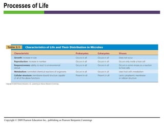
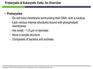
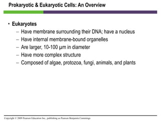
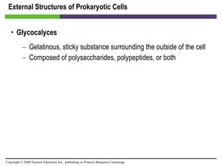
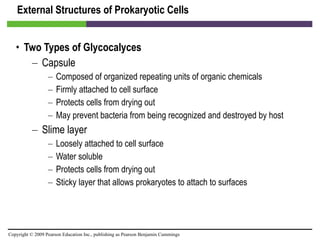
This document provides an overview of cell structure and function, comparing prokaryotic and eukaryotic cells. It describes that prokaryotes lack membrane-bound organelles and a nucleus, while eukaryotes have these structures. The document outlines the external structures of prokaryotic cells such as glycocalyces, flagella, fimbriae and pili. It also describes prokaryotic cell walls, cytoplasmic membranes and internal cell structures. For eukaryotic cells, it summarizes cell walls, cytoplasmic membranes and various membrane-bound and non-membrane bound organelles.
![Prokaryotic & Eukaryotic Cells: An Overview [INSERT FIGURE 3.1]](https://image.slidesharecdn.com/startherech03lecture-100519101818-phpapp02/85/Start-here_ch03_lecture-4-320.jpg)
![Prokaryotic & Eukaryotic Cells: An Overview [INSERT FIGURE 3.2]](https://image.slidesharecdn.com/startherech03lecture-100519101818-phpapp02/85/Start-here_ch03_lecture-6-320.jpg)
![Prokaryotic & Eukaryotic Cells: An Overview [INSERT FIGURE 3.3]](https://image.slidesharecdn.com/startherech03lecture-100519101818-phpapp02/85/Start-here_ch03_lecture-8-320.jpg)
![Prokaryotic & Eukaryotic Cells: An Overview [INSERT FIGURE 3.4]](https://image.slidesharecdn.com/startherech03lecture-100519101818-phpapp02/85/Start-here_ch03_lecture-9-320.jpg)
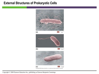
![External Structures of Prokaryotic Cells [INSERT FIGURE 3.5]](https://image.slidesharecdn.com/startherech03lecture-100519101818-phpapp02/85/Start-here_ch03_lecture-12-320.jpg)
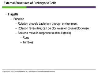
![External Structures of Prokaryotic Cells [INSERT FIGURE 3.6]](https://image.slidesharecdn.com/startherech03lecture-100519101818-phpapp02/85/Start-here_ch03_lecture-17-320.jpg)

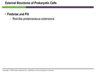
![External Structures of Prokaryotic Cells [INSERT FIGURE 3.8]](https://image.slidesharecdn.com/startherech03lecture-100519101818-phpapp02/85/Start-here_ch03_lecture-20-320.jpg)
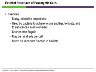
![External Structures of Prokaryotic Cells [INSERT FIGURE 3.9]](https://image.slidesharecdn.com/startherech03lecture-100519101818-phpapp02/85/Start-here_ch03_lecture-22-320.jpg)

![External Structures of Prokaryotic Cells [INSERT FIGURE 3.10]](https://image.slidesharecdn.com/startherech03lecture-100519101818-phpapp02/85/Start-here_ch03_lecture-26-320.jpg)
![External Structures of Prokaryotic Cells [INSERT FIGURE 3.11]](https://image.slidesharecdn.com/startherech03lecture-100519101818-phpapp02/85/Start-here_ch03_lecture-28-320.jpg)
![External Structures of Prokaryotic Cells [INSERT FIGURE 3.12]](https://image.slidesharecdn.com/startherech03lecture-100519101818-phpapp02/85/Start-here_ch03_lecture-31-320.jpg)
![External Structures of Prokaryotic Cells [INSERT FIGURE 3.13]](https://image.slidesharecdn.com/startherech03lecture-100519101818-phpapp02/85/Start-here_ch03_lecture-32-320.jpg)
![Prokaryotic Cell Walls [INSERT FIGURE 3.14a]](https://image.slidesharecdn.com/startherech03lecture-100519101818-phpapp02/85/Start-here_ch03_lecture-34-320.jpg)
![Prokaryotic Cell Walls [INSERT FIGURE 3.14b]](https://image.slidesharecdn.com/startherech03lecture-100519101818-phpapp02/85/Start-here_ch03_lecture-36-320.jpg)

![Prokaryotic Cytoplasmic Membranes [INSERT FIGURE 3.15]](https://image.slidesharecdn.com/startherech03lecture-100519101818-phpapp02/85/Start-here_ch03_lecture-39-320.jpg)
![Prokaryotic Cytoplasmic Membranes [INSERT FIGURE 3.16]](https://image.slidesharecdn.com/startherech03lecture-100519101818-phpapp02/85/Start-here_ch03_lecture-41-320.jpg)
![Prokaryotic Cytoplasmic Membranes [INSERT FIGURE 3.17]](https://image.slidesharecdn.com/startherech03lecture-100519101818-phpapp02/85/Start-here_ch03_lecture-43-320.jpg)
![Prokaryotic Cytoplasmic Membranes [INSERT FIGURE 3.18]](https://image.slidesharecdn.com/startherech03lecture-100519101818-phpapp02/85/Start-here_ch03_lecture-44-320.jpg)
![Prokaryotic Cytoplasmic Membranes [INSERT FIGURE 3.19]](https://image.slidesharecdn.com/startherech03lecture-100519101818-phpapp02/85/Start-here_ch03_lecture-45-320.jpg)
![Prokaryotic Cytoplasmic Membranes [INSERT FIGURE 3.20]](https://image.slidesharecdn.com/startherech03lecture-100519101818-phpapp02/85/Start-here_ch03_lecture-47-320.jpg)
![Prokaryotic Cytoplasmic Membranes [INSERT FIGURE 3.21]](https://image.slidesharecdn.com/startherech03lecture-100519101818-phpapp02/85/Start-here_ch03_lecture-50-320.jpg)
![Prokaryotic Cytoplasmic Membranes [INSERT TABLE 3.2]](https://image.slidesharecdn.com/startherech03lecture-100519101818-phpapp02/85/Start-here_ch03_lecture-51-320.jpg)
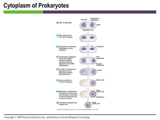
![Cytoplasm of Prokaryotes [INSERT FIGURE 3.23]](https://image.slidesharecdn.com/startherech03lecture-100519101818-phpapp02/85/Start-here_ch03_lecture-55-320.jpg)
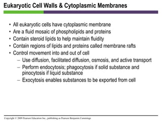
![Eukaryotic Cell Walls & Cytoplasmic Membranes [INSERT FIGURE 3.24]](https://image.slidesharecdn.com/startherech03lecture-100519101818-phpapp02/85/Start-here_ch03_lecture-58-320.jpg)
![Eukaryotic Cell Walls & Cytoplasmic Membranes [INSERT FIGURE 3.25]](https://image.slidesharecdn.com/startherech03lecture-100519101818-phpapp02/85/Start-here_ch03_lecture-60-320.jpg)
![Eukaryotic Cell Walls & Cytoplasmic Membranes [INSERT TABLE 3.3]](https://image.slidesharecdn.com/startherech03lecture-100519101818-phpapp02/85/Start-here_ch03_lecture-61-320.jpg)
![Eukaryotic Cell Walls & Cytoplasmic Membranes [INSERT FIGURE 3.26]](https://image.slidesharecdn.com/startherech03lecture-100519101818-phpapp02/85/Start-here_ch03_lecture-62-320.jpg)
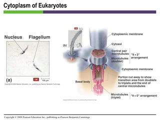
![Cytoplasm of Eukaryotes [INSERT FIGURE 3.28a & b]](https://image.slidesharecdn.com/startherech03lecture-100519101818-phpapp02/85/Start-here_ch03_lecture-66-320.jpg)
![Cytoplasm of Eukaryotes [INSERT FIGURE 3.27c]](https://image.slidesharecdn.com/startherech03lecture-100519101818-phpapp02/85/Start-here_ch03_lecture-68-320.jpg)
![Cytoplasm of Eukaryotes [INSERT FIGURE 3.29]](https://image.slidesharecdn.com/startherech03lecture-100519101818-phpapp02/85/Start-here_ch03_lecture-70-320.jpg)
![Cytoplasm of Eukaryotes [INSERT FIGURE 3.30]](https://image.slidesharecdn.com/startherech03lecture-100519101818-phpapp02/85/Start-here_ch03_lecture-72-320.jpg)
![Cytoplasm of Eukaryotes [INSERT FIGURE 3.31]](https://image.slidesharecdn.com/startherech03lecture-100519101818-phpapp02/85/Start-here_ch03_lecture-74-320.jpg)
![Cytoplasm of Eukaryotes [INSERT FIGURE 3.32]](https://image.slidesharecdn.com/startherech03lecture-100519101818-phpapp02/85/Start-here_ch03_lecture-76-320.jpg)
![Cytoplasm of Eukaryotes [INSERT FIGURE 3.33]](https://image.slidesharecdn.com/startherech03lecture-100519101818-phpapp02/85/Start-here_ch03_lecture-78-320.jpg)
![Cytoplasm of Eukaryotes [INSERT FIGURE 3.34]](https://image.slidesharecdn.com/startherech03lecture-100519101818-phpapp02/85/Start-here_ch03_lecture-80-320.jpg)
![Cytoplasm of Eukaryotes [INSERT FIGURE 3.35]](https://image.slidesharecdn.com/startherech03lecture-100519101818-phpapp02/85/Start-here_ch03_lecture-81-320.jpg)
![Cytoplasm of Eukaryotes [INSERT FIGURE 3.36]](https://image.slidesharecdn.com/startherech03lecture-100519101818-phpapp02/85/Start-here_ch03_lecture-83-320.jpg)
![Cytoplasm of Eukaryotes [INSERT FIGURE 3.37]](https://image.slidesharecdn.com/startherech03lecture-100519101818-phpapp02/85/Start-here_ch03_lecture-85-320.jpg)
![Cytoplasm of Eukaryotes [INSERT TABLE 3.4]](https://image.slidesharecdn.com/startherech03lecture-100519101818-phpapp02/85/Start-here_ch03_lecture-86-320.jpg)
![Cytoplasm of Eukaryotes [INSERT TABLE 3.5]](https://image.slidesharecdn.com/startherech03lecture-100519101818-phpapp02/85/Start-here_ch03_lecture-88-320.jpg)