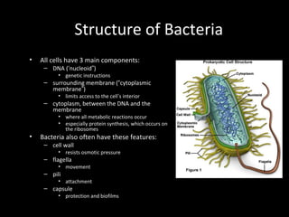
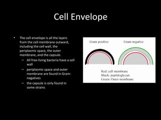
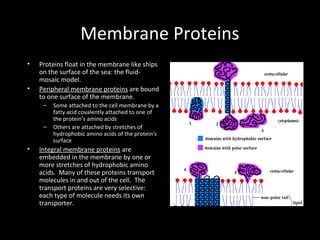
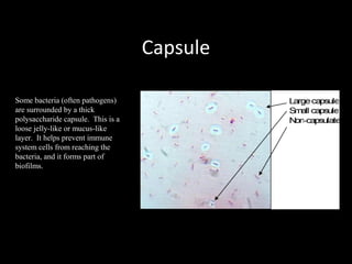
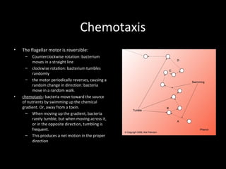
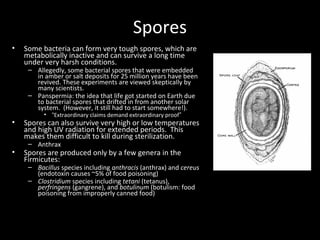
SlideShare a Scribd company logo
Bacterial Structure
Structure of Bacteria
• All cells have 3 main components:
– DNA (‘nucleoid”)
• genetic instructions
– surrounding membrane (“cytoplasmic
membrane”)
• limits access to the cell’s interior
– cytoplasm, between the DNA and the
membrane
• where all metabolic reactions occur
• especially protein synthesis, which occurs on
the ribosomes
• Bacteria also often have these features:
– cell wall
• resists osmotic pressure
– flagella
• movement
– pili
• attachment
– capsule
• protection and biofilms
Cell Envelope
• The cell envelope is all the layers
from the cell membrane outward,
including the cell wall, the
periplasmic space, the outer
membrane, and the capsule.
– All free-living bacteria have a cell
wall
– periplasmic space and outer
membrane are found in Gram-
negatives
– the capsule is only found in
some strains
Cell Membrane
• The cell membrane (often called the plasma
membrane) is composed of 2 layers of
phospholipids.
• Phospholipids have polar heads and non-polar
tails.
– “Polar” implies that the heads are hydrophilic:
they like to stay in an aqueous environment:
facing the outside world and the inside of the
cell.
– “non-polar” means that the tails are
hydrophobic: they want to be away from
water, in an oily environment. The tails are in
the center of the membrane
• A pure phospholipid membrane only allows
water, gasses, and a few small molecules to
move freely through it.
Membrane Proteins
• Proteins float in the membrane like ships
on the surface of the sea: the fluid-
mosaic model.
• Peripheral membrane proteins are bound
to one surface of the membrane.
– Some attached to the cell membrane by a
fatty acid covalently attached to one of
the protein’s amino acids
– Others are attached by stretches of
hydrophobic amino acids of the protein’s
surface
• Integral membrane proteins are
embedded in the membrane by one or
more stretches of hydrophobic amino
acids. Many of these proteins transport
molecules in and out of the cell. The
transport proteins are very selective:
each type of molecule needs its own
transporter.
Transport Across the Cell Membrane
• Basic rule: things spontaneously move
from high concentration to low
concentration (downhill). This process is
called diffusion.
– Getting many molecules into the cell is
simply a matter of opening up a protein
channel of the proper size and shape. The
molecules then move into the cell by
diffusing down the concentration gradient.
Passive transport, or facilitated diffusion.
• To get things to move from low to high
(uphill), you need to add energy: the
molecules must be pumped into the cell.
Pumps are driven by ATP energy. Active
transport.
More Membrane Transport
Often 2 molecules are transported
together, with one moving by
diffusion down its concentration
gradient and the other carried
along up its concentration
gradient.
– If the two molecules move in
the same direction, the
protein channel is a
symporter. See the diagram of
the sodium-glucose symport
mechanism
– If the two molecules move in
opposite directions, the
channel is an antiporter.
Cell Wall
• Osmotic pressure is the force generated by water
attempting to move into the cell.
– Water can go through the cell membrane freely
– The contents of the cell are very concentrated
– Like all things, water moves from areas of high
concentration to areas of low concentration. This
means, water will move from outside the cell
(dilute environment) to inside (concentrated
environment).
– Osmotic pressure can easily cause a cell to swell
up and burst.
• Bacteria, along with plants and fungi, resist
osmotic pressure by surrounding the cell in a
rigid box, the cell wall.
– Composed of peptidoglycan (also called
proteoglycan or murein)
– Long chains of polysaccharide cross-linked by
short peptides (amino acid chains).
• The peptides contain the unusual mirror-image
amino acids D-alanine and D-glutamate
• polysaccharide is composed of alternating “amino
sugars”: N-acetylglucosamine and N-acetylmuramic
acid
More Cell Wall
• Gram-positive vs Gram-negative are defined by the
structure of the cell wall
– the Gram stain binds to peptidoglycan
• Gram-positive: many layers of peptidoglycan, which
is anchored to the cell membrane by teichoic acid.
• Gram-negative: 1-2 layers of peptidoglycan = thin
– The periplasmic space is between the cell membrane
and the cell wall. It contains enzymes and other
proteins, such as chemoreceptors for sensing the
environment.
– Outside the peptidglycan layer is the “outer
membrane”. It is pierced by porins: protein channels,
and its out surface is covered with lipopolysaccharides
(sugars linked to membrane lipids), which are often
antigenic and or toxic.
Capsule
Some bacteria (often pathogens)
are surrounded by a thick
polysaccharide capsule. This is a
loose jelly-like or mucus-like
layer. It helps prevent immune
system cells from reaching the
bacteria, and it forms part of
biofilms.
Membrane Structures
• Pili (singular = pilus) are hairs projecting from the
surface. They are composed of pilin protein. There are
several types:
– DNA can be transferred between bacteria by conjugation,
which is initiated when sex pili on the donor cell attach to
and draw in the recipient cell.
– Fimbriae (singular = fimbria) are pili used to attach the
bacteria to target cells ( in infection) or to surfaces, where
they form a biofilm.
• Flagella are long hairs used to propel the cells. They are
composed of flagellin protein.
– At the base of each flagellum is a motor embedded in the
membrane and cell wall. It turns in a rotary motion,
driven by proton-motive force (the flow of protons i.e. H+
ions across the cell membrane).
– The suffix “-trichous” is used to describe the placement of
flagella: e.g. lophotrichous = several flagella all clustered
at one end.
Chemotaxis
• The flagellar motor is reversible:
– Counterclockwise rotation: bacterium
moves in a straight line
– clockwise rotation: bacterium tumbles
randomly
– the motor periodically reverses, causing a
random change in direction: bacteria
move in a random walk.
• chemotaxis: bacteria move toward the source
of nutrients by swimming up the chemical
gradient. Or, away from a toxin.
– When moving up the gradient, bacteria
rarely tumble, but when moving across it,
or in the opposite direction, tumbling is
frequent.
– This produces a net motion in the proper
direction
Spores
• Some bacteria can form very tough spores, which are
metabolically inactive and can survive a long time
under very harsh conditions.
– Allegedly, some bacterial spores that were embedded
in amber or salt deposits for 25 million years have been
revived. These experiments are viewed skeptically by
many scientists.
– Panspermia: the idea that life got started on Earth due
to bacterial spores that drifted in from another solar
system. (However, it still had to start somewhere!).
• “Extraordinary claims demand extraordinary proof”
• Spores can also survive very high or low temperatures
and high UV radiation for extended periods. This
makes them difficult to kill during sterilization.
– Anthrax
• Spores are produced only by a few genera in the
Firmicutes:
– Bacillus species including anthracis (anthrax) and cereus
(endotoxin causes ~5% of food poisoning)
– Clostridium species including tetani (tetanus),
perfringens (gangrene), and botulinum (botulism: food
poisoning from improperly canned food)

More Related Content

What's hot

IMMUNITY TYPE (ACQUIRED IMMUNITY)
IMMUNITY TYPE (ACQUIRED IMMUNITY)IMMUNITY TYPE (ACQUIRED IMMUNITY)
IMMUNITY TYPE (ACQUIRED IMMUNITY)
GAUDINE PRESNTATION
 
Bacterial morphology
Bacterial morphologyBacterial morphology
Immunity, immune system
Immunity, immune systemImmunity, immune system
Immunity, immune system
Amen Ullah
 
Bacteria, Bacteria Structure
Bacteria, Bacteria StructureBacteria, Bacteria Structure
Bacteria, Bacteria Structure
Umesh Maskare
 
Introduction to Immunology
Introduction to ImmunologyIntroduction to Immunology
Introduction to Immunology
Dr. Amer Ali Khaleel /HMU
 
Bacterial morphology & anatomy
Bacterial morphology & anatomyBacterial morphology & anatomy
Bacterial morphology & anatomy
Kalpesh Zunjarrao
 
Organs of the immune system
Organs of the immune systemOrgans of the immune system
Structure of bacterial cell
Structure of bacterial cellStructure of bacterial cell
Structure of bacterial cell
gohil sanjay bhagvanji
 
Cell wall:Bacteria
Cell wall:BacteriaCell wall:Bacteria
Cell wall:Bacteria
Mohit Hinsu
 
Structure and Functions of the Immune System
Structure and Functions of the Immune SystemStructure and Functions of the Immune System
Structure and Functions of the Immune System
autumnpianist
 
Immunity
ImmunityImmunity
Lymphatic immune system
Lymphatic immune systemLymphatic immune system
Lymphatic immune system
som allul
 
Immune response
Immune responseImmune response
Immune response
Pulipati Sowjanya
 
Hypersensitivity and autoimmunity
Hypersensitivity and autoimmunityHypersensitivity and autoimmunity
Hypersensitivity and autoimmunity
Lubna Abu Alrub,DDS
 
Classification microorganism
Classification microorganismClassification microorganism
Classification microorganism
Dr.Sabeeha M.A
 
Identification of microbes
Identification of microbesIdentification of microbes
Identification of microbes
Dr. sreeremya S
 
Gram negative cocci
Gram negative cocciGram negative cocci
Gram negative cocci
reena condez
 
Bacterial growth curve
Bacterial growth curveBacterial growth curve
Bacterial growth curve
Anup Bajracharya
 
ANTIBODY
ANTIBODYANTIBODY
ANTIBODY
SaiBadri2
 

What's hot (20)

IMMUNITY TYPE (ACQUIRED IMMUNITY)
IMMUNITY TYPE (ACQUIRED IMMUNITY)IMMUNITY TYPE (ACQUIRED IMMUNITY)
IMMUNITY TYPE (ACQUIRED IMMUNITY)
 
Bacterial morphology
Bacterial morphologyBacterial morphology
Bacterial morphology
 
Immunity, immune system
Immunity, immune systemImmunity, immune system
Immunity, immune system
 
Bacteria, Bacteria Structure
Bacteria, Bacteria StructureBacteria, Bacteria Structure
Bacteria, Bacteria Structure
 
Introduction to Immunology
Introduction to ImmunologyIntroduction to Immunology
Introduction to Immunology
 
Bacterial morphology & anatomy
Bacterial morphology & anatomyBacterial morphology & anatomy
Bacterial morphology & anatomy
 
Organs of the immune system
Organs of the immune systemOrgans of the immune system
Organs of the immune system
 
Structure of bacterial cell
Structure of bacterial cellStructure of bacterial cell
Structure of bacterial cell
 
Cell wall:Bacteria
Cell wall:BacteriaCell wall:Bacteria
Cell wall:Bacteria
 
Structure and Functions of the Immune System
Structure and Functions of the Immune SystemStructure and Functions of the Immune System
Structure and Functions of the Immune System
 
Immunity
ImmunityImmunity
Immunity
 
Immune response
Immune responseImmune response
Immune response
 
Lymphatic immune system
Lymphatic immune systemLymphatic immune system
Lymphatic immune system
 
Immune response
Immune responseImmune response
Immune response
 
Hypersensitivity and autoimmunity
Hypersensitivity and autoimmunityHypersensitivity and autoimmunity
Hypersensitivity and autoimmunity
 
Classification microorganism
Classification microorganismClassification microorganism
Classification microorganism
 
Identification of microbes
Identification of microbesIdentification of microbes
Identification of microbes
 
Gram negative cocci
Gram negative cocciGram negative cocci
Gram negative cocci
 
Bacterial growth curve
Bacterial growth curveBacterial growth curve
Bacterial growth curve
 
ANTIBODY
ANTIBODYANTIBODY
ANTIBODY
 

Similar to Bacterial structures

Plasma membrane : cell biology
Plasma membrane : cell biologyPlasma membrane : cell biology
Plasma membrane : cell biology
Gauri Haval
 
Plasma membrane or cell membrane (2).pptx
Plasma membrane or cell membrane (2).pptxPlasma membrane or cell membrane (2).pptx
Plasma membrane or cell membrane (2).pptx
aminagulshan526
 
Biological membranes and transport
Biological membranes and transportBiological membranes and transport
Biological membranes and transport
Bahauddin zakariya university,Multan
 
Ultra structure of bateria
Ultra structure of bateriaUltra structure of bateria
Ultra structure of bateria
HemantKurmi3
 
Chapter 3
Chapter 3Chapter 3
Chapter 3
Yukti Sharma
 
Biological membrane and transport BY Mohammadali
Biological membrane and transport BY Mohammadali Biological membrane and transport BY Mohammadali
Biological membrane and transport BY Mohammadali
mohammadali783
 
Cell & cell membrane
Cell &  cell membraneCell &  cell membrane
Cell & cell membrane
Drmohamed Badr
 
Biological membrane and transport
Biological membrane and transportBiological membrane and transport
Biological membrane and transport
ArchanaSoni3
 
Biological membrane and transport
Biological membrane and transportBiological membrane and transport
Biological membrane and transport
SSMV2016
 
Biological membrane and transport
Biological membrane and transportBiological membrane and transport
Biological membrane and transport
Shri Shankaracharya College, Bhilai,Junwani
 
Cell Membrane Structure-1.pptx cell outer layer
Cell Membrane Structure-1.pptx cell outer layerCell Membrane Structure-1.pptx cell outer layer
Cell Membrane Structure-1.pptx cell outer layer
SalsabeelArif
 
Properties of cells & cell membrane
Properties of cells & cell membraneProperties of cells & cell membrane
Properties of cells & cell membrane
Pulipati Sowjanya
 
Structure of a prokaryotic cell
Structure of a prokaryotic cellStructure of a prokaryotic cell
Structure of a prokaryotic cell
Bahauddin Zakariya University lahore
 
Cell Membrane & ion transport.pptx
Cell Membrane & ion transport.pptxCell Membrane & ion transport.pptx
Cell Membrane & ion transport.pptx
Mira426412
 
Plasma membrane
Plasma membranePlasma membrane
Plasma membrane
SoniaBajaj10
 
Plasma membrane
Plasma membranePlasma membrane
Plasma membrane
SSMV2016
 
Cell membrane.pptx
Cell membrane.pptxCell membrane.pptx
Cell membrane.pptx
Dr. Sarita Sharma
 
Cell and their function.pptx
Cell and their function.pptxCell and their function.pptx
Cell and their function.pptx
Jagruti Marathe
 
BMP 111 LECTURE 3&4.pptx
BMP 111 LECTURE 3&4.pptxBMP 111 LECTURE 3&4.pptx
BMP 111 LECTURE 3&4.pptx
SaadMohdBello
 
A&P Chapter 03
A&P Chapter 03A&P Chapter 03
A&P Chapter 03
Michael Walls
 

Similar to Bacterial structures (20)

Plasma membrane : cell biology
Plasma membrane : cell biologyPlasma membrane : cell biology
Plasma membrane : cell biology
 
Plasma membrane or cell membrane (2).pptx
Plasma membrane or cell membrane (2).pptxPlasma membrane or cell membrane (2).pptx
Plasma membrane or cell membrane (2).pptx
 
Biological membranes and transport
Biological membranes and transportBiological membranes and transport
Biological membranes and transport
 
Ultra structure of bateria
Ultra structure of bateriaUltra structure of bateria
Ultra structure of bateria
 
Chapter 3
Chapter 3Chapter 3
Chapter 3
 
Biological membrane and transport BY Mohammadali
Biological membrane and transport BY Mohammadali Biological membrane and transport BY Mohammadali
Biological membrane and transport BY Mohammadali
 
Cell & cell membrane
Cell &  cell membraneCell &  cell membrane
Cell & cell membrane
 
Biological membrane and transport
Biological membrane and transportBiological membrane and transport
Biological membrane and transport
 
Biological membrane and transport
Biological membrane and transportBiological membrane and transport
Biological membrane and transport
 
Biological membrane and transport
Biological membrane and transportBiological membrane and transport
Biological membrane and transport
 
Cell Membrane Structure-1.pptx cell outer layer
Cell Membrane Structure-1.pptx cell outer layerCell Membrane Structure-1.pptx cell outer layer
Cell Membrane Structure-1.pptx cell outer layer
 
Properties of cells & cell membrane
Properties of cells & cell membraneProperties of cells & cell membrane
Properties of cells & cell membrane
 
Structure of a prokaryotic cell
Structure of a prokaryotic cellStructure of a prokaryotic cell
Structure of a prokaryotic cell
 
Cell Membrane & ion transport.pptx
Cell Membrane & ion transport.pptxCell Membrane & ion transport.pptx
Cell Membrane & ion transport.pptx
 
Plasma membrane
Plasma membranePlasma membrane
Plasma membrane
 
Plasma membrane
Plasma membranePlasma membrane
Plasma membrane
 
Cell membrane.pptx
Cell membrane.pptxCell membrane.pptx
Cell membrane.pptx
 
Cell and their function.pptx
Cell and their function.pptxCell and their function.pptx
Cell and their function.pptx
 
BMP 111 LECTURE 3&4.pptx
BMP 111 LECTURE 3&4.pptxBMP 111 LECTURE 3&4.pptx
BMP 111 LECTURE 3&4.pptx
 
A&P Chapter 03
A&P Chapter 03A&P Chapter 03
A&P Chapter 03
 

Bacterial structures

  • 2. Structure of Bacteria • All cells have 3 main components: – DNA (‘nucleoid”) • genetic instructions – surrounding membrane (“cytoplasmic membrane”) • limits access to the cell’s interior – cytoplasm, between the DNA and the membrane • where all metabolic reactions occur • especially protein synthesis, which occurs on the ribosomes • Bacteria also often have these features: – cell wall • resists osmotic pressure – flagella • movement – pili • attachment – capsule • protection and biofilms
  • 3. Cell Envelope • The cell envelope is all the layers from the cell membrane outward, including the cell wall, the periplasmic space, the outer membrane, and the capsule. – All free-living bacteria have a cell wall – periplasmic space and outer membrane are found in Gram- negatives – the capsule is only found in some strains
  • 4. Cell Membrane • The cell membrane (often called the plasma membrane) is composed of 2 layers of phospholipids. • Phospholipids have polar heads and non-polar tails. – “Polar” implies that the heads are hydrophilic: they like to stay in an aqueous environment: facing the outside world and the inside of the cell. – “non-polar” means that the tails are hydrophobic: they want to be away from water, in an oily environment. The tails are in the center of the membrane • A pure phospholipid membrane only allows water, gasses, and a few small molecules to move freely through it.
  • 5. Membrane Proteins • Proteins float in the membrane like ships on the surface of the sea: the fluid- mosaic model. • Peripheral membrane proteins are bound to one surface of the membrane. – Some attached to the cell membrane by a fatty acid covalently attached to one of the protein’s amino acids – Others are attached by stretches of hydrophobic amino acids of the protein’s surface • Integral membrane proteins are embedded in the membrane by one or more stretches of hydrophobic amino acids. Many of these proteins transport molecules in and out of the cell. The transport proteins are very selective: each type of molecule needs its own transporter.
  • 6. Transport Across the Cell Membrane • Basic rule: things spontaneously move from high concentration to low concentration (downhill). This process is called diffusion. – Getting many molecules into the cell is simply a matter of opening up a protein channel of the proper size and shape. The molecules then move into the cell by diffusing down the concentration gradient. Passive transport, or facilitated diffusion. • To get things to move from low to high (uphill), you need to add energy: the molecules must be pumped into the cell. Pumps are driven by ATP energy. Active transport.
  • 7. More Membrane Transport Often 2 molecules are transported together, with one moving by diffusion down its concentration gradient and the other carried along up its concentration gradient. – If the two molecules move in the same direction, the protein channel is a symporter. See the diagram of the sodium-glucose symport mechanism – If the two molecules move in opposite directions, the channel is an antiporter.
  • 8. Cell Wall • Osmotic pressure is the force generated by water attempting to move into the cell. – Water can go through the cell membrane freely – The contents of the cell are very concentrated – Like all things, water moves from areas of high concentration to areas of low concentration. This means, water will move from outside the cell (dilute environment) to inside (concentrated environment). – Osmotic pressure can easily cause a cell to swell up and burst. • Bacteria, along with plants and fungi, resist osmotic pressure by surrounding the cell in a rigid box, the cell wall. – Composed of peptidoglycan (also called proteoglycan or murein) – Long chains of polysaccharide cross-linked by short peptides (amino acid chains). • The peptides contain the unusual mirror-image amino acids D-alanine and D-glutamate • polysaccharide is composed of alternating “amino sugars”: N-acetylglucosamine and N-acetylmuramic acid
  • 9. More Cell Wall • Gram-positive vs Gram-negative are defined by the structure of the cell wall – the Gram stain binds to peptidoglycan • Gram-positive: many layers of peptidoglycan, which is anchored to the cell membrane by teichoic acid. • Gram-negative: 1-2 layers of peptidoglycan = thin – The periplasmic space is between the cell membrane and the cell wall. It contains enzymes and other proteins, such as chemoreceptors for sensing the environment. – Outside the peptidglycan layer is the “outer membrane”. It is pierced by porins: protein channels, and its out surface is covered with lipopolysaccharides (sugars linked to membrane lipids), which are often antigenic and or toxic.
  • 10. Capsule Some bacteria (often pathogens) are surrounded by a thick polysaccharide capsule. This is a loose jelly-like or mucus-like layer. It helps prevent immune system cells from reaching the bacteria, and it forms part of biofilms.
  • 11. Membrane Structures • Pili (singular = pilus) are hairs projecting from the surface. They are composed of pilin protein. There are several types: – DNA can be transferred between bacteria by conjugation, which is initiated when sex pili on the donor cell attach to and draw in the recipient cell. – Fimbriae (singular = fimbria) are pili used to attach the bacteria to target cells ( in infection) or to surfaces, where they form a biofilm. • Flagella are long hairs used to propel the cells. They are composed of flagellin protein. – At the base of each flagellum is a motor embedded in the membrane and cell wall. It turns in a rotary motion, driven by proton-motive force (the flow of protons i.e. H+ ions across the cell membrane). – The suffix “-trichous” is used to describe the placement of flagella: e.g. lophotrichous = several flagella all clustered at one end.
  • 12. Chemotaxis • The flagellar motor is reversible: – Counterclockwise rotation: bacterium moves in a straight line – clockwise rotation: bacterium tumbles randomly – the motor periodically reverses, causing a random change in direction: bacteria move in a random walk. • chemotaxis: bacteria move toward the source of nutrients by swimming up the chemical gradient. Or, away from a toxin. – When moving up the gradient, bacteria rarely tumble, but when moving across it, or in the opposite direction, tumbling is frequent. – This produces a net motion in the proper direction
  • 13. Spores • Some bacteria can form very tough spores, which are metabolically inactive and can survive a long time under very harsh conditions. – Allegedly, some bacterial spores that were embedded in amber or salt deposits for 25 million years have been revived. These experiments are viewed skeptically by many scientists. – Panspermia: the idea that life got started on Earth due to bacterial spores that drifted in from another solar system. (However, it still had to start somewhere!). • “Extraordinary claims demand extraordinary proof” • Spores can also survive very high or low temperatures and high UV radiation for extended periods. This makes them difficult to kill during sterilization. – Anthrax • Spores are produced only by a few genera in the Firmicutes: – Bacillus species including anthracis (anthrax) and cereus (endotoxin causes ~5% of food poisoning) – Clostridium species including tetani (tetanus), perfringens (gangrene), and botulinum (botulism: food poisoning from improperly canned food)