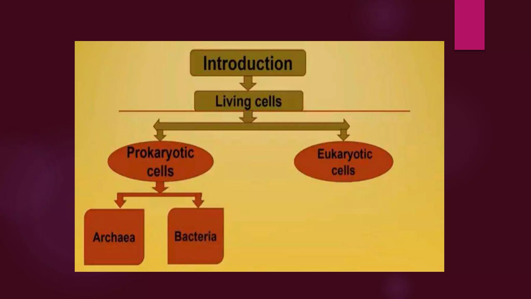
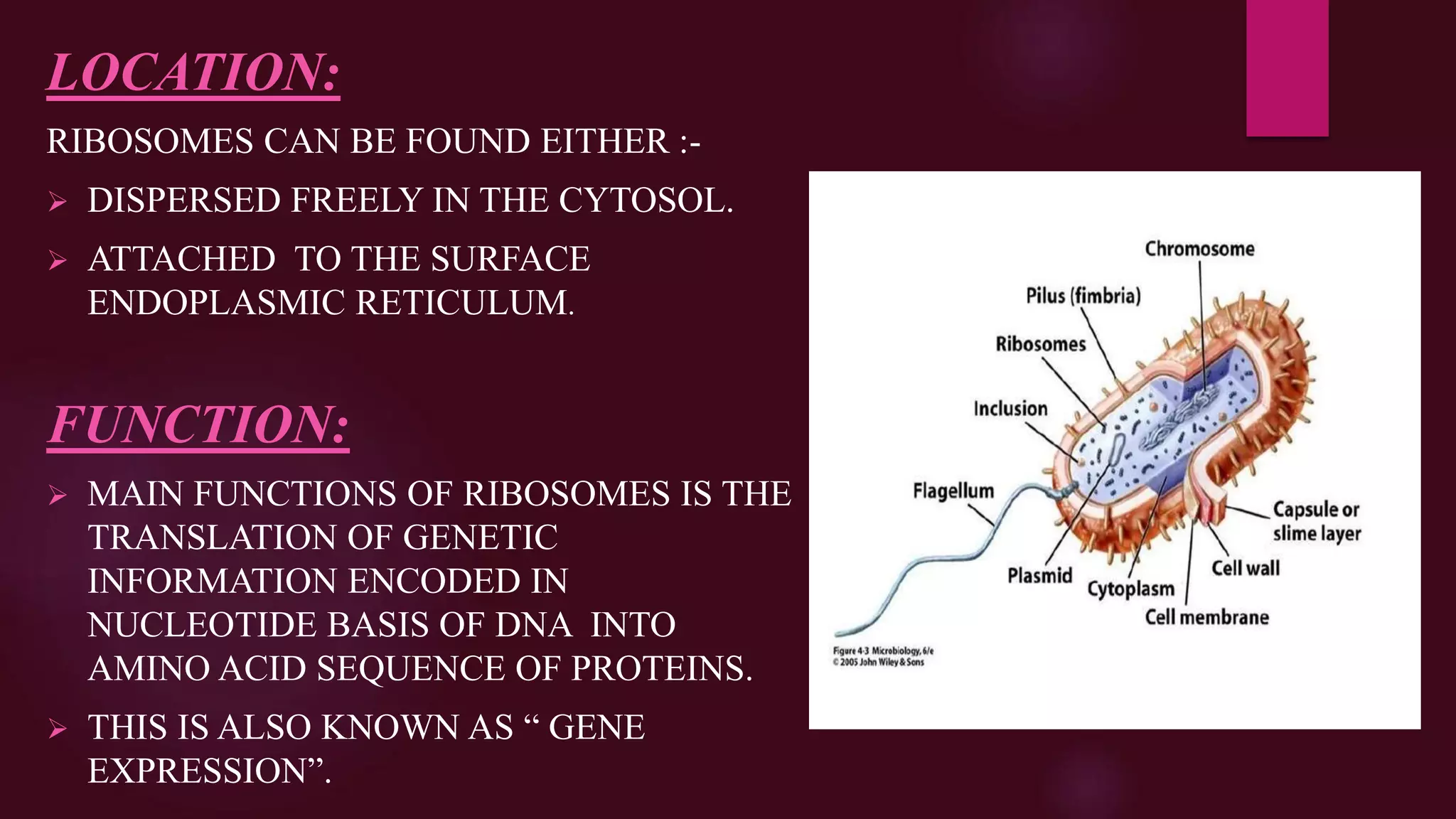
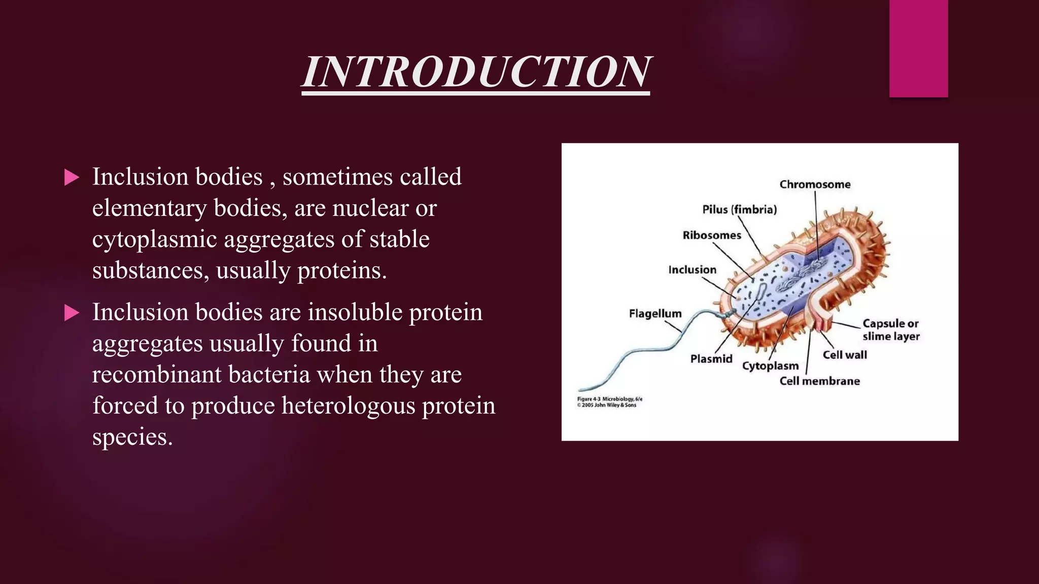
Prokaryotic cell
BY:
SARAH HAMMAD
INTRODUCTION
 The word prokaryotes is derived from Greek words
Pro means before or primitive and Karyon means nucleus.
 Cells containing false nucleus or lack an organized nucleus are
called as Prokaryotes. The organisms containing prokaryotic cells
are called as Prokaryotes.
 Prokaryotic cells are mainly classified into
1. Bacteria
2. Archae
 Ancient type of cells.
 Prokaryotic cells are almost similar in morphology with eukaryotic
cells but the major difference is in genetics.
STRUCTURE OF PROKARYOTIC CELLS
 Lacks membrane bound organelles.
 Chloroplast, Golgi bodies, Mitochondria,
Endoplasmic reticulum and Lysosomes are absent.
 Respiratory system is present in plasma membrane.
 Mesosomes, endospores and magnetosomes may or
may not be present in prokaryotic cells.
 Ribosomes are of 70S size and are dispersed in
cytoplasm.
 Protein synthesis is carried out in cytoplasm.
 Plasma membrane lack sterols.
THE NUCLEUS
 Lack of nuclear membrane, histone proteins and
nucleolus is also absent.
 Genetic material is located in nuclear region or
nucleoid.
 Nucleoid contains single chromosome and piece of
circular double stranded DNA. The chromosome is
circular as well.
 Genes are present in form of clusters.
 The G+C content is about 20 to 70%.
 Mitosis and meosis mechanism is absent
 Reproduction through binary fission mechanism.
 Have ability to fix atmospheric nitrogen.
 Shows wide variety of anaerobic energy yielding
mechanism.
 The cell wall composed of:
1. Peptidoglycan
2. Polysaccharide
3. Glycoprotein
 Divided into gram +ve and gram –ve
 Contains flagella, pili, capsule on its surface.
Size of prokaryotic cell
 Average size of prokaryotic cells ranges from 0.2 to 0.5 micro
meters in diameter.
 Size ranges in nano and ultra micro meters as well.
 The small size of prokaryotic cells allows ions and organic
molecules that enter them to quickly spread to other parts of cell.
 The largest prokaryote discovered till now is Thiomargarita
namibiensis which is gram –ve bacteria ranges from 0.1 to
0.33mm (100-300 micrometrer) sometimes ataining size upto
0.75mm.
Plasma Membrane
CONTENTS
 What is plasma membrane ?
 How could youe define plasma membrane ?
 Structure
 Fluide mosaic model
 Overview
 Membrane protein
 Chemical composition
 Functions and Movement
What is plasma membrane ?
1. Outermost layer of animal cell
2. Thickness is 5-8 Nm
3. Selectively permeable membrane
4. Serve as outer boundary
5. Allow some substances to cross more easily than others
6. Made of phospholipids, proteins and conjugated molecules
7. Protect cell from external environment
8. Also called cell membrane
How could you define plasma
membrane ?
“An outermost envelope surrounding the cell that
separate
and protects the cell from external environment and
provides
a connecting system between the cell and its
environment.”
Structure of Plasma Membrane
The plasma membrane is
Composed of two layers Of
“phospholipids back to back”.
Phospholipids are lipids attached
to them.
Phospholipids
 Fatty acid tails hydrophobic
 Phosphate group head hydrophilic
 Arranged as bilayer
“Phospholipids, cholesterol and glycolipids
are amphiphatic lipids composed of Hydrophobic and
Hydrophilic ends.”
Phospholipid Bilayer :
Fluid Mosaic Model
 In 1972 S.J. Singer and G. Nicolson proposed Fluid Mosaic model
What is Fluid Mosaic Model ?
“Cell membrane is lipid bilayer in which
proteins are partially embeded like
floating Iceburge in sea.
“The fluid mosaic model desribes the
plasma membrane as a flexible boundary
of a cell. The phospholipid moves within
the cells “
Membrane Proteins
Membrane proteins are categorized into following
1. Extrinsic (peripheral ) membrane
2. Intrinsic (internal) membrane
Extrinsic Membrane protein
 Membrane loosely associated with membrane Surface
 Located entirely outside of the lipid bilayer
 Either on the extracellular or Cytoplasmic surface
 Examples
1. Cytochrome C of Mitochondria
2. Cell surface identity marker (antigens )
Intrinsic Membrane Protein
 Directly incorporated within the lipid bilayer
 Tightly bound to the lipid bilayer
 Provide channel for water soluble substances
Examples
1. Transmembrane proteins
2. Transport proteins
3. Channels , pumps
Plasma Membrane protein
(Transmembrane)
Chemical Composition
 Composed of lipid , protein and carbohydrates
 Actual component differs from tissue to tissue
Lipids of cell membrane are :
1. Phospholipids
2. Glycolipids
3. Sterols
4. Cholesterol
Functions
Movement
 Moving large molecules in
And out of the cell through vesicles
And vacuoles.
Endocytosis
1. Phagocytosis
2. Pinocytosis
Exocytosis
RIBOSOMES
DEFINITION:
RIBOSOMES COMPRISE A COMPLEX MACROMOLECULAR MACHINE
FOUND WITHIN ALL LIVING CELLS THAT SERVES AS A SITE OF
BIOLOGICAL PROTEIN SYNTHESIS.
 RIBOSOMES LINK AMINO ACIDS TOGETHER IN THE ORDER
SPECIFIED BY MESSENGER RNA.
DISCOVERY:
 RIBOSOMES WERE FIRST OBSERVED IN MID 1950S BY
ROMANIAN-AMERICAN CELL BIOLOGIST GEORGE EMIL PALADE
USING AN ELECTRON MICROSCOPE ; AS DENSE PARTICLES OR
GRANULES.
 TERM “RIBOSOMES” WAS PROPOSED BY RICHARD B . ROBERTS
IN END OF 1950S.
STRUCTURE:
 HIGHLY COMPLEXED STRUCTURE CELLULAR MACHINE.
 IT IS LARGELY MADE UP OF TWO MAIN COMPONENTS :-
 PROTEINS = 25-40%
 RRNA =37-62%
 TWO MAIN SUBUNITS ARE PRESENT.
A SMALLER SUBUNIT (CONSISTS OF SINGLE RNA STRAND).
A LARGER SUBUNIT (CONSISTS OF TWO STRANDS, A LONGER
AND A SHORTER STRAND WRAP UPON EACH OTHER).
 EUKARYOTIC SUBUNIT CONTAINS LARGER SUBUNIT 60S
AND SMALLER SUBUNITS 40S , TOTAL RIBOSOMAL
COMPLEX=80S
 PROKARYOTIC SUBUNIT CONTAINS LARGER SUBUNIT 50S
AND SMALLER SUBUNIT 30S AND TOTAL RIBOSOMAL
COMPLEX =70S
LOCATION:
RIBOSOMES CAN BE FOUND EITHER :-
 DISPERSED FREELY IN THE CYTOSOL.
 ATTACHED TO THE SURFACE
ENDOPLASMIC RETICULUM.
FUNCTION:
 MAIN FUNCTIONS OF RIBOSOMES IS THE
TRANSLATION OF GENETIC
INFORMATION ENCODED IN
NUCLEOTIDE BASIS OF DNA INTO
AMINO ACID SEQUENCE OF PROTEINS.
 THIS IS ALSO KNOWN AS “ GENE
EXPRESSION”.
RIBOSOME ASSOCIATED DISEASES:-
 DURING STRESS CONDITIONS , RIBOSOMES ON
ENDOPLASMIC RETICULUM FUNCTION IMPROPERLY.
 IMPROPER FUNCTIONING IS CHARACTERIZED BY
IMPROPER FOLDING OF PROTEINS.
SOME OF SUCH DISEASES ARE :-
 DIABETES
 CYSTIC FIBROSIS
INCLUSION BODIES IN BACTERIA
INTRODUCTION
 Inclusion bodies , sometimes called
elementary bodies, are nuclear or
cytoplasmic aggregates of stable
substances, usually proteins.
 Inclusion bodies are insoluble protein
aggregates usually found in
recombinant bacteria when they are
forced to produce heterologous protein
species.
INCLUSION BODIES
 Inclusion bodies are basically used to store variety of carbon
compounds, inorganic substances and energy.
 They also reduce osmotic pressure by tying up molecules in
particular manner.
EXAPMLES:
 Poly-hydroxybutyrate granules.
 Gas vacuoles.
 Sulphur Granules.
 Carboxysomes.
 Glycogens
TYPES OF INCLUSION BODIES IN
BACTERIA
Cyanophycins Granules:
 Present in Cyanobacteria.
 Are of large polypeptides.
 Contain equal amount of Amino acids i.e. Arginine
and Aspartic Acid.
Carboxysomes:
 Present in Cyanobacteria, Nitrifying bacteria,
Thiobacilli.
 Contain enzyme Ribulose-1,5-bisphosphate
carboxylase.
 Serve as reserve of this enzyme and site for C02
Fixation.
Gas Vacuoles:
 They are organic inclusion bodies.
 Present in Cyanobacteria, Purple and
Green photosynthetic bacteria.
 They are aggregates of enormous
number of small hollow, cylindrical gas
vesicles.
 They are composed of single small
protein.
 It is impermeable to water but freely
permeable to atmospheric gases.

Prokaryotic cell Of Bacteria

  • 1.
  • 3.
    INTRODUCTION  The wordprokaryotes is derived from Greek words Pro means before or primitive and Karyon means nucleus.  Cells containing false nucleus or lack an organized nucleus are called as Prokaryotes. The organisms containing prokaryotic cells are called as Prokaryotes.  Prokaryotic cells are mainly classified into 1. Bacteria 2. Archae  Ancient type of cells.  Prokaryotic cells are almost similar in morphology with eukaryotic cells but the major difference is in genetics.
  • 5.
    STRUCTURE OF PROKARYOTICCELLS  Lacks membrane bound organelles.  Chloroplast, Golgi bodies, Mitochondria, Endoplasmic reticulum and Lysosomes are absent.  Respiratory system is present in plasma membrane.  Mesosomes, endospores and magnetosomes may or may not be present in prokaryotic cells.  Ribosomes are of 70S size and are dispersed in cytoplasm.  Protein synthesis is carried out in cytoplasm.  Plasma membrane lack sterols.
  • 7.
    THE NUCLEUS  Lackof nuclear membrane, histone proteins and nucleolus is also absent.  Genetic material is located in nuclear region or nucleoid.  Nucleoid contains single chromosome and piece of circular double stranded DNA. The chromosome is circular as well.  Genes are present in form of clusters.  The G+C content is about 20 to 70%.  Mitosis and meosis mechanism is absent  Reproduction through binary fission mechanism.
  • 9.
     Have abilityto fix atmospheric nitrogen.  Shows wide variety of anaerobic energy yielding mechanism.  The cell wall composed of: 1. Peptidoglycan 2. Polysaccharide 3. Glycoprotein  Divided into gram +ve and gram –ve  Contains flagella, pili, capsule on its surface.
  • 11.
    Size of prokaryoticcell  Average size of prokaryotic cells ranges from 0.2 to 0.5 micro meters in diameter.  Size ranges in nano and ultra micro meters as well.  The small size of prokaryotic cells allows ions and organic molecules that enter them to quickly spread to other parts of cell.  The largest prokaryote discovered till now is Thiomargarita namibiensis which is gram –ve bacteria ranges from 0.1 to 0.33mm (100-300 micrometrer) sometimes ataining size upto 0.75mm.
  • 14.
  • 15.
    CONTENTS  What isplasma membrane ?  How could youe define plasma membrane ?  Structure  Fluide mosaic model  Overview  Membrane protein  Chemical composition  Functions and Movement
  • 16.
    What is plasmamembrane ? 1. Outermost layer of animal cell 2. Thickness is 5-8 Nm 3. Selectively permeable membrane 4. Serve as outer boundary 5. Allow some substances to cross more easily than others 6. Made of phospholipids, proteins and conjugated molecules 7. Protect cell from external environment 8. Also called cell membrane
  • 17.
    How could youdefine plasma membrane ? “An outermost envelope surrounding the cell that separate and protects the cell from external environment and provides a connecting system between the cell and its environment.”
  • 18.
    Structure of PlasmaMembrane The plasma membrane is Composed of two layers Of “phospholipids back to back”. Phospholipids are lipids attached to them.
  • 19.
    Phospholipids  Fatty acidtails hydrophobic  Phosphate group head hydrophilic  Arranged as bilayer “Phospholipids, cholesterol and glycolipids are amphiphatic lipids composed of Hydrophobic and Hydrophilic ends.”
  • 20.
  • 21.
    Fluid Mosaic Model In 1972 S.J. Singer and G. Nicolson proposed Fluid Mosaic model
  • 22.
    What is FluidMosaic Model ? “Cell membrane is lipid bilayer in which proteins are partially embeded like floating Iceburge in sea. “The fluid mosaic model desribes the plasma membrane as a flexible boundary of a cell. The phospholipid moves within the cells “
  • 23.
    Membrane Proteins Membrane proteinsare categorized into following 1. Extrinsic (peripheral ) membrane 2. Intrinsic (internal) membrane
  • 24.
    Extrinsic Membrane protein Membrane loosely associated with membrane Surface  Located entirely outside of the lipid bilayer  Either on the extracellular or Cytoplasmic surface  Examples 1. Cytochrome C of Mitochondria 2. Cell surface identity marker (antigens )
  • 25.
    Intrinsic Membrane Protein Directly incorporated within the lipid bilayer  Tightly bound to the lipid bilayer  Provide channel for water soluble substances Examples 1. Transmembrane proteins 2. Transport proteins 3. Channels , pumps
  • 26.
  • 27.
    Chemical Composition  Composedof lipid , protein and carbohydrates  Actual component differs from tissue to tissue Lipids of cell membrane are : 1. Phospholipids 2. Glycolipids 3. Sterols 4. Cholesterol
  • 28.
  • 29.
    Movement  Moving largemolecules in And out of the cell through vesicles And vacuoles. Endocytosis 1. Phagocytosis 2. Pinocytosis Exocytosis
  • 30.
  • 31.
    DEFINITION: RIBOSOMES COMPRISE ACOMPLEX MACROMOLECULAR MACHINE FOUND WITHIN ALL LIVING CELLS THAT SERVES AS A SITE OF BIOLOGICAL PROTEIN SYNTHESIS.  RIBOSOMES LINK AMINO ACIDS TOGETHER IN THE ORDER SPECIFIED BY MESSENGER RNA. DISCOVERY:  RIBOSOMES WERE FIRST OBSERVED IN MID 1950S BY ROMANIAN-AMERICAN CELL BIOLOGIST GEORGE EMIL PALADE USING AN ELECTRON MICROSCOPE ; AS DENSE PARTICLES OR GRANULES.  TERM “RIBOSOMES” WAS PROPOSED BY RICHARD B . ROBERTS IN END OF 1950S.
  • 32.
    STRUCTURE:  HIGHLY COMPLEXEDSTRUCTURE CELLULAR MACHINE.  IT IS LARGELY MADE UP OF TWO MAIN COMPONENTS :-  PROTEINS = 25-40%  RRNA =37-62%  TWO MAIN SUBUNITS ARE PRESENT. A SMALLER SUBUNIT (CONSISTS OF SINGLE RNA STRAND). A LARGER SUBUNIT (CONSISTS OF TWO STRANDS, A LONGER AND A SHORTER STRAND WRAP UPON EACH OTHER).  EUKARYOTIC SUBUNIT CONTAINS LARGER SUBUNIT 60S AND SMALLER SUBUNITS 40S , TOTAL RIBOSOMAL COMPLEX=80S  PROKARYOTIC SUBUNIT CONTAINS LARGER SUBUNIT 50S AND SMALLER SUBUNIT 30S AND TOTAL RIBOSOMAL COMPLEX =70S
  • 33.
    LOCATION: RIBOSOMES CAN BEFOUND EITHER :-  DISPERSED FREELY IN THE CYTOSOL.  ATTACHED TO THE SURFACE ENDOPLASMIC RETICULUM. FUNCTION:  MAIN FUNCTIONS OF RIBOSOMES IS THE TRANSLATION OF GENETIC INFORMATION ENCODED IN NUCLEOTIDE BASIS OF DNA INTO AMINO ACID SEQUENCE OF PROTEINS.  THIS IS ALSO KNOWN AS “ GENE EXPRESSION”.
  • 34.
    RIBOSOME ASSOCIATED DISEASES:- DURING STRESS CONDITIONS , RIBOSOMES ON ENDOPLASMIC RETICULUM FUNCTION IMPROPERLY.  IMPROPER FUNCTIONING IS CHARACTERIZED BY IMPROPER FOLDING OF PROTEINS. SOME OF SUCH DISEASES ARE :-  DIABETES  CYSTIC FIBROSIS
  • 35.
  • 36.
    INTRODUCTION  Inclusion bodies, sometimes called elementary bodies, are nuclear or cytoplasmic aggregates of stable substances, usually proteins.  Inclusion bodies are insoluble protein aggregates usually found in recombinant bacteria when they are forced to produce heterologous protein species.
  • 37.
    INCLUSION BODIES  Inclusionbodies are basically used to store variety of carbon compounds, inorganic substances and energy.  They also reduce osmotic pressure by tying up molecules in particular manner. EXAPMLES:  Poly-hydroxybutyrate granules.  Gas vacuoles.  Sulphur Granules.  Carboxysomes.  Glycogens
  • 39.
    TYPES OF INCLUSIONBODIES IN BACTERIA Cyanophycins Granules:  Present in Cyanobacteria.  Are of large polypeptides.  Contain equal amount of Amino acids i.e. Arginine and Aspartic Acid.
  • 40.
    Carboxysomes:  Present inCyanobacteria, Nitrifying bacteria, Thiobacilli.  Contain enzyme Ribulose-1,5-bisphosphate carboxylase.  Serve as reserve of this enzyme and site for C02 Fixation.
  • 41.
    Gas Vacuoles:  Theyare organic inclusion bodies.  Present in Cyanobacteria, Purple and Green photosynthetic bacteria.  They are aggregates of enormous number of small hollow, cylindrical gas vesicles.  They are composed of single small protein.  It is impermeable to water but freely permeable to atmospheric gases.