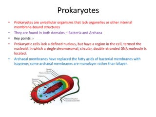
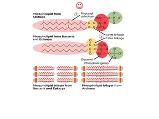
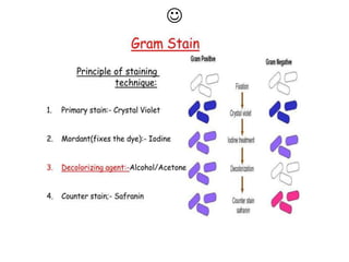
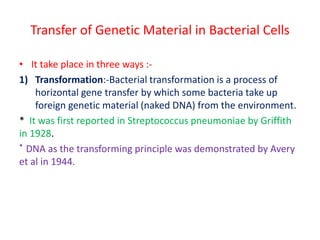
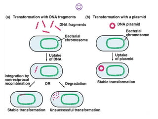
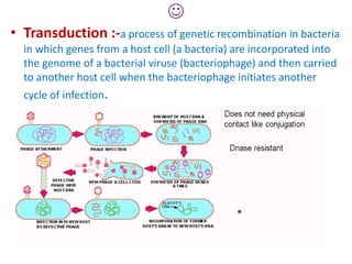
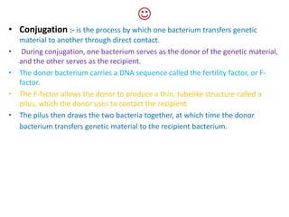
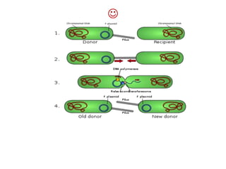
Prokaryotes are unicellular organisms that lack internal membrane-bound structures. They are divided into bacteria and archaea. Prokaryotic cells lack a nucleus but contain a nucleoid region where DNA is located. Archaeal membranes contain isoprene lipids instead of fatty acids found in bacteria. Prokaryotes reproduce through binary fission and exchange genetic material through transformation, transduction, and conjugation.
Cell as basic unit of life ppt 88 slidesICHHA PURAK
This Power point presentation describes Cell as basic unit of life. The slides provide information about Discovery of cell,cell theory,number,size,shape and cell types .Differentiates prokaryotic and eukaryotic cell types and point out major differences in plant and animal cell and also about structure and function of cell organelles
A fimbria (Latin for 'fringe', plural fimbriae), also referred to as an "attachment pilus" by some scientists, is an appendage that can be found on many Gram-negative and some Gram-positive bacteria, that is thinner and shorter than a flagellum. This appendage ranges from 3–10 nanometers in diameter and can be up to several micrometers long. Fimbriae are used by bacteria to adhere to one another and to adhere to animal cells and some inanimate objects. A bacterium can have as many as 1,000 fimbriae. Fimbriae are only visible with the use of an electron microscope. They may be straight or flexible.
A pilus (Latin for 'hair'; plural: pili) is a hair-like appendage found on the surface of many bacteria and archaea.[1] The terms pilus and fimbria (Latin for 'fringe'; plural: fimbriae) can be used interchangeably, although some researchers reserve the term pilus for the appendage required for bacterial conjugation. All pili in the latter sense are primarily composed of pilin proteins, which are oligomeric.
FOLLOW US ON YOUTUBE # BIOTECH SIMPLIFIED #
The archaebacteria
group members
Rameen nadeem
Syeda iqra hussain
Hina zamir
Mahnoor khan
Maleeha inayat
Background
Biologists have long organized living things into large groups called kingdoms.
There are six of them:
Archaebacteria
Eubacteria
Protista
Fungi
Plantae
Animalia
Some recent findings…
In 1996, scientists decided to split Monera into two groups of bacteria:
Archaebacteria and Eubacteria
Because these two groups of bacteria were different in many ways scientists created a new level of classification called a DOMAIN.
Now we have 3 domains
Bacteria
Archaea
Eukarya
KingdomArchaebacteria
Any of a large group of primitive bacteria having unusual cell walls, membrane lipids, ribosomes, and RNA sequences, and having the ability to produce methane and to live in anaerobic, extremely hot, salty, or acidic conditions
The Domain Archaea
“ancient” bacteria
Some of the first archaebacteria were discovered in Yellowstone National Park’s hot springs
Prokaryotes are structurally simple, but biochemically complex
Basic Facts
They live in extreme environments (like hot springs or salty lakes) and normal environments (like soil and ocean water).
All are unicellular (each individual is only one cell).
No peptidoglycan in their cell wall.
Some have a flagella that aids in their locomotion.
Most don’t need oxygen to survive
They can produce ATP (energy) from sunlight
They can survive enormous temperature extremes
They can survive under rocks and in ocean floor vents deep below the ocean’s surface
They can tolerate huge pressure differences
STRUCTURE
Size
Archaea are slightly less than 1 micron long.
A micron is 1/1,000 of a millimeter.
In order to see their cellular features, scientists use powerful electron microscopes.
Shape
Shapes can be spherical or ball shaped and are called coccus.
Others are rod shaped, long and thin, and labeled bacillus.
Variations of cells have been discovered in square and triangular shapes.
STRUCTURE
Locomotion
Some archaea have flagella, hair-like structures that assist in movement.
There can be one or many attached to the cell's outer membrane. Protein networks can also be found on the cell membrane, which allow cells to attach themselves in groups.
Cell Features
Within the cell membrane, the archaea cell contains cytoplasm and DNA, which are in single-looped forms called plasmids.
Most archaeal cells also have a semi-rigid cell wall that helps it to maintain its shape and chemical balance.
This protects the cytoplasm, which is the semi-liquid gel that fills the cell and enables the various parts to function.
STRUCTURE
Phospholipids
The molecules that make up cell membranes are called phospholipids, which act as building blocks for the cell.
In archaea, these molecules are made of glycerol-ether lipids.
Ether Bonding
The ether bonding makes it possible for archaea to survive in environments that are extremely acidic or al
Fungi are a kingdom of usually multicellular eukaryotic organisms that are heterotrophs (cannot make their own food) and have important roles in nutrient cycling in an ecosystem. Fungi reproduce both sexually and asexually, and they also have symbiotic associations with plants and bacteria.
Powerpoint on viruses, bacteria, protists and Fungi. Intended for the SA Grade 11 Life Sciences syllabus. Includes information on HIV, virus reproduction, malaria, TB, thrush, characteristics of microbes etc. Hope it helps!
Bacteria are unicellular, procaryotic microorganisms which have diverse shape size and structures. Bacteria are found almost everywhere on Earth. Even the human body is full of bacteria, and in fact is estimated to contain more bacterial cells than human cells. Most bacteria in the body are harmless, and some are even helpful. A relatively small number of species cause disease.
Bacteria- Bacteria, the oldest and most diversified creatures on our planet, have a structure that is both basic and interesting.
Key points-
cell envelope- Investigate the bacterial cell's outermost layers, including the cell wall, cell membrane, and any other components that defend and preserve cell integrity.
cytoplasm and nucleotide- Discover the inner workings of bacterial cells, where genetic material is stored, metabolism occurs, and critical functions are organised.
Appandages and Flagella-Learn about the many appendages that bacteria can have, such as flagella, pili, and fimbriae, and how they help in motility and adherence.
Inclusions and Granules:Learn how bacteria adapt to their surroundings by storing energy and critical chemicals in the form of inclusions and granules.
Structural variation-Explore the variety of bacterial structure across various species and how these changes contribute to their adaptation and success.
Interactions and Ecological Importance: Investigate how bacteria's structure effects their interactions with other species and their significance in ecosystems.
This slide is presented by
Deepti Negi
Assistant professor
Pharmacology
Shri Guru Ram Rai University
Dehradun
This presentation explores a brief idea about the structural and functional attributes of nucleotides, the structure and function of genetic materials along with the impact of UV rays and pH upon them.
Slide 1: Title Slide
Extrachromosomal Inheritance
Slide 2: Introduction to Extrachromosomal Inheritance
Definition: Extrachromosomal inheritance refers to the transmission of genetic material that is not found within the nucleus.
Key Components: Involves genes located in mitochondria, chloroplasts, and plasmids.
Slide 3: Mitochondrial Inheritance
Mitochondria: Organelles responsible for energy production.
Mitochondrial DNA (mtDNA): Circular DNA molecule found in mitochondria.
Inheritance Pattern: Maternally inherited, meaning it is passed from mothers to all their offspring.
Diseases: Examples include Leber’s hereditary optic neuropathy (LHON) and mitochondrial myopathy.
Slide 4: Chloroplast Inheritance
Chloroplasts: Organelles responsible for photosynthesis in plants.
Chloroplast DNA (cpDNA): Circular DNA molecule found in chloroplasts.
Inheritance Pattern: Often maternally inherited in most plants, but can vary in some species.
Examples: Variegation in plants, where leaf color patterns are determined by chloroplast DNA.
Slide 5: Plasmid Inheritance
Plasmids: Small, circular DNA molecules found in bacteria and some eukaryotes.
Features: Can carry antibiotic resistance genes and can be transferred between cells through processes like conjugation.
Significance: Important in biotechnology for gene cloning and genetic engineering.
Slide 6: Mechanisms of Extrachromosomal Inheritance
Non-Mendelian Patterns: Do not follow Mendel’s laws of inheritance.
Cytoplasmic Segregation: During cell division, organelles like mitochondria and chloroplasts are randomly distributed to daughter cells.
Heteroplasmy: Presence of more than one type of organellar genome within a cell, leading to variation in expression.
Slide 7: Examples of Extrachromosomal Inheritance
Four O’clock Plant (Mirabilis jalapa): Shows variegated leaves due to different cpDNA in leaf cells.
Petite Mutants in Yeast: Result from mutations in mitochondrial DNA affecting respiration.
Slide 8: Importance of Extrachromosomal Inheritance
Evolution: Provides insight into the evolution of eukaryotic cells.
Medicine: Understanding mitochondrial inheritance helps in diagnosing and treating mitochondrial diseases.
Agriculture: Chloroplast inheritance can be used in plant breeding and genetic modification.
Slide 9: Recent Research and Advances
Gene Editing: Techniques like CRISPR-Cas9 are being used to edit mitochondrial and chloroplast DNA.
Therapies: Development of mitochondrial replacement therapy (MRT) for preventing mitochondrial diseases.
Slide 10: Conclusion
Summary: Extrachromosomal inheritance involves the transmission of genetic material outside the nucleus and plays a crucial role in genetics, medicine, and biotechnology.
Future Directions: Continued research and technological advancements hold promise for new treatments and applications.
Slide 11: Questions and Discussion
Invite Audience: Open the floor for any questions or further discussion on the topic.
Comparing Evolved Extractive Text Summary Scores of Bidirectional Encoder Rep...University of Maribor
Slides from:
11th International Conference on Electrical, Electronics and Computer Engineering (IcETRAN), Niš, 3-6 June 2024
Track: Artificial Intelligence
https://www.etran.rs/2024/en/home-english/
(May 29th, 2024) Advancements in Intravital Microscopy- Insights for Preclini...Scintica Instrumentation
Intravital microscopy (IVM) is a powerful tool utilized to study cellular behavior over time and space in vivo. Much of our understanding of cell biology has been accomplished using various in vitro and ex vivo methods; however, these studies do not necessarily reflect the natural dynamics of biological processes. Unlike traditional cell culture or fixed tissue imaging, IVM allows for the ultra-fast high-resolution imaging of cellular processes over time and space and were studied in its natural environment. Real-time visualization of biological processes in the context of an intact organism helps maintain physiological relevance and provide insights into the progression of disease, response to treatments or developmental processes.
In this webinar we give an overview of advanced applications of the IVM system in preclinical research. IVIM technology is a provider of all-in-one intravital microscopy systems and solutions optimized for in vivo imaging of live animal models at sub-micron resolution. The system’s unique features and user-friendly software enables researchers to probe fast dynamic biological processes such as immune cell tracking, cell-cell interaction as well as vascularization and tumor metastasis with exceptional detail. This webinar will also give an overview of IVM being utilized in drug development, offering a view into the intricate interaction between drugs/nanoparticles and tissues in vivo and allows for the evaluation of therapeutic intervention in a variety of tissues and organs. This interdisciplinary collaboration continues to drive the advancements of novel therapeutic strategies.
Introduction:
RNA interference (RNAi) or Post-Transcriptional Gene Silencing (PTGS) is an important biological process for modulating eukaryotic gene expression.
It is highly conserved process of posttranscriptional gene silencing by which double stranded RNA (dsRNA) causes sequence-specific degradation of mRNA sequences.
dsRNA-induced gene silencing (RNAi) is reported in a wide range of eukaryotes ranging from worms, insects, mammals and plants.
This process mediates resistance to both endogenous parasitic and exogenous pathogenic nucleic acids, and regulates the expression of protein-coding genes.
What are small ncRNAs?
micro RNA (miRNA)
short interfering RNA (siRNA)
Properties of small non-coding RNA:
Involved in silencing mRNA transcripts.
Called “small” because they are usually only about 21-24 nucleotides long.
Synthesized by first cutting up longer precursor sequences (like the 61nt one that Lee discovered).
Silence an mRNA by base pairing with some sequence on the mRNA.
Discovery of siRNA?
The first small RNA:
In 1993 Rosalind Lee (Victor Ambros lab) was studying a non- coding gene in C. elegans, lin-4, that was involved in silencing of another gene, lin-14, at the appropriate time in the
development of the worm C. elegans.
Two small transcripts of lin-4 (22nt and 61nt) were found to be complementary to a sequence in the 3' UTR of lin-14.
Because lin-4 encoded no protein, she deduced that it must be these transcripts that are causing the silencing by RNA-RNA interactions.
Types of RNAi ( non coding RNA)
MiRNA
Length (23-25 nt)
Trans acting
Binds with target MRNA in mismatch
Translation inhibition
Si RNA
Length 21 nt.
Cis acting
Bind with target Mrna in perfect complementary sequence
Piwi-RNA
Length ; 25 to 36 nt.
Expressed in Germ Cells
Regulates trnasposomes activity
MECHANISM OF RNAI:
First the double-stranded RNA teams up with a protein complex named Dicer, which cuts the long RNA into short pieces.
Then another protein complex called RISC (RNA-induced silencing complex) discards one of the two RNA strands.
The RISC-docked, single-stranded RNA then pairs with the homologous mRNA and destroys it.
THE RISC COMPLEX:
RISC is large(>500kD) RNA multi- protein Binding complex which triggers MRNA degradation in response to MRNA
Unwinding of double stranded Si RNA by ATP independent Helicase
Active component of RISC is Ago proteins( ENDONUCLEASE) which cleave target MRNA.
DICER: endonuclease (RNase Family III)
Argonaute: Central Component of the RNA-Induced Silencing Complex (RISC)
One strand of the dsRNA produced by Dicer is retained in the RISC complex in association with Argonaute
ARGONAUTE PROTEIN :
1.PAZ(PIWI/Argonaute/ Zwille)- Recognition of target MRNA
2.PIWI (p-element induced wimpy Testis)- breaks Phosphodiester bond of mRNA.)RNAse H activity.
MiRNA:
The Double-stranded RNAs are naturally produced in eukaryotic cells during development, and they have a key role in regulating gene expression .
This pdf is about the Schizophrenia.
For more details visit on YouTube; @SELF-EXPLANATORY;
https://www.youtube.com/channel/UCAiarMZDNhe1A3Rnpr_WkzA/videos
Thanks...!
In silico drugs analogue design: novobiocin analogues.pptx
Prokaryotic cell
1. Prokaryotes
• Prokaryotes are unicellular organisms that lack organelles or other internal
membrane-bound structures
• They are found in both domains – Bacteria and Archaea
• Key points :-
• Prokaryotic cells lack a defined nucleus, but have a region in the cell, termed the
nucleoid, in which a single chromosomal, circular, double-stranded DNA molecule is
located.
• Archaeal membranes have replaced the fatty acids of bacterial membranes with
isoprene; some archaeal membranes are monolayer rather than bilayer.
2.
• Key terms :-
• nucleoid: the irregularly-shaped region within a prokaryote cell
where the genetic material is localized
• plasmid: a circle of double-stranded DNA that is separate from
the chromosomes, which is found in bacteria and protozoa
• osmotic pressure: the hydrostatic pressure exerted by a solution
across a semipermeable membrane from a pure solvent
• The Plasma Membrane :-
• In archaeal cell membranes, isoprene (phytanyl) chains linked to
glycerol replace the fatty acids linked to glycerol in bacterial
membranes.
• Some archaeal membranes are lipid monolayers instead of
bilayers.
4.
• Archaeal phospholipids differ from those found in Bacteria and
Eukarya in two ways.
• First, they have branched phytanyl(isoperene) sidechains
instead of linear ones.
• Second, an ether bond instead of an ester bond connects the
lipid to the glycerol.
• The compound ester possesses a carbon-carbonyl-oxygen bond,
while the ether compound has a carbon-oxygen-carbon bond.
6.
• The P.M. also provide specific site at which the single DNA
remain attached. It is the point from where DNA replication
start.
7. P.M. Intrusion
• Infolding of the p.m. of all gram + and gram – bacteria give rise
two main types of structures :
• Mesosomes ( chondriods) :- an organelle of bacteria that
appears as an invagination of the plasma membrane and
functions either in DNA replication and cell division or excretion
of exoenzymes
• They are seen in Nitrosomonas(chemoautotrophic) and
Rhodopseudomonas (photosynthetic pigment) bacteria.
8.
• Mesosome are also involved in cross-wall (septum)
formation during the division of cell.
• Chromatophores :-
• these are photosynthetic pigment-bearing membranous
structure of photosynthetic bacteria.
• they vary in form as vesicles, tubes, bundled tubes, stacks
or thylakoid ( as in cynobacteria)
• Rhodospirillum rubrum
9.
• Cell wall :-
• The cytoplasm of prokaryotic cells has a high concentration of
dissolved solutes. Therefore, the osmotic pressure within the
cell is relatively high. The cell wall is a protective layer that
surrounds some cells and gives them shape and rigidity.
• The cell wall is absent in Mycoplasma
• Bacterial cell walls contain peptidoglycan composed of
polysaccharide chains that are cross-linked by unusual peptides
containing both L- and D-amino acids, including D-glutamic acid
and D-alanine.
• Proteins normally have only L-amino acids; as a consequence,
many of our antibiotics work by mimicking D-amino acids and,
therefore, have specific effects on bacterial cell wall
development.
10.
• Peptidoglycan layer contain carbohydrates and proteins
• Bacteria are divided into two major groups: gram-positive and
gram-negative, based on their reaction to gram staining.
• Note that all gram-positive bacteria belong to one phylum
• bacteria in the other phyla (Proteobacteria, Chlamydias,
Spirochetes, Cyanobacteria, and others) are gram-negative.
• Gram-positive bacteria can be divided into two major
subdivisions:
• the phylum Actinobacteria, also described as the high-G+C
gram-positives.
• the phylum Firmicutes, also known as the low-G+C gram-
positives, a group that includes such well-known genera
as Bacillus and Clostridium.
11.
• Gram-positive bacteria typically have a cell wall consisting of a
single thick layer of peptidoglycan.
• the most thoroughly studied gram-positive bacteria include
human pathogens (e.g., Mycobacterium tuberculosis, Bacillus
anthracis)
• Gram postive cell wall consist of : peptidoglycan and teichoic acid
• Peptidoglycan is complex of oligosaccharide and short peptide
• Oligosaccharide are N-acetylglucosamine & N-acetylmuramic acid
• Teichoic acid are coploymer of glycerol phosphate or ribitol
phosphate and carbohydrate linked via phosphodiester bond
• They are covalently linked to N-acetylmuramic acid or a terminal
D-alanine in the tetrapeptide
• The main function of teichoic acids is to provide rigidity to the cell-
wall by attracting cations such as magnesium and sodium.
13.
• Teichoic acids may be covalently linked to lipids in the plasma
membrane to form lipoteichoic acids.
• Lipoteichoic acids anchor the cell wall to the cell membrane.
14.
• Gram negative :- Escherichia coli
• Cell wall consist of three layers Peptidoglycan, Periplasmic space,
Outer membrane
• Peptidoglycan layer is thin lacks teichoic acid and lies next to
P.M.
• The periplasmic space lies outside the peptidoglycan layer and
contains proteins secreted by cell
• The outer layer consists of lipid bilayer and contains
lipopolysaccharide
• It is permeable and also contain proteins called porin which lines
channels large enough to let many chemicals pass
15.
• Peptidoglycan are chemically similar in all prokaryotes and
differ only in the types of AA present in peptide portion of
molecule
18.
• Capsule :- in some bacteria, the cell wall is surrounded by an
additional slime or gel layer called capsule.
• It is thick, gummy, mucilaginous and is secreted by the P.M.
• The capsule serves mainly as a protective layer against attack by
phagocytes and by viruses
• It also help regulating the concentration and uptake of essential
ions and water.
• Capsule (also known as K antigen) is a major virulence factor of
bacteria, e.g. all of the principal pathogens which cause
pneumonia and meningitis, including Streptococcus
pneumoniae, Haemophilus influenzae, Neisseria meningitidis,
Klebsiella pneumoniae, Escherichia coli, and group B
streptococci have polysaccharide capsules on their surface
20. Forms of Bacteria
• Cocci:- these bacteria are spherical or round in shape
• E.g. Diplococcus pneumonia(diplococci), streptococus
pyrogenes(bead like chain, cause sore throat)
21.
• Bacilli :- rod like bacteria
• Bacillus cause certain disease of man like
• Tuberculosis – mycobacterium or bacillus tuberculosis
• Tetanus – clostridium tetani, Typhoid – salmonella typhous
• Diptheria – corynebacterium diptheriae,
• Leprosy – mycobacterium leprae
• Dysentery or food poisoning – closteridium botylinum
• Anthrax – bacillus anthracis
22.
• Spirilla :- these are also called spirochetes
• these are spiral shaped and motile bacteria
• Spirilla cause human disease such as syphilis(treponema
pallidium)
• Vibrio :- these are comma-shaped or bent-rod like bacteria
• Cause cholera – vibrio cholerae
24.
• The flagellum is attached at its base, by a short flexible hook
that is rotated like a propeller of ship, By the flagellar rotatory
motor
• The flagellar motor comprises four distinct parts :-
• The Rotor(M ring), Stator, Bearing ( S ring ) and Rod
• The Rotor is a protein disc integrated into the P.M.
• Rotor is driven by energy stored in the trans-membrane proton
H+ gradient & rotate rapidly in the lipid bilayer against another
protein disc called Stator
• A rod links the rotor to a hook & flagellum, thereby causing
them to rotate
• The protein Bearing serves as a seal the outer membrane of the
cell wall as the rotating rod passes through it
• The stator & bearing remains stationary
25.
• The shaft bears a pair of ring in Gram + and two pairs of ring in
Gram negative
• The bacterial flagellum usually does not have any bounding
membranes and it grows at the tip
• Eukaryotic flagellum grows at the base adjacent to the basal
body
27.
• Fimbriae or pili :- are non-motile but adhesive structure
• they enable the bacteria to stick firmly to other cells, to a
surface
• Pili help in conjugation ( sex pili)
• E.g. Gonorrhea- causing coccus, Neiseria gonorrhoeae
28. Transfer of Genetic Material in Bacterial Cells
• It take place in three ways :-
1) Transformation:-Bacterial transformation is a process of
horizontal gene transfer by which some bacteria take up
foreign genetic material (naked DNA) from the environment.
* It was first reported in Streptococcus pneumoniae by Griffith
in 1928.
* DNA as the transforming principle was demonstrated by Avery
et al in 1944.
30.
• Transduction :-a process of genetic recombination in bacteria
in which genes from a host cell (a bacteria) are incorporated into
the genome of a bacterial viruse (bacteriophage) and then carried
to another host cell when the bacteriophage initiates another
cycle of infection.
31.
• Conjugation :- is the process by which one bacterium transfers genetic
material to another through direct contact.
• During conjugation, one bacterium serves as the donor of the genetic material,
and the other serves as the recipient.
• The donor bacterium carries a DNA sequence called the fertility factor, or F-
factor.
• The F-factor allows the donor to produce a thin, tubelike structure called a
pilus, which the donor uses to contact the recipient.
• The pilus then draws the two bacteria together, at which time the donor
bacterium transfers genetic material to the recipient bacterium.