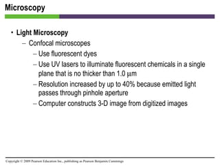
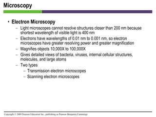
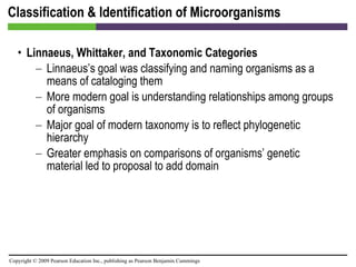
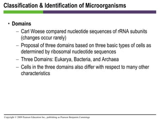
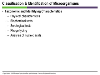
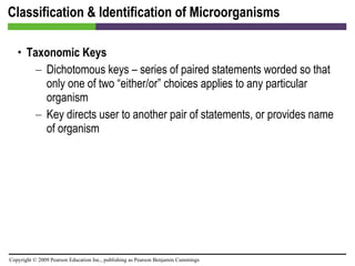
Chapter 4 Microscopy, Staining, and Classification
Units of Measurement Animation: Microscopy and Staining
Units of Measurement [INSERT TABLE 4.1]
Microscopy General Principles of Microscopy Wavelength of radiation Magnification Resolution Contrast
Microscopy [INSERT FIGURE 4.1]
Microscopy [INSERT FIGURE 4.2]
Microscopy [INSERT FIGURE 4.3]
Microscopy General Principles of Microscopy Contrast Differences in intensity between two objects, or between an object and background Important in determining resolution Staining increases contrast Use of light that is in phase increases contrast
Microscopy Light Microscopy Bright-field microscopes Simple Contain a single magnifying lens Similar to magnifying glass Leeuwenhoek used simple microscope to observe microorganisms
Microscopy Light Microscopy Bright-field microscopes Compound Use a series of lenses for magnification Light rays pass through specimen and into objective lens (one of a series of objective lenses) Oil immersion lens increases resolution because light does not refract Have one or two ocular lenses Total magnification = magnification of objective lens X magnification of ocular lens Most have condenser lens to direct light through specimen
Microscopy [INSERT FIGURE 4.4]
Microscopy [INSERT FIGURE 4.5]
Microscopy Light Microscopy Dark-field microscopes Best for observing pale objects Only light rays scattered by specimen enter objective lens Specimen appears light against dark background Increases contrast and enables observation of more details
Microscopy [INSERT FIGURE 4.6]
Microscopy Light Microscopy Phase microscopes Used to examine living organisms or specimens that would be damaged or altered by attaching them to slides or staining them Treat one set of light rays differently from another set Light rays in phase produce brighter image, while light rays out of phase produce darker image Contrast is created because light waves are 1/2 wavelength out of phase Two types Phase-contrast microscope  Differential interference contrast microscope
Microscopy [INSERT FIGURE 4.7]
Microscopy [INSERT FIGURE 4.8]
Microscopy Light Microscopy Fluorescent microscopes Direct UV light source at specimen; causes the specimen to radiate energy back as a longer, visible wavelength UV light increases resolution and contrast  Some cells and molecules are naturally fluorescent, while others must be stained  Used in immunofluorescence to identify pathogens and to locate and make visible a variety of proteins
Microscopy [INSERT FIGURE 4.9]
Microscopy [INSERT FIGURE 4.10]
Microscopy Light Microscopy Confocal microscopes Use fluorescent dyes Use UV lasers to illuminate fluorescent chemicals in a single plane that is no thicker than 1.0   m Resolution increased by up to 40% because emitted light passes through pinhole aperture Computer constructs 3-D image from digitized images
Microscopy Animation: Light Microscopy
Microscopy Electron Microscopy Light microscopes cannot resolve structures closer than 200 nm because shortest wavelength of visible light is 400 nm Electrons have wavelengths of 0.01 nm to 0.001 nm, so electron microscopes have greater resolving power and greater magnification Magnifies objects 10,000X to 100,000X Gives detailed views of bacteria, viruses, internal cellular structures, molecules, and large atoms Two types Transmission electron microscopes Scanning electron microscopes
Microscopy [INSERT FIGURE 4.11]
Microscopy [INSERT FIGURE 4.12]
Microscopy [INSERT FIGURE 4.13]
Microscopy Animation: Electron Microscopy
Microscopy Probe Microscopy Uses minuscule, pointed, electronic probes to magnify more than 100,000,000 times Two types Scanning tunneling microscopes Atomic force microscopes
Microscopy [INSERT FIGURE 4.14]
Microscopy [INSERT TABLE 4.2]
Staining Increases contrast and resolution by coloring specimens with stains/dyes Smear of microorganisms (thin film) air dried to slide and then fixed to surface by heat or chemical fixation Microbiological stains usually salts composed of cation and anion, with one colored (chromophore) Acidic dyes stain alkaline structures; more commonly, basic dyes stain acidic structures
Staining [INSERT FIGURE 4.15]
Staining Simple stains  Differential stains  Gram stain Acid-fast stain Endospore stain Special stains  Negative (capsule) stain Flagellar stain
Staining [INSERT FIGURE 4.16]
Staining [INSERT FIGURE 4.17]
Staining [INSERT FIGURE 4.18]
Staining [INSERT FIGURE 4.19]
Staining [INSERT FIGURE 4.20]
Staining [INSERT FIGURE 4.21]
Staining [INSERT TABLE 4.3]
Staining Staining for Electron Microscopy Chemicals containing heavy metals used for transmission electron microscopy  Stains may bind molecules in specimens or the background
Staining Animation: Staining
Classification & Identification of Microorganisms Taxonomy consists of classification, nomenclature, and identification Enables scientists to organize large amounts of information about organisms and make predictions based on knowledge of similar organisms
Classification & Identification of Microorganisms Linnaeus, Whittaker, and Taxonomic Categories Linnaeus Provided system that standardized the naming and classification of organisms based on characteristics in common Grouped similar organisms that can successfully interbreed into categories called species Used binomial nomenclature in his system
Classification & Identification of Microorganisms
Classification & Identification of Microorganisms Linnaeus, Whittaker, and Taxonomic Categories Whittaker Linnaeus proposed only two kingdoms Whittaker proposed a widely accepted taxonomic approach based on five kingdoms: Animalia, Plantae, Fungi, Protista, and Prokaryotae
Classification & Identification of Microorganisms [INSERT FIGURE 4.23]
Classification & Identification of Microorganisms Linnaeus, Whittaker, and Taxonomic Categories Linnaeus’s goal was classifying and naming organisms as a means of cataloging them More modern goal is understanding relationships among groups of organisms Major goal of modern taxonomy is to reflect phylogenetic hierarchy  Greater emphasis on comparisons of organisms’ genetic material led to proposal to add domain
Classification & Identification of Microorganisms Domains Carl Woese compared nucleotide sequences of rRNA subunits (changes occur rarely) Proposal of three domains based on three basic types of cells as determined by ribosomal nucleotide sequences Three Domains: Eukarya, Bacteria, and Archaea Cells in the three domains also differ with respect to many other characteristics
Classification & Identification of Microorganisms Taxonomic and Identifying Characteristics Physical characteristics Biochemical tests Serological tests Phage typing Analysis of nucleic acids
Classification & Identification of Microorganisms [INSERT FIGURE 4.24]
Classification & Identification of Microorganisms [INSERT FIGURE 4.25]
Classification & Identification of Microorganisms [INSERT FIGURE 4.26]
Classification & Identification of Microorganisms [INSERT FIGURE 4.27]
Classification & Identification of Microorganisms Taxonomic Keys Dichotomous keys – series of paired statements worded so that only one of two “either/or” choices applies to any particular organism Key directs user to another pair of statements, or provides name of organism
Classification & Identification of Microorganisms [INSERT FIGURE 4.28]
Classification & Identification of Microorganisms Animation: Dichotomous Keys

Start here_ch04_lecture

  • 1.
    Chapter 4 Microscopy,Staining, and Classification
  • 2.
    Units of MeasurementAnimation: Microscopy and Staining
  • 3.
    Units of Measurement[INSERT TABLE 4.1]
  • 4.
    Microscopy General Principlesof Microscopy Wavelength of radiation Magnification Resolution Contrast
  • 5.
  • 6.
  • 7.
  • 8.
    Microscopy General Principlesof Microscopy Contrast Differences in intensity between two objects, or between an object and background Important in determining resolution Staining increases contrast Use of light that is in phase increases contrast
  • 9.
    Microscopy Light MicroscopyBright-field microscopes Simple Contain a single magnifying lens Similar to magnifying glass Leeuwenhoek used simple microscope to observe microorganisms
  • 10.
    Microscopy Light MicroscopyBright-field microscopes Compound Use a series of lenses for magnification Light rays pass through specimen and into objective lens (one of a series of objective lenses) Oil immersion lens increases resolution because light does not refract Have one or two ocular lenses Total magnification = magnification of objective lens X magnification of ocular lens Most have condenser lens to direct light through specimen
  • 11.
  • 12.
  • 13.
    Microscopy Light MicroscopyDark-field microscopes Best for observing pale objects Only light rays scattered by specimen enter objective lens Specimen appears light against dark background Increases contrast and enables observation of more details
  • 14.
  • 15.
    Microscopy Light MicroscopyPhase microscopes Used to examine living organisms or specimens that would be damaged or altered by attaching them to slides or staining them Treat one set of light rays differently from another set Light rays in phase produce brighter image, while light rays out of phase produce darker image Contrast is created because light waves are 1/2 wavelength out of phase Two types Phase-contrast microscope Differential interference contrast microscope
  • 16.
  • 17.
  • 18.
    Microscopy Light MicroscopyFluorescent microscopes Direct UV light source at specimen; causes the specimen to radiate energy back as a longer, visible wavelength UV light increases resolution and contrast Some cells and molecules are naturally fluorescent, while others must be stained Used in immunofluorescence to identify pathogens and to locate and make visible a variety of proteins
  • 19.
  • 20.
  • 21.
    Microscopy Light MicroscopyConfocal microscopes Use fluorescent dyes Use UV lasers to illuminate fluorescent chemicals in a single plane that is no thicker than 1.0  m Resolution increased by up to 40% because emitted light passes through pinhole aperture Computer constructs 3-D image from digitized images
  • 22.
  • 23.
    Microscopy Electron MicroscopyLight microscopes cannot resolve structures closer than 200 nm because shortest wavelength of visible light is 400 nm Electrons have wavelengths of 0.01 nm to 0.001 nm, so electron microscopes have greater resolving power and greater magnification Magnifies objects 10,000X to 100,000X Gives detailed views of bacteria, viruses, internal cellular structures, molecules, and large atoms Two types Transmission electron microscopes Scanning electron microscopes
  • 24.
  • 25.
  • 26.
  • 27.
  • 28.
    Microscopy Probe MicroscopyUses minuscule, pointed, electronic probes to magnify more than 100,000,000 times Two types Scanning tunneling microscopes Atomic force microscopes
  • 29.
  • 30.
  • 31.
    Staining Increases contrastand resolution by coloring specimens with stains/dyes Smear of microorganisms (thin film) air dried to slide and then fixed to surface by heat or chemical fixation Microbiological stains usually salts composed of cation and anion, with one colored (chromophore) Acidic dyes stain alkaline structures; more commonly, basic dyes stain acidic structures
  • 32.
  • 33.
    Staining Simple stains Differential stains Gram stain Acid-fast stain Endospore stain Special stains Negative (capsule) stain Flagellar stain
  • 34.
  • 35.
  • 36.
  • 37.
  • 38.
  • 39.
  • 40.
  • 41.
    Staining Staining forElectron Microscopy Chemicals containing heavy metals used for transmission electron microscopy Stains may bind molecules in specimens or the background
  • 42.
  • 43.
    Classification & Identificationof Microorganisms Taxonomy consists of classification, nomenclature, and identification Enables scientists to organize large amounts of information about organisms and make predictions based on knowledge of similar organisms
  • 44.
    Classification & Identificationof Microorganisms Linnaeus, Whittaker, and Taxonomic Categories Linnaeus Provided system that standardized the naming and classification of organisms based on characteristics in common Grouped similar organisms that can successfully interbreed into categories called species Used binomial nomenclature in his system
  • 45.
  • 46.
    Classification & Identificationof Microorganisms Linnaeus, Whittaker, and Taxonomic Categories Whittaker Linnaeus proposed only two kingdoms Whittaker proposed a widely accepted taxonomic approach based on five kingdoms: Animalia, Plantae, Fungi, Protista, and Prokaryotae
  • 47.
    Classification & Identificationof Microorganisms [INSERT FIGURE 4.23]
  • 48.
    Classification & Identificationof Microorganisms Linnaeus, Whittaker, and Taxonomic Categories Linnaeus’s goal was classifying and naming organisms as a means of cataloging them More modern goal is understanding relationships among groups of organisms Major goal of modern taxonomy is to reflect phylogenetic hierarchy Greater emphasis on comparisons of organisms’ genetic material led to proposal to add domain
  • 49.
    Classification & Identificationof Microorganisms Domains Carl Woese compared nucleotide sequences of rRNA subunits (changes occur rarely) Proposal of three domains based on three basic types of cells as determined by ribosomal nucleotide sequences Three Domains: Eukarya, Bacteria, and Archaea Cells in the three domains also differ with respect to many other characteristics
  • 50.
    Classification & Identificationof Microorganisms Taxonomic and Identifying Characteristics Physical characteristics Biochemical tests Serological tests Phage typing Analysis of nucleic acids
  • 51.
    Classification & Identificationof Microorganisms [INSERT FIGURE 4.24]
  • 52.
    Classification & Identificationof Microorganisms [INSERT FIGURE 4.25]
  • 53.
    Classification & Identificationof Microorganisms [INSERT FIGURE 4.26]
  • 54.
    Classification & Identificationof Microorganisms [INSERT FIGURE 4.27]
  • 55.
    Classification & Identificationof Microorganisms Taxonomic Keys Dichotomous keys – series of paired statements worded so that only one of two “either/or” choices applies to any particular organism Key directs user to another pair of statements, or provides name of organism
  • 56.
    Classification & Identificationof Microorganisms [INSERT FIGURE 4.28]
  • 57.
    Classification & Identificationof Microorganisms Animation: Dichotomous Keys