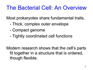
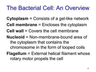
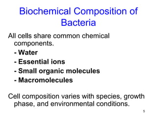
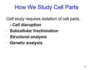
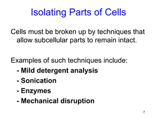
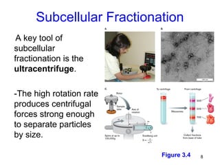
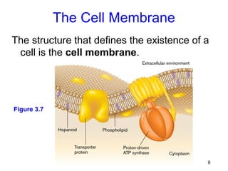
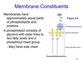
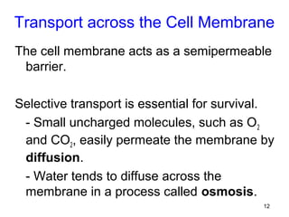
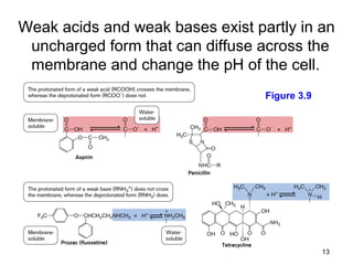
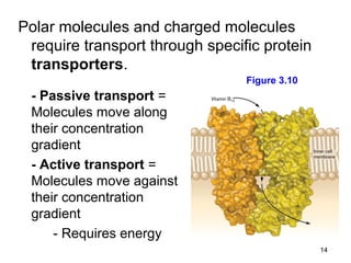
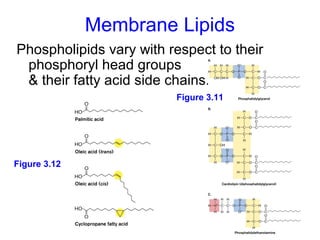
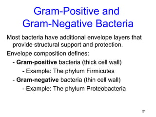
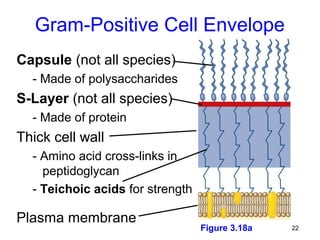
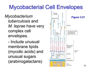
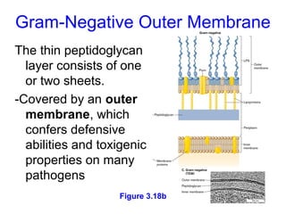
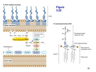
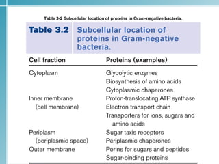
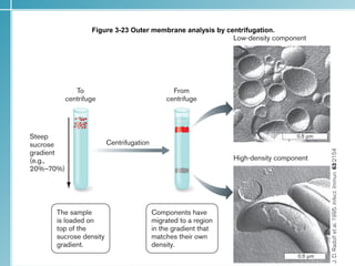
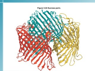
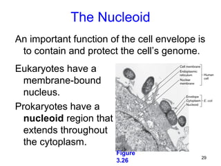
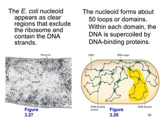
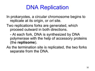
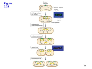
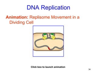
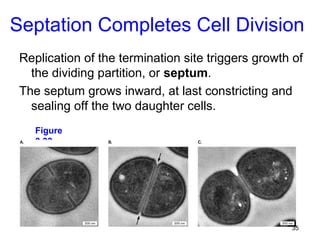
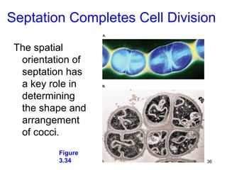
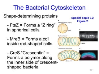
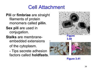
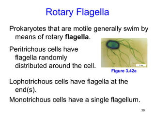
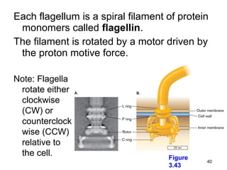
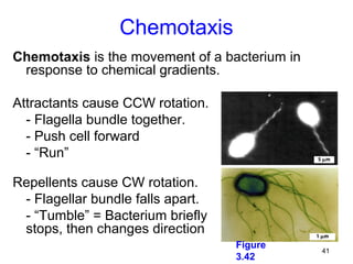
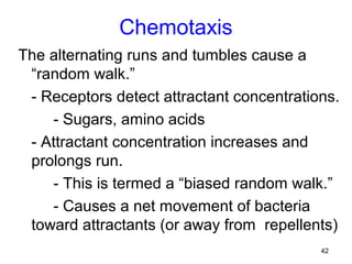
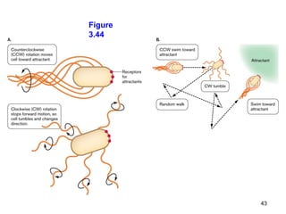
Chapter Overview
â—Ź A synopsis of the bacterial cell
â—Ź How cell parts are studied

â—Ź The plasma membrane and transport

â—Ź The cell wall and other outer layers

â—Ź The nucleoid: structure and expression

â—Ź How bacterial cells divide

â—Ź Specialized structures, including pili & stalks

â—Ź Bacterial flagella and chemotaxis

                                              1
The Bacterial Cell: An Overview
Most prokaryotes share fundamental traits.
 - Thick, complex outer envelope
 - Compact genome
 - Tightly coordinated cell functions

Modern research shows that the cell’s parts
 fit together in a structure that is ordered,
 though flexible.
                                             2
Figure 3.1


             3
The Bacterial Cell: An Overview
Cytoplasm = Consists of a gel-like network
Cell membrane = Encloses the cytoplasm
Cell wall = Covers the cell membrane
Nucleoid = Non-membrane-bound area of
  the cytoplasm that contains the
  chromosome in the form of looped coils
Flagellum = External helical filament whose
  rotary motor propels the cell

                                         4
Biochemical Composition of
           Bacteria
All cells share common chemical
  components.
  - Water
  - Essential ions
  - Small organic molecules
  - Macromolecules

Cell composition varies with species, growth
 phase, and environmental conditions.
                                           5
How We Study Cell Parts
Cell study requires isolation of cell parts.
  - Cell disruption
- Subcellular fractionation

- Structural analysis

- Genetic analysis


                                               6
Isolating Parts of Cells
Cells must be broken up by techniques that
 allow subcellular parts to remain intact.

Examples of such techniques include:
 - Mild detergent analysis
 - Sonication
 - Enzymes
 - Mechanical disruption
                                         7
Subcellular Fractionation
 A key tool of
subcellular
fractionation is the
ultracentrifuge.

-The high rotation rate
produces centrifugal
forces strong enough
to separate particles
by size.

                           Figure 3.4   8
The Cell Membrane
The structure that defines the existence of a
 cell is the cell membrane.


Figure 3.7


                                            9
Membrane Constituents
Membranes have
  approximately equal parts      Figure 3.8
  of phospholipids and
  proteins.
A phospholipid consists of
  glycerol with ester links to
  two fatty acids and a
  phosphoryl head group.
  - May have side chain


                                        10
Membrane Constituents
Membrane proteins serve numerous functions,
 including:
 - Structural support
 - Detection of environmental signals
 - Secretion of virulence factors and
 communication signals
 - Ion transport and energy storage

Have hydrophilic and hydrophobic regions that
 lock the protein in the membrane

                                                11
Transport across the Cell Membrane
The cell membrane acts as a semipermeable
 barrier.

Selective transport is essential for survival.
 - Small uncharged molecules, such as O 2
 and CO2, easily permeate the membrane by
 diffusion.
 - Water tends to diffuse across the
 membrane in a process called osmosis.
                                            12
Weak acids and weak bases exist partly in an
 uncharged form that can diffuse across the
 membrane and change the pH of the cell.
                                  Figure 3.9


                                               13
Polar molecules and charged molecules
 require transport through specific protein
 transporters.
                                 Figure 3.10
 - Passive transport =
 Molecules move along
 their concentration
 gradient
 - Active transport =
 Molecules move against
 their concentration
 gradient
     - Requires energy
                                               14
Membrane Lipids
Phospholipids vary with respect to their
 phosphoryl head groups
 & their fatty acid side chains.
                         Figure 3.11


Figure 3.12


                                           15
Membranes also include planar molecules that fill
  gaps between hydrocarbon chains.
In eukaryotic membranes, the reinforcing agents
  are sterols, such as cholesterol.
In bacteria, the same function is filled by
  hopanoids, or hopanes.


 Figure 3.13


                                                16
Archaea have the most extreme variations in
  phospholipid side-chain structures.
 - Ether links between glycerol and fatty acids
 - Hydrocarbon chains are branched terpenoids.


                                       Figure 3.14


                                                  17
The Cell Wall
The cell wall confers shape and rigidity to the
 cell, and helps it withstand turgor pressure.
The bacterial cell wall, or the sacculus,
 consists of a single interlinked molecule.


                   Figure 3.16               18
Peptidoglycan Structure
Most bacterial cell walls are made up of
 peptidoglycan (or murein).
The molecule consists of:
 - Long polymers of two disaccharides called
 N-acetylglucosamine and N-acetylmuramic
 acid, bound to a peptide of 4-6 amino acids
    - The peptides can form cross-bridges
 connecting the parallel glycan strands.

                                          19
Figure
3.17


         20
Gram-Positive and
     Gram-Negative Bacteria
Most bacteria have additional envelope layers that
  provide structural support and protection.
Envelope composition defines:
  - Gram-positive bacteria (thick cell wall)
      - Example: The phylum Firmicutes
  - Gram-negative bacteria (thin cell wall)
      - Example: The phylum Proteobacteria

                           Figure 1.1
                                                     21
Gram-Positive Cell Envelope
Capsule (not all species)
  - Made of polysaccharides
S-Layer (not all species)
  - Made of protein
Thick cell wall
  - Amino acid cross-links in
    peptidoglycan
  - Teichoic acids for strength

Plasma membrane
                                  Figure 3.18a   22
Mycobacterial Cell Envelopes
Mycobacterium            Figure 3.21
 tuberculosis and
 M. leprae have very
 complex cell
 envelopes.
 - Include unusual
 membrane lipids
 (mycolic acids) and
 unusual sugars
 (arabinogalactans)
                                   23
Gram-Negative Outer Membrane
The thin peptidoglycan
  layer consists of one
  or two sheets.
-Covered by an outer
  membrane, which
  confers defensive
  abilities and toxigenic
  properties on many
  pathogens
                   Figure 3.18b
                                  24
Figure
3.22


         25
Table 3-2 Subcellular location of proteins in Gram-negative bacteria.


                                                                        26
Figure 3-23 Outer membrane analysis by centrifugation.


                                                         27
Figure 3-24 Sucrose porin.


                             28
The Nucleoid
An important function of the cell envelope is
 to contain and protect the cell’s genome.
Eukaryotes have a
  membrane-bound
  nucleus.
Prokaryotes have a
  nucleoid region that
  extends throughout
  the cytoplasm.
                         Figure
                                            29
                         3.26
The E. coli nucleoid     The nucleoid forms about
  appears as clear         50 loops or domains.
  regions that exclude     Within each domain, the
  the ribosome and
  contain the DNA          DNA is supercoiled by
  strands.                 DNA-binding proteins.


     Figure                       Figure
     3.27                         3.28         30
Cell Division
Cell division, or cell fission, requires highly
  coordinated growth and expansion of all the
  cell’s parts.
Unlike eukaryotes, prokaryotes synthesize RNA
  and proteins continually while the cell’s DNA
  undergoes replication.
Bacterial DNA replication is coordinated with the
  cell wall expansion and ultimately the separation
  of the two daughter cells.


                                                 31
DNA Replication
In prokaryotes, a circular chromosome begins to
  replicate at its origin, or ori site.
Two replications forks are generated, which
  proceed outward in both directions.
  - At each fork, DNA is synthesized by DNA
  polymerase with the help of accessory proteins
  (the replisome).
As the termination site is replicated, the two forks
  separate from the DNA.


                                                   32
Figure
3.32


         33
DNA Replication
Animation: Replisome Movement in a
 Dividing Cell


           Click box to launch animation
                                           34
Septation Completes Cell Division
 Replication of the termination site triggers growth of
   the dividing partition, or septum.
 The septum grows inward, at last constricting and
   sealing off the two daughter cells.
    Figure
    3.33


                                                    35
Septation Completes Cell Division

 The spatial
  orientation of
  septation has
  a key role in
  determining
  the shape and
  arrangement
  of cocci.
          Figure
          3.34                 36
The Bacterial Cytoskeleton
Shape-determining proteins     Special Topic 3.2
                                  Figure 2
 - FtsZ = Forms a “Z ring”
 in spherical cells

 - MreB = Forms a coil
 inside rod-shaped cells

 - CreS “Crescentin” =
 Forms a polymer along
 the inner side of crescent-
 shaped bacteria
                                             37
Cell Attachment
Pili or fimbriae are straight
  filaments of protein
  monomers called pilin.
Sex pili are used in
  conjugation.
                                Figure
Stalks are membrane-            3.40
  embedded extensions
  of the cytoplasm.
  - Tips secrete adhesion
  factors called holdfasts.
                                Figure 3.41

                                              38
Rotary Flagella
Prokaryotes that are motile generally swim by
  means of rotary flagella.
Peritrichous cells have
 flagella randomly
 distributed around the cell.
                                 Figure 3.42a

Lophotrichous cells have flagella at the
  end(s).
Monotrichous cells have a single flagellum.
                                                39
Each flagellum is a spiral filament of protein
 monomers called flagellin.
The filament is rotated by a motor driven by
 the proton motive force.

Note: Flagella
 rotate either
 clockwise
 (CW) or
 counterclock
 wise (CCW)
 relative to
 the cell.                          Figure
                                             40
                                    3.43
Chemotaxis
Chemotaxis is the movement of a bacterium in
 response to chemical gradients.

Attractants cause CCW rotation.
  - Flagella bundle together.
  - Push cell forward
  - “Run”

Repellents cause CW rotation.
 - Flagellar bundle falls apart.
 - “Tumble” = Bacterium briefly
 stops, then changes direction
                                   Figure
                                               41
                                   3.42
Chemotaxis
The alternating runs and tumbles cause a
 “random walk.”
 - Receptors detect attractant concentrations.
    - Sugars, amino acids
 - Attractant concentration increases and
 prolongs run.
    - This is termed a “biased random walk.”
    - Causes a net movement of bacteria
 toward attractants (or away from repellents)
                                           42
Figure
3.44


         43
Chemotaxis
Animation: Chemotaxis: Molecular Events


           Click box to launch animation
                                           44
Chapter Summary
â—Ź   While prokaryotes are diverse, they share certain
    fundamental traits and biochemistry.
â—Ź   The study of cells employs various methods
    including subcellular fractionation, structural
    analysis, and genetic analysis.
â—Ź   The cell membrane consists of a phospholipid
    bilayer containing proteins.
    - Bacterial phospholipids are ester-linked, while
    those of Archaea contain ether linkages.
â—Ź   The Gram-negative cell envelope is much more
    complex than that of Gram-positive cells.
                                                    45
Chapter Summary
â—Ź   The DNA of prokaryotes is organized into loops in
    the nucleoid.
    - Transcription and translation are coupled.
â—Ź   Most bacteria divide by binary fission.
    - Cell growth and DNA replication are coordinated.
â—Ź   Bacteria may have specialized structures, including
    thylakoids, storage granules, and magnetosomes.
â—Ź   Pili and stalks are used for attachment.
â—Ź   Flagella are rotary appendages used for movement
    and chemotaxis.

                                                    46
Concept Quiz
Which one of these membranes is not
found in Gram-negative bacteria?
a) Plasma membrane
b) Inner membrane
c) Nuclear membrane
d) Outer membrane


                                      47
Concept Quiz

Peptidoglycan is composed primarily of
a) sugars and amino acids.
b) sugars and nucleic acids.
c) nucleic acids and lipids.
d) amino acids and lipids.


                                         48
Concept Quiz

An extension of the cytoplasm that attaches
bacteria to a surface is called a
a) pilus.
b) flagellum.
c) fimbrium.
d) stalk.


                                          49
Concept Quiz

The structure in prokaryotes that performs
the same function as mitochondria in
eukaryotes is the
a) cell membrane.
b) chloroplast.
c) outer membrane.
d) cell wall.


                                             50
Concept Quiz

In archaeal membranes, the glycerol is
linked to the hydrocarbon chains by _____
bonds.
a) ether
b) ester
c) glycan
d) peptide

                                            51
Concept Quiz
All of the following statements about prokaryotic
   flagella are correct except
a)they are driven by the proton motive force.
b)they are found in both Gram-positive and
   Gram-negative bacteria.
c) they move with a whiplike motion.
d)they are used for chemotaxis.


                                                    52

Presentation3 - Microbio

  • 1.
    Chapter Overview â—Ź Asynopsis of the bacterial cell â—Ź How cell parts are studied â—Ź The plasma membrane and transport â—Ź The cell wall and other outer layers â—Ź The nucleoid: structure and expression â—Ź How bacterial cells divide â—Ź Specialized structures, including pili & stalks â—Ź Bacterial flagella and chemotaxis 1
  • 2.
    The Bacterial Cell:An Overview Most prokaryotes share fundamental traits. - Thick, complex outer envelope - Compact genome - Tightly coordinated cell functions Modern research shows that the cell’s parts fit together in a structure that is ordered, though flexible. 2
  • 3.
  • 4.
    The Bacterial Cell:An Overview Cytoplasm = Consists of a gel-like network Cell membrane = Encloses the cytoplasm Cell wall = Covers the cell membrane Nucleoid = Non-membrane-bound area of the cytoplasm that contains the chromosome in the form of looped coils Flagellum = External helical filament whose rotary motor propels the cell 4
  • 5.
    Biochemical Composition of Bacteria All cells share common chemical components. - Water - Essential ions - Small organic molecules - Macromolecules Cell composition varies with species, growth phase, and environmental conditions. 5
  • 6.
    How We StudyCell Parts Cell study requires isolation of cell parts. - Cell disruption - Subcellular fractionation - Structural analysis - Genetic analysis 6
  • 7.
    Isolating Parts ofCells Cells must be broken up by techniques that allow subcellular parts to remain intact. Examples of such techniques include: - Mild detergent analysis - Sonication - Enzymes - Mechanical disruption 7
  • 8.
    Subcellular Fractionation Akey tool of subcellular fractionation is the ultracentrifuge. -The high rotation rate produces centrifugal forces strong enough to separate particles by size. Figure 3.4 8
  • 9.
    The Cell Membrane Thestructure that defines the existence of a cell is the cell membrane. Figure 3.7 9
  • 10.
    Membrane Constituents Membranes have approximately equal parts Figure 3.8 of phospholipids and proteins. A phospholipid consists of glycerol with ester links to two fatty acids and a phosphoryl head group. - May have side chain 10
  • 11.
    Membrane Constituents Membrane proteinsserve numerous functions, including: - Structural support - Detection of environmental signals - Secretion of virulence factors and communication signals - Ion transport and energy storage Have hydrophilic and hydrophobic regions that lock the protein in the membrane 11
  • 12.
    Transport across theCell Membrane The cell membrane acts as a semipermeable barrier. Selective transport is essential for survival. - Small uncharged molecules, such as O 2 and CO2, easily permeate the membrane by diffusion. - Water tends to diffuse across the membrane in a process called osmosis. 12
  • 13.
    Weak acids andweak bases exist partly in an uncharged form that can diffuse across the membrane and change the pH of the cell. Figure 3.9 13
  • 14.
    Polar molecules andcharged molecules require transport through specific protein transporters. Figure 3.10 - Passive transport = Molecules move along their concentration gradient - Active transport = Molecules move against their concentration gradient - Requires energy 14
  • 15.
    Membrane Lipids Phospholipids varywith respect to their phosphoryl head groups & their fatty acid side chains. Figure 3.11 Figure 3.12 15
  • 16.
    Membranes also includeplanar molecules that fill gaps between hydrocarbon chains. In eukaryotic membranes, the reinforcing agents are sterols, such as cholesterol. In bacteria, the same function is filled by hopanoids, or hopanes. Figure 3.13 16
  • 17.
    Archaea have themost extreme variations in phospholipid side-chain structures. - Ether links between glycerol and fatty acids - Hydrocarbon chains are branched terpenoids. Figure 3.14 17
  • 18.
    The Cell Wall Thecell wall confers shape and rigidity to the cell, and helps it withstand turgor pressure. The bacterial cell wall, or the sacculus, consists of a single interlinked molecule. Figure 3.16 18
  • 19.
    Peptidoglycan Structure Most bacterialcell walls are made up of peptidoglycan (or murein). The molecule consists of: - Long polymers of two disaccharides called N-acetylglucosamine and N-acetylmuramic acid, bound to a peptide of 4-6 amino acids - The peptides can form cross-bridges connecting the parallel glycan strands. 19
  • 20.
  • 21.
    Gram-Positive and Gram-Negative Bacteria Most bacteria have additional envelope layers that provide structural support and protection. Envelope composition defines: - Gram-positive bacteria (thick cell wall) - Example: The phylum Firmicutes - Gram-negative bacteria (thin cell wall) - Example: The phylum Proteobacteria Figure 1.1 21
  • 22.
    Gram-Positive Cell Envelope Capsule(not all species) - Made of polysaccharides S-Layer (not all species) - Made of protein Thick cell wall - Amino acid cross-links in peptidoglycan - Teichoic acids for strength Plasma membrane Figure 3.18a 22
  • 23.
    Mycobacterial Cell Envelopes Mycobacterium Figure 3.21 tuberculosis and M. leprae have very complex cell envelopes. - Include unusual membrane lipids (mycolic acids) and unusual sugars (arabinogalactans) 23
  • 24.
    Gram-Negative Outer Membrane Thethin peptidoglycan layer consists of one or two sheets. -Covered by an outer membrane, which confers defensive abilities and toxigenic properties on many pathogens Figure 3.18b 24
  • 25.
  • 26.
    Table 3-2 Subcellularlocation of proteins in Gram-negative bacteria. 26
  • 27.
    Figure 3-23 Outermembrane analysis by centrifugation. 27
  • 28.
  • 29.
    The Nucleoid An importantfunction of the cell envelope is to contain and protect the cell’s genome. Eukaryotes have a membrane-bound nucleus. Prokaryotes have a nucleoid region that extends throughout the cytoplasm. Figure 29 3.26
  • 30.
    The E. colinucleoid The nucleoid forms about appears as clear 50 loops or domains. regions that exclude Within each domain, the the ribosome and contain the DNA DNA is supercoiled by strands. DNA-binding proteins. Figure Figure 3.27 3.28 30
  • 31.
    Cell Division Cell division,or cell fission, requires highly coordinated growth and expansion of all the cell’s parts. Unlike eukaryotes, prokaryotes synthesize RNA and proteins continually while the cell’s DNA undergoes replication. Bacterial DNA replication is coordinated with the cell wall expansion and ultimately the separation of the two daughter cells. 31
  • 32.
    DNA Replication In prokaryotes,a circular chromosome begins to replicate at its origin, or ori site. Two replications forks are generated, which proceed outward in both directions. - At each fork, DNA is synthesized by DNA polymerase with the help of accessory proteins (the replisome). As the termination site is replicated, the two forks separate from the DNA. 32
  • 33.
  • 34.
    DNA Replication Animation: ReplisomeMovement in a Dividing Cell Click box to launch animation 34
  • 35.
    Septation Completes CellDivision Replication of the termination site triggers growth of the dividing partition, or septum. The septum grows inward, at last constricting and sealing off the two daughter cells. Figure 3.33 35
  • 36.
    Septation Completes CellDivision The spatial orientation of septation has a key role in determining the shape and arrangement of cocci. Figure 3.34 36
  • 37.
    The Bacterial Cytoskeleton Shape-determiningproteins Special Topic 3.2 Figure 2 - FtsZ = Forms a “Z ring” in spherical cells - MreB = Forms a coil inside rod-shaped cells - CreS “Crescentin” = Forms a polymer along the inner side of crescent- shaped bacteria 37
  • 38.
    Cell Attachment Pili orfimbriae are straight filaments of protein monomers called pilin. Sex pili are used in conjugation. Figure Stalks are membrane- 3.40 embedded extensions of the cytoplasm. - Tips secrete adhesion factors called holdfasts. Figure 3.41 38
  • 39.
    Rotary Flagella Prokaryotes thatare motile generally swim by means of rotary flagella. Peritrichous cells have flagella randomly distributed around the cell. Figure 3.42a Lophotrichous cells have flagella at the end(s). Monotrichous cells have a single flagellum. 39
  • 40.
    Each flagellum isa spiral filament of protein monomers called flagellin. The filament is rotated by a motor driven by the proton motive force. Note: Flagella rotate either clockwise (CW) or counterclock wise (CCW) relative to the cell. Figure 40 3.43
  • 41.
    Chemotaxis Chemotaxis is themovement of a bacterium in response to chemical gradients. Attractants cause CCW rotation. - Flagella bundle together. - Push cell forward - “Run” Repellents cause CW rotation. - Flagellar bundle falls apart. - “Tumble” = Bacterium briefly stops, then changes direction Figure 41 3.42
  • 42.
    Chemotaxis The alternating runsand tumbles cause a “random walk.” - Receptors detect attractant concentrations. - Sugars, amino acids - Attractant concentration increases and prolongs run. - This is termed a “biased random walk.” - Causes a net movement of bacteria toward attractants (or away from repellents) 42
  • 43.
  • 44.
    Chemotaxis Animation: Chemotaxis: MolecularEvents Click box to launch animation 44
  • 45.
    Chapter Summary â—Ź While prokaryotes are diverse, they share certain fundamental traits and biochemistry. â—Ź The study of cells employs various methods including subcellular fractionation, structural analysis, and genetic analysis. â—Ź The cell membrane consists of a phospholipid bilayer containing proteins. - Bacterial phospholipids are ester-linked, while those of Archaea contain ether linkages. â—Ź The Gram-negative cell envelope is much more complex than that of Gram-positive cells. 45
  • 46.
    Chapter Summary â—Ź The DNA of prokaryotes is organized into loops in the nucleoid. - Transcription and translation are coupled. â—Ź Most bacteria divide by binary fission. - Cell growth and DNA replication are coordinated. â—Ź Bacteria may have specialized structures, including thylakoids, storage granules, and magnetosomes. â—Ź Pili and stalks are used for attachment. â—Ź Flagella are rotary appendages used for movement and chemotaxis. 46
  • 47.
    Concept Quiz Which oneof these membranes is not found in Gram-negative bacteria? a) Plasma membrane b) Inner membrane c) Nuclear membrane d) Outer membrane 47
  • 48.
    Concept Quiz Peptidoglycan iscomposed primarily of a) sugars and amino acids. b) sugars and nucleic acids. c) nucleic acids and lipids. d) amino acids and lipids. 48
  • 49.
    Concept Quiz An extensionof the cytoplasm that attaches bacteria to a surface is called a a) pilus. b) flagellum. c) fimbrium. d) stalk. 49
  • 50.
    Concept Quiz The structurein prokaryotes that performs the same function as mitochondria in eukaryotes is the a) cell membrane. b) chloroplast. c) outer membrane. d) cell wall. 50
  • 51.
    Concept Quiz In archaealmembranes, the glycerol is linked to the hydrocarbon chains by _____ bonds. a) ether b) ester c) glycan d) peptide 51
  • 52.
    Concept Quiz All ofthe following statements about prokaryotic flagella are correct except a)they are driven by the proton motive force. b)they are found in both Gram-positive and Gram-negative bacteria. c) they move with a whiplike motion. d)they are used for chemotaxis. 52

Editor's Notes