SlideShare a Scribd company logo
PAIN
PATHOPHYSIOLOGY
BY SURAJ DHARA
MMCH, WEST BENGAL
The provinceof medicine is to preserve and restorehealth and to relieve suffering.
Understanding pain is essential to both of these goals. Because pain is universally
understoodasa signalof disease, it is themostcommon symptomthatbrings a patient
to a physician’s attention. The function of the pain sensory system is to protect the
body and maintain homeostasis. It does this by detecting, localizing, and identifying
potential or actual tissue-damaging processes. Because different diseases produce
characteristic patterns of tissue damage, the quality, time course, and location of a
patient’s pain lend important diagnostic clues. It is the physician’s responsibility to
assess each patient promptly for any remediable cause underlying the pain and to
provide rapid and effective pain relief whenever possible.
THE PAIN SENSORY SYSTEM
Pain is an unpleasantsensation localized to a part of the body. Itis often described
in terms of a penetrating or tissue-destructive process (e.g., stabbing, burning,
twisting, tearing, squeezing) and/or of a bodily or emotional reaction (e.g., terrifying,
nauseating, sickening). Furthermore, any pain of moderate or higher intensity is
accompanied by anxiety and the urge to escape or terminate the feeling. These
properties illustrate the duality of pain: it is both sensation and emotion. When it is
acute, pain is characteristically associated with behavioral arousal and a stress
response consisting of increased blood pressure, heart rate, pupil diameter, and
plasma cortisol levels. In addition, local muscle contraction (e.g., limb flexion,
abdominal wall rigidity) is often present.
PERIPHERAL MECHANISMS
The Primary Afferent Nociceptor - A peripheralnerveconsists of the axons of three
different types of neurons: primary sensory afferents, motor neurons, and
sympathetic postganglionic neurons (Fig. 10-1). The cell bodies of primary sensory
afferents are located in the dorsal root ganglia within the vertebral foramina. The
primaryafferentaxon hastwo branches:oneprojectscentrally into thespinalcordand
the other projects peripherally to innervate tissues. Primary afferents areclassified by
their diameter, degree of myelination, and conduction velocity. The largest diameter
afferent fibers, A-beta (Aβ), respond maximally to light touch and/or moving stimuli;
they are present primarily in nerves that innervate the skin. In normal individuals, the
activity of these fibers does not produce pain. There are two other classes of primary
afferent nerve fibers: the small diameter myelinated A-delta (Aδ) and the
unmyelinated (C) axons (Fig. 10-1). These fibers are present in nerves to the skin and
to deep somatic and visceral structures. Some tissues, such as the cornea, are
innervated only by Aδ and C fiber afferents. Most Aδ and C fiber afferents respond
maximally only to intense (painful) stimuli and produce the subjective experience of
pain when they are electrically stimulated; this defines them as primary afferent
nociceptors (pain receptors). The ability to detect painful stimuli is completely
abolished when conduction in Aδ and C fiber axons is blocked.
Individual primary afferent nociceptors can respond to several different types of
noxious stimuli. For example, most nociceptors respond to heat; intense cold; intense
mechanical distortion, such as a pinch; changes in pH, particularly an acidic
environment; and application of chemical irritants including adenosine triphosphate
(ATP), serotonin, bradykinin (BK), and histamine. The transient receptor potential
cation channel subfamily V member 1 (TrpV1), also known as the vanilloid receptor,
mediates perception of some noxious stimuli, especially heat sensations, by
nociceptive neurons; it is activated by acidic pH, endogenous mediators and by
capsaicin, a component of hot chili peppers.
Sensitization-When intense, repeated, or prolonged stimuli are applied to damaged
or inflamed tissues, the threshold for activating primary afferent nociceptors is
lowered, and the frequency of firing is higher for all stimulus intensities. Inflammatory
mediators such as BK, nerve-growth factor, some prostaglandins (PGs), and
leukotrienes contribute to this process, which is called sensitization. Sensitization
occurs atthe level of the peripheralnerveterminal (peripheralsensitization) as wellas
at the level of the dorsal horn of the spinal cord (central sensitization). Peripheral
sensitization occurs in damaged or inflamed tissues, when inflammatory mediators
activate intracellular signal transduction in nociceptors, prompting an increase in the
production, transport, and membraneinsertion of chemically gated and voltage-gated
ion channels.These changesincreasetheexcitability of nociceptor terminals and lower
their threshold for activation by mechanical, thermal, and chemical stimuli. Central
sensitization occurs when activity, generated by nociceptors during inflammation,
enhances the excitability of nerve cells in the dorsalhorn of the spinal cord. Following
injury and resultant sensitization, normally innocuous stimuli can produce pain
(termed allodynia). Sensitization is a clinically important process that contributes to
tenderness, soreness, and hyperalgesia (increased pain intensity in response to the
same noxious stimulus; e.g., pinprick causes severe pain). A striking example of
sensitization is sunburned skin, in which severe pain can be produced by a gentle slap
on the back or a warm shower.
Sensitization is of particular importance for pain and tenderness in deep tissues.
Viscera are normally relatively insensitive to noxious mechanical and thermal stimuli,
although hollow viscera do generate significant discomfort when distended. In
contrast, when affected a disease process with an inflammatory component, deep
structures such as joints or hollow viscera characteristically become exquisitely
sensitive to mechanical stimulation.
A large proportion of Aδ and C fiber afferents innervating viscera are completely
insensitivein normalnoninjured, noninflamed tissue. Thatis, they cannotbe activated
by known mechanical or thermal stimuli and are not spontaneously active. However,
in the presence of inflammatory mediators, these afferents become sensitive to
mechanical stimuli. Such afferents have been termed silent nociceptors, and their
characteristic properties may explain how, under pathologic conditions, the relatively
insensitivedeep structures can become the sourceof severe and debilitating pain and
tenderness. Low pH, PGs, leukotrienes, and other inflammatory mediators such as BK
play a significant role in sensitization.
Nociceptor-Induced Inflammation Primary afferent nociceptors also have a
neuroeffector function. Most nociceptors contain polypeptide mediators that are
released from their peripheral terminals when they are activated (Fig. 10-2). An
example is substance P, an 11-amino-acid peptide. Substance P is released from
primary afferent nociceptors and has multiple biologic activities. It is a potent
vasodilator, causes mast cell degranulation, is a chemoattractant for leukocytes, and
increases the production and release of inflammatory mediators. Interestingly,
depletion of substance P from joints reduces the severity of experimental arthritis.
Primary afferent nociceptors are not simply passive messengers of threats to tissue
injury but also play an active role in tissue protection through these neuroeffector
functions.
CENTRAL MECHANISMS
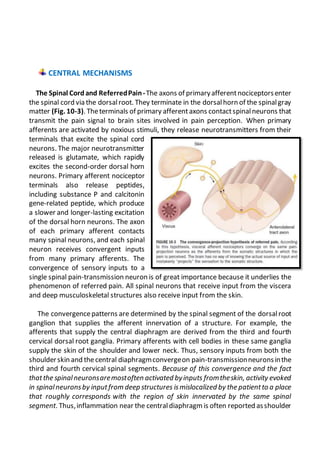
The Spinal Cordand ReferredPain-The axons of primaryafferentnociceptorsenter
the spinal cord via the dorsalroot. They terminate in the dorsalhorn of the spinalgray
matter (Fig. 10-3). Theterminals of primary afferentaxons contactspinalneurons that
transmit the pain signal to brain sites involved in pain perception. When primary
afferents are activated by noxious stimuli, they release neurotransmitters from their
terminals that excite the spinal cord
neurons. The major neurotransmitter
released is glutamate, which rapidly
excites the second-order dorsal horn
neurons. Primary afferent nociceptor
terminals also release peptides,
including substance P and calcitonin
gene-related peptide, which produce
a slower and longer-lasting excitation
of the dorsal horn neurons. The axon
of each primary afferent contacts
many spinal neurons, and each spinal
neuron receives convergent inputs
from many primary afferents. The
convergence of sensory inputs to a
single spinal pain-transmission neuron is of great importance because it underlies the
phenomenon of referred pain. All spinal neurons that receive input from the viscera
and deep musculoskeletal structures also receive input from the skin.
The convergencepatterns are determined by the spinal segment of the dorsalroot
ganglion that supplies the afferent innervation of a structure. For example, the
afferents that supply the central diaphragm are derived from the third and fourth
cervical dorsal root ganglia. Primary afferents with cell bodies in these same ganglia
supply the skin of the shoulder and lower neck. Thus, sensory inputs from both the
shoulderskin and thecentral diaphragmconvergeon pain-transmissionneuronsinthe
third and fourth cervical spinal segments. Because of this convergence and the fact
thatthe spinalneuronsaremostoften activated byinputs fromtheskin, activity evoked
in spinalneuronsby inputfrom deep structures ismislocalized by the patientto a place
that roughly corresponds with the region of skin innervated by the same spinal
segment. Thus,inflammation near the centraldiaphragm is often reported asshoulder
discomfort. This spatial displacement of pain sensation from the site of the injury that
produces it is known as referred pain.
Ascending Pathways for Pain - A majority of spinal neurons contacted by primary
afferentnociceptors send their axons to the contralateralthalamus. These axons form
the contralateral spinothalamic tract, which lies in the anterolateral white matter of
the spinalcord,the lateral edgeof the medulla, and the lateral ponsand midbrain. The
spinothalamic pathway is crucial for
pain sensation in humans.
Interruption of this pathway produces
permanent deficits in pain and
temperature discrimination.
Spinothalamic tract axons ascend to
severalregions of the thalamus. There
is tremendous divergence of the pain
signal from these thalamic sites to
several distinct areas of the cerebral
cortex that subservedifferent aspects
of the pain experience (Fig. 10-4). One
of the thalamic projections is to the
somatosensory cortex. This projection
mediates the purely sensory aspects
of pain, i.e., its location, intensity, and
quality. Other thalamic neurons
project to cortical regions that are
linked to emotional responses,suchas
the cingulate gyrus and other areas of
the frontal lobes, including the insular
cortex. These pathways to the frontal
cortex subserve the affective or
unpleasant emotional dimension of
pain. This affective dimension of pain produces suffering and exerts potent control of
behavior. Because of this dimension, fear is a constant companion of pain. As a
consequence, injury or surgical lesions to areas of the frontal cortex activated by
painful stimuli can diminish the emotional impact of pain while largely preserving the
individual’s ability to recognize noxious stimuli as painful.
PAIN MODULATION
Thepain producedby injuriesofsimilar magnitudeis remarkablyvariablein different
situations and in different individuals. For example, athletes have been known to
sustain serious fractures with only minor pain, and Beecher’s classic World War II
survey revealed that many soldiers in battle were unbothered by injuries that would
have produced agonizing pain in civilian patients. Furthermore, even the suggestion
that a treatment will relieve pain can have a significant analgesic effect (the placebo
effect). On the other hand, many patients
find even minor injuriessuch as venipuncture
frightening and unbearable, and the
expectation of pain can induce pain even
without a noxious stimulus. The suggestion
that pain will worsen following
administration of an inert substance can
increase its perceived intensity (the nocebo
effect).
The powerful effect of expectation and
other psychological variables on the
perceived intensity of pain is explained by
brain circuitsthatmodulate theactivity of the
pain-transmission pathways. One of these
circuits has links to the hypothalamus,
midbrain, and medulla, and it selectively
controls spinal pain-transmission neurons
through a descending pathway (Fig. 10-4).
Human brain-imaging studies have
implicated this pain-modulating circuit in the
pain-relieving effect of attention, suggestion,
and opioid analgesic medications (Fig. 10-5).
Furthermore, each of the component
structures of the pathway contains opioid
receptors and is sensitive to the direct application of opioid drugs. In animals, lesions
of this descending modulatory system reduce the analgesic effect of systemically
administered opioids such as morphine. Along with the opioid receptor, the
component nuclei of this pain-modulating circuit contain endogenous opioid peptides
such as the enkephalins and β-endorphin.
The most reliable way to activate this endogenous opioid-mediated modulating
system is by suggestion of pain relief or by intense emotion directed away from the
pain-causing injury (e.g., during severethreator an athletic competition). In fact, pain-
relieving endogenous opioids are released following surgical procedures and in
patients given a placebo for pain relief.
Pain-modulating circuits can enhance as well as suppress pain. Both pain-inhibiting
and pain-facilitating neurons in the medulla project to and control spinal pain-
transmission neurons. Because pain-transmission neurons can be activated by
modulatory neurons, it is theoretically possible to generate a pain signal with no
peripheral noxious stimulus. In fact, human functional imaging studies have
demonstrated increased activity in this circuit during migraine headaches. A central
circuit that facilitates pain could account for the finding that pain can be induced by
suggestion or enhanced by expectation and provides a framework for understanding
how psychological factors can contribute to chronic pain.
NEUROPATHIC PAIN
Lesions of the peripheral or central nociceptive pathways typically resultin a loss or
impairment of pain sensation. Paradoxically, damage to or dysfunction of these
pathways can also producepain. For example, damage to peripheral nerves, as occurs
in diabetic neuropathy,orto primaryafferents,as in herpes zosterinfection, can result
in pain that is referredto the bodyregion innervated by the damaged nerves.Pain may
also beproducedby damageto the centralnervoussystem(CNS),forexample, in some
patients following trauma or vascular injury to the spinal cord, brainstem, or thalamic
areas that contain central nociceptive pathways. Such pains are termed neuropathic
and are often severe and are typically resistant to standard treatments for pain.
Neuropathic pain typically has an unusual burning, tingling, or electric shock-like
quality and may occur spontaneously, without any stimulus, or be triggered by very
light touch. These features are rare in other types of pain. On examination, a sensory
deficit is characteristically present in the area of the patient’s pain. Hyperpathia, a
greatly exaggerated pain responseto innocuous or mild nociceptive stimuli, especially
when applied repeatedly, is also characteristic of neuropathic pain; patients often
complain that the very lightest moving stimulus evokes exquisite pain (allodynia). In
this regard, it is of clinical interest that a topical preparation of 5% lidocaine in patch
form is effective for patients with postherpetic neuralgia who have prominent
allodynia.
A variety of mechanisms contribute to neuropathic pain. As with sensitized primary
afferent nociceptors, damaged primary afferents, including nociceptors, become
highly sensitive to mechanical stimulation and may generate impulses in the absence
of stimulation. Increased sensitivity and spontaneous activity are due, in part, to an
increased density of sodium channels in the damaged nerve fiber. Damaged primary
afferents may also develop sensitivity to norepinephrine. Interestingly, spinal cord
pain-transmission neurons cut off from their normal input may also become
spontaneously active. Thus, both central and peripheral nervous system hyperactivity
contribute to neuropathic pain.
Sympathetically MaintainedPain Patients with peripheral nerve injury occasionally
develop spontaneous pain in the region innervated by the nerve. This pain is often
described as having a burning quality. The pain typically begins after a delay of hours
to days or even weeks and is accompanied by swelling of the extremity, periarticular
bone loss, and arthritic changes in the distaljoints. The pain may be relieved by a local
anesthetic block of the sympathetic innervation to the affected extremity. Damaged
primary afferent nociceptors acquire adrenergic sensitivity and can be activated by
stimulation of the sympathetic outflow. This constellation of spontaneous pain and
signs of sympathetic dysfunction following injury has been termed complex regional
pain syndrome (CRPS). When this occurs after an identifiable nerveinjury, it is termed
CRPS type II (also known as posttraumatic neuralgia or, if severe, causalgia). When a
similar clinical picture appears without obvious nerve injury, it is termed CRPS type I
(also known as reflex sympathetic dystrophy). CRPS can be produced by a variety of
injuries, including fractures of bone, soft tissue trauma, myocardial infarction, and
stroke. CRPS type I typically resolves with symptomatic treatment; however, when it
persists, detailed examination often reveals evidence of peripheral nerve injury.
Although the pathophysiology of CRPS is poorly understood, the pain and the signs of
inflammation, when acute, can be rapidly relieved by blocking the sympathetic
nervous system. This implies that sympathetic activity can activate undamaged
nociceptors when inflammation is present. Signs of sympathetic hyperactivity should
be soughtin patients with posttraumatic pain and inflammation and no other obvious
explanation.

More Related Content

What's hot

PATHOPHYSIOLOGY AND MANAGEMEMENT OF PAIN
PATHOPHYSIOLOGY AND   MANAGEMEMENT	OF PAINPATHOPHYSIOLOGY AND   MANAGEMEMENT	OF PAIN
PATHOPHYSIOLOGY AND MANAGEMEMENT OF PAIN
PGIMER,DR.RML HOSPITAL
 
Pain pathways
Pain pathwaysPain pathways
Pain pathways
Meghana Devulapalli
 
Pain.
Pain.Pain.
Pain.
Naresh sah
 
Pain pathway theories of pain
Pain pathway theories of painPain pathway theories of pain
Pain pathway theories of pain
DrKamini Dadsena
 
Pain presentation
Pain presentationPain presentation
Pain presentationvacagodx
 
Pain management
Pain managementPain management
Pain management
ainakadir
 
pain physiology Y2S1 2014
pain physiology Y2S1 2014pain physiology Y2S1 2014
pain physiology Y2S1 2014vajira54
 
Pain management
Pain managementPain management
Pain management
Dr. Ankit Gaur
 
Pain
PainPain
PainAnan
 
Pain pathway physiology- Dr. Anil babu Swarna
Pain pathway physiology- Dr. Anil babu SwarnaPain pathway physiology- Dr. Anil babu Swarna
Pain pathway physiology- Dr. Anil babu Swarna
Dr Anilbabu Swarna
 
Pain pathways
Pain pathwaysPain pathways
Pain pathways
Anusha416872
 
Pain anatomy and physiology
Pain anatomy and physiologyPain anatomy and physiology
Pain anatomy and physiology
MUHAMMAD ANEEQUE KHAN
 
Pathway , physiology , perception of pain
Pathway , physiology , perception of painPathway , physiology , perception of pain
Pathway , physiology , perception of pain
Maharashtra University of Health sciences ,Nashik
 
PAIN MANAGEMENT
PAIN MANAGEMENTPAIN MANAGEMENT
PAIN MANAGEMENT
April Allison Trasmonte
 
Pathophysiology of pain
Pathophysiology of painPathophysiology of pain
Pathophysiology of pain
Dr. Ujjwala Rastogi
 
Anatomy of pain
Anatomy of painAnatomy of pain
Anatomy of pain
drdeepti14
 
Neurophysiology of pain
Neurophysiology of pain Neurophysiology of pain
Neurophysiology of pain
Indian dental academy
 

What's hot (20)

PATHOPHYSIOLOGY AND MANAGEMEMENT OF PAIN
PATHOPHYSIOLOGY AND   MANAGEMEMENT	OF PAINPATHOPHYSIOLOGY AND   MANAGEMEMENT	OF PAIN
PATHOPHYSIOLOGY AND MANAGEMEMENT OF PAIN
 
Pain pathways
Pain pathwaysPain pathways
Pain pathways
 
Pain pathway
Pain pathwayPain pathway
Pain pathway
 
Pain.
Pain.Pain.
Pain.
 
Anatomy of pain pathway
Anatomy of pain pathwayAnatomy of pain pathway
Anatomy of pain pathway
 
Pain pathway theories of pain
Pain pathway theories of painPain pathway theories of pain
Pain pathway theories of pain
 
Pain presentation
Pain presentationPain presentation
Pain presentation
 
Pain management
Pain managementPain management
Pain management
 
pain physiology Y2S1 2014
pain physiology Y2S1 2014pain physiology Y2S1 2014
pain physiology Y2S1 2014
 
Classification of Pain
Classification of PainClassification of Pain
Classification of Pain
 
Pain management
Pain managementPain management
Pain management
 
Pain
PainPain
Pain
 
Pain pathway physiology- Dr. Anil babu Swarna
Pain pathway physiology- Dr. Anil babu SwarnaPain pathway physiology- Dr. Anil babu Swarna
Pain pathway physiology- Dr. Anil babu Swarna
 
Pain pathways
Pain pathwaysPain pathways
Pain pathways
 
Pain anatomy and physiology
Pain anatomy and physiologyPain anatomy and physiology
Pain anatomy and physiology
 
Pathway , physiology , perception of pain
Pathway , physiology , perception of painPathway , physiology , perception of pain
Pathway , physiology , perception of pain
 
PAIN MANAGEMENT
PAIN MANAGEMENTPAIN MANAGEMENT
PAIN MANAGEMENT
 
Pathophysiology of pain
Pathophysiology of painPathophysiology of pain
Pathophysiology of pain
 
Anatomy of pain
Anatomy of painAnatomy of pain
Anatomy of pain
 
Neurophysiology of pain
Neurophysiology of pain Neurophysiology of pain
Neurophysiology of pain
 

Similar to PAIN : PATHOPHYSIOLOGY

Pain pathways
Pain pathwaysPain pathways
Pain pathways
Ishani Sharma
 
03 pain neorology
03 pain neorology03 pain neorology
03 pain neorology
Hasan Sultan
 
14. pain 08-09
14. pain 08-0914. pain 08-09
14. pain 08-09Nasir Koko
 
Anatomy And Physiology Of Pain
Anatomy And Physiology Of PainAnatomy And Physiology Of Pain
Anatomy And Physiology Of Pain
Steven Wallach
 
Analgesics in oral surgery/rotary endodontic courses by indian dental academy
Analgesics in oral surgery/rotary endodontic courses by indian dental academyAnalgesics in oral surgery/rotary endodontic courses by indian dental academy
Analgesics in oral surgery/rotary endodontic courses by indian dental academyIndian dental academy
 
Analgesics in oral surgery
Analgesics in oral surgeryAnalgesics in oral surgery
Analgesics in oral surgery
Indian dental academy
 
Pain physiology
Pain physiologyPain physiology
Pain physiology
Richa Kumar
 
pain
painpain
PAIN PHYSIOLOGY.ppt
PAIN PHYSIOLOGY.pptPAIN PHYSIOLOGY.ppt
PAIN PHYSIOLOGY.ppt
MercyHombe
 
Pain
PainPain
Pain
IAU Dent
 
Patho physiology of pain
Patho physiology of painPatho physiology of pain
Patho physiology of pain
sunenasomani
 
PAIN.ppt
PAIN.pptPAIN.ppt
PAIN.ppt
Asadkhattak16
 
ANALGESIC
ANALGESICANALGESIC
ANALGESIC
DeeptiWaghmare2
 
Physiology of Pain
Physiology of PainPhysiology of Pain
Neurophysiology of pain
Neurophysiology of painNeurophysiology of pain
Neurophysiology of pain
Dr Ravneet Kour
 
Nociception.pptx
Nociception.pptxNociception.pptx
Nociception.pptx
Neeraj1980
 
Stimulus & Pain.pptx
Stimulus & Pain.pptxStimulus & Pain.pptx
Stimulus & Pain.pptx
Neeraj1980
 
pain pathway
pain pathwaypain pathway
Pain Management.pptx
Pain Management.pptxPain Management.pptx
Pain Management.pptx
Neeraj1980
 
Pain Management in Dentistry.pptx
Pain Management in Dentistry.pptxPain Management in Dentistry.pptx
Pain Management in Dentistry.pptx
Neeraj1980
 

Similar to PAIN : PATHOPHYSIOLOGY (20)

Pain pathways
Pain pathwaysPain pathways
Pain pathways
 
03 pain neorology
03 pain neorology03 pain neorology
03 pain neorology
 
14. pain 08-09
14. pain 08-0914. pain 08-09
14. pain 08-09
 
Anatomy And Physiology Of Pain
Anatomy And Physiology Of PainAnatomy And Physiology Of Pain
Anatomy And Physiology Of Pain
 
Analgesics in oral surgery/rotary endodontic courses by indian dental academy
Analgesics in oral surgery/rotary endodontic courses by indian dental academyAnalgesics in oral surgery/rotary endodontic courses by indian dental academy
Analgesics in oral surgery/rotary endodontic courses by indian dental academy
 
Analgesics in oral surgery
Analgesics in oral surgeryAnalgesics in oral surgery
Analgesics in oral surgery
 
Pain physiology
Pain physiologyPain physiology
Pain physiology
 
pain
painpain
pain
 
PAIN PHYSIOLOGY.ppt
PAIN PHYSIOLOGY.pptPAIN PHYSIOLOGY.ppt
PAIN PHYSIOLOGY.ppt
 
Pain
PainPain
Pain
 
Patho physiology of pain
Patho physiology of painPatho physiology of pain
Patho physiology of pain
 
PAIN.ppt
PAIN.pptPAIN.ppt
PAIN.ppt
 
ANALGESIC
ANALGESICANALGESIC
ANALGESIC
 
Physiology of Pain
Physiology of PainPhysiology of Pain
Physiology of Pain
 
Neurophysiology of pain
Neurophysiology of painNeurophysiology of pain
Neurophysiology of pain
 
Nociception.pptx
Nociception.pptxNociception.pptx
Nociception.pptx
 
Stimulus & Pain.pptx
Stimulus & Pain.pptxStimulus & Pain.pptx
Stimulus & Pain.pptx
 
pain pathway
pain pathwaypain pathway
pain pathway
 
Pain Management.pptx
Pain Management.pptxPain Management.pptx
Pain Management.pptx
 
Pain Management in Dentistry.pptx
Pain Management in Dentistry.pptxPain Management in Dentistry.pptx
Pain Management in Dentistry.pptx
 

More from Suraj Dhara

SHOCK
SHOCKSHOCK
ACUTE INFLAMMATION
ACUTE INFLAMMATIONACUTE INFLAMMATION
ACUTE INFLAMMATION
Suraj Dhara
 
EXAMINATION OF SWELLING
EXAMINATION OF SWELLING EXAMINATION OF SWELLING
EXAMINATION OF SWELLING
Suraj Dhara
 
PAIN : HISTORY TAKING
PAIN : HISTORY TAKINGPAIN : HISTORY TAKING
PAIN : HISTORY TAKING
Suraj Dhara
 
MANAGEMENT OF BREAST CARCINOMA
MANAGEMENT OF BREAST CARCINOMAMANAGEMENT OF BREAST CARCINOMA
MANAGEMENT OF BREAST CARCINOMA
Suraj Dhara
 
DISEASE AND DENGUE
DISEASE AND DENGUEDISEASE AND DENGUE
DISEASE AND DENGUE
Suraj Dhara
 
THE VESTIGIALS
THE VESTIGIALSTHE VESTIGIALS
THE VESTIGIALS
Suraj Dhara
 
STAPHYLOCOCCUS
STAPHYLOCOCCUSSTAPHYLOCOCCUS
STAPHYLOCOCCUS
Suraj Dhara
 
PNEUMOCOCCUS
PNEUMOCOCCUSPNEUMOCOCCUS
PNEUMOCOCCUS
Suraj Dhara
 
SALMONELLA
SALMONELLASALMONELLA
SALMONELLA
Suraj Dhara
 
EPISTAXIS
EPISTAXISEPISTAXIS
EPISTAXIS
Suraj Dhara
 
NASAL POLYP
NASAL POLYPNASAL POLYP
NASAL POLYP
Suraj Dhara
 
FORENSIC MEDICINE MCQS
FORENSIC MEDICINE MCQSFORENSIC MEDICINE MCQS
FORENSIC MEDICINE MCQS
Suraj Dhara
 
AMYLOIDOSIS
AMYLOIDOSISAMYLOIDOSIS
AMYLOIDOSIS
Suraj Dhara
 
RABIES (RHABDOVIRUS)
RABIES (RHABDOVIRUS)RABIES (RHABDOVIRUS)
RABIES (RHABDOVIRUS)
Suraj Dhara
 
ANATOMY OF EXTERNAL EAR
ANATOMY OF EXTERNAL EARANATOMY OF EXTERNAL EAR
ANATOMY OF EXTERNAL EAR
Suraj Dhara
 
ANATOMY OF LARYNX
ANATOMY OF LARYNXANATOMY OF LARYNX
ANATOMY OF LARYNX
Suraj Dhara
 
CONCEPTS OF HEALTH AND DISEASE
CONCEPTS OF HEALTH AND DISEASECONCEPTS OF HEALTH AND DISEASE
CONCEPTS OF HEALTH AND DISEASE
Suraj Dhara
 
LOW BIRTH WEIGHT (ACTIONS TAKEN BY BMOH)
LOW BIRTH WEIGHT (ACTIONS TAKEN BY BMOH)LOW BIRTH WEIGHT (ACTIONS TAKEN BY BMOH)
LOW BIRTH WEIGHT (ACTIONS TAKEN BY BMOH)
Suraj Dhara
 
VIROLOGY
VIROLOGYVIROLOGY
VIROLOGY
Suraj Dhara
 

More from Suraj Dhara (20)

SHOCK
SHOCKSHOCK
SHOCK
 
ACUTE INFLAMMATION
ACUTE INFLAMMATIONACUTE INFLAMMATION
ACUTE INFLAMMATION
 
EXAMINATION OF SWELLING
EXAMINATION OF SWELLING EXAMINATION OF SWELLING
EXAMINATION OF SWELLING
 
PAIN : HISTORY TAKING
PAIN : HISTORY TAKINGPAIN : HISTORY TAKING
PAIN : HISTORY TAKING
 
MANAGEMENT OF BREAST CARCINOMA
MANAGEMENT OF BREAST CARCINOMAMANAGEMENT OF BREAST CARCINOMA
MANAGEMENT OF BREAST CARCINOMA
 
DISEASE AND DENGUE
DISEASE AND DENGUEDISEASE AND DENGUE
DISEASE AND DENGUE
 
THE VESTIGIALS
THE VESTIGIALSTHE VESTIGIALS
THE VESTIGIALS
 
STAPHYLOCOCCUS
STAPHYLOCOCCUSSTAPHYLOCOCCUS
STAPHYLOCOCCUS
 
PNEUMOCOCCUS
PNEUMOCOCCUSPNEUMOCOCCUS
PNEUMOCOCCUS
 
SALMONELLA
SALMONELLASALMONELLA
SALMONELLA
 
EPISTAXIS
EPISTAXISEPISTAXIS
EPISTAXIS
 
NASAL POLYP
NASAL POLYPNASAL POLYP
NASAL POLYP
 
FORENSIC MEDICINE MCQS
FORENSIC MEDICINE MCQSFORENSIC MEDICINE MCQS
FORENSIC MEDICINE MCQS
 
AMYLOIDOSIS
AMYLOIDOSISAMYLOIDOSIS
AMYLOIDOSIS
 
RABIES (RHABDOVIRUS)
RABIES (RHABDOVIRUS)RABIES (RHABDOVIRUS)
RABIES (RHABDOVIRUS)
 
ANATOMY OF EXTERNAL EAR
ANATOMY OF EXTERNAL EARANATOMY OF EXTERNAL EAR
ANATOMY OF EXTERNAL EAR
 
ANATOMY OF LARYNX
ANATOMY OF LARYNXANATOMY OF LARYNX
ANATOMY OF LARYNX
 
CONCEPTS OF HEALTH AND DISEASE
CONCEPTS OF HEALTH AND DISEASECONCEPTS OF HEALTH AND DISEASE
CONCEPTS OF HEALTH AND DISEASE
 
LOW BIRTH WEIGHT (ACTIONS TAKEN BY BMOH)
LOW BIRTH WEIGHT (ACTIONS TAKEN BY BMOH)LOW BIRTH WEIGHT (ACTIONS TAKEN BY BMOH)
LOW BIRTH WEIGHT (ACTIONS TAKEN BY BMOH)
 
VIROLOGY
VIROLOGYVIROLOGY
VIROLOGY
 

Recently uploaded

For Better Surat #ℂall #Girl Service ❤85270-49040❤ Surat #ℂall #Girls
For Better Surat #ℂall #Girl Service ❤85270-49040❤ Surat #ℂall #GirlsFor Better Surat #ℂall #Girl Service ❤85270-49040❤ Surat #ℂall #Girls
For Better Surat #ℂall #Girl Service ❤85270-49040❤ Surat #ℂall #Girls
Savita Shen $i11
 
ARTHROLOGY PPT NCISM SYLLABUS AYURVEDA STUDENTS
ARTHROLOGY PPT NCISM SYLLABUS AYURVEDA STUDENTSARTHROLOGY PPT NCISM SYLLABUS AYURVEDA STUDENTS
ARTHROLOGY PPT NCISM SYLLABUS AYURVEDA STUDENTS
Dr. Vinay Pareek
 
NVBDCP.pptx Nation vector borne disease control program
NVBDCP.pptx Nation vector borne disease control programNVBDCP.pptx Nation vector borne disease control program
NVBDCP.pptx Nation vector borne disease control program
Sapna Thakur
 
The Normal Electrocardiogram - Part I of II
The Normal Electrocardiogram - Part I of IIThe Normal Electrocardiogram - Part I of II
The Normal Electrocardiogram - Part I of II
MedicoseAcademics
 
Tom Selleck Health: A Comprehensive Look at the Iconic Actor’s Wellness Journey
Tom Selleck Health: A Comprehensive Look at the Iconic Actor’s Wellness JourneyTom Selleck Health: A Comprehensive Look at the Iconic Actor’s Wellness Journey
Tom Selleck Health: A Comprehensive Look at the Iconic Actor’s Wellness Journey
greendigital
 
How STIs Influence the Development of Pelvic Inflammatory Disease.pptx
How STIs Influence the Development of Pelvic Inflammatory Disease.pptxHow STIs Influence the Development of Pelvic Inflammatory Disease.pptx
How STIs Influence the Development of Pelvic Inflammatory Disease.pptx
FFragrant
 
Maxilla, Mandible & Hyoid Bone & Clinical Correlations by Dr. RIG.pptx
Maxilla, Mandible & Hyoid Bone & Clinical Correlations by Dr. RIG.pptxMaxilla, Mandible & Hyoid Bone & Clinical Correlations by Dr. RIG.pptx
Maxilla, Mandible & Hyoid Bone & Clinical Correlations by Dr. RIG.pptx
Dr. Rabia Inam Gandapore
 
263778731218 Abortion Clinic /Pills In Harare ,
263778731218 Abortion Clinic /Pills In Harare ,263778731218 Abortion Clinic /Pills In Harare ,
263778731218 Abortion Clinic /Pills In Harare ,
sisternakatoto
 
ACUTE SCROTUM.....pdf. ACUTE SCROTAL CONDITIOND
ACUTE SCROTUM.....pdf. ACUTE SCROTAL CONDITIONDACUTE SCROTUM.....pdf. ACUTE SCROTAL CONDITIOND
ACUTE SCROTUM.....pdf. ACUTE SCROTAL CONDITIOND
DR SETH JOTHAM
 
BENIGN PROSTATIC HYPERPLASIA.BPH. BPHpdf
BENIGN PROSTATIC HYPERPLASIA.BPH. BPHpdfBENIGN PROSTATIC HYPERPLASIA.BPH. BPHpdf
BENIGN PROSTATIC HYPERPLASIA.BPH. BPHpdf
DR SETH JOTHAM
 
Physiology of Special Chemical Sensation of Taste
Physiology of Special Chemical Sensation of TastePhysiology of Special Chemical Sensation of Taste
Physiology of Special Chemical Sensation of Taste
MedicoseAcademics
 
New Directions in Targeted Therapeutic Approaches for Older Adults With Mantl...
New Directions in Targeted Therapeutic Approaches for Older Adults With Mantl...New Directions in Targeted Therapeutic Approaches for Older Adults With Mantl...
New Directions in Targeted Therapeutic Approaches for Older Adults With Mantl...
i3 Health
 
Report Back from SGO 2024: What’s the Latest in Cervical Cancer?
Report Back from SGO 2024: What’s the Latest in Cervical Cancer?Report Back from SGO 2024: What’s the Latest in Cervical Cancer?
Report Back from SGO 2024: What’s the Latest in Cervical Cancer?
bkling
 
Ocular injury ppt Upendra pal optometrist upums saifai etawah
Ocular injury  ppt  Upendra pal  optometrist upums saifai etawahOcular injury  ppt  Upendra pal  optometrist upums saifai etawah
Ocular injury ppt Upendra pal optometrist upums saifai etawah
pal078100
 
micro teaching on communication m.sc nursing.pdf
micro teaching on communication m.sc nursing.pdfmicro teaching on communication m.sc nursing.pdf
micro teaching on communication m.sc nursing.pdf
Anurag Sharma
 
Pharynx and Clinical Correlations BY Dr.Rabia Inam Gandapore.pptx
Pharynx and Clinical Correlations BY Dr.Rabia Inam Gandapore.pptxPharynx and Clinical Correlations BY Dr.Rabia Inam Gandapore.pptx
Pharynx and Clinical Correlations BY Dr.Rabia Inam Gandapore.pptx
Dr. Rabia Inam Gandapore
 
KDIGO 2024 guidelines for diabetologists
KDIGO 2024 guidelines for diabetologistsKDIGO 2024 guidelines for diabetologists
KDIGO 2024 guidelines for diabetologists
د.محمود نجيب
 
basicmodesofventilation2022-220313203758.pdf
basicmodesofventilation2022-220313203758.pdfbasicmodesofventilation2022-220313203758.pdf
basicmodesofventilation2022-220313203758.pdf
aljamhori teaching hospital
 
Pulmonary Thromboembolism - etilogy, types, medical- Surgical and nursing man...
Pulmonary Thromboembolism - etilogy, types, medical- Surgical and nursing man...Pulmonary Thromboembolism - etilogy, types, medical- Surgical and nursing man...
Pulmonary Thromboembolism - etilogy, types, medical- Surgical and nursing man...
VarunMahajani
 
New Drug Discovery and Development .....
New Drug Discovery and Development .....New Drug Discovery and Development .....
New Drug Discovery and Development .....
NEHA GUPTA
 

Recently uploaded (20)

For Better Surat #ℂall #Girl Service ❤85270-49040❤ Surat #ℂall #Girls
For Better Surat #ℂall #Girl Service ❤85270-49040❤ Surat #ℂall #GirlsFor Better Surat #ℂall #Girl Service ❤85270-49040❤ Surat #ℂall #Girls
For Better Surat #ℂall #Girl Service ❤85270-49040❤ Surat #ℂall #Girls
 
ARTHROLOGY PPT NCISM SYLLABUS AYURVEDA STUDENTS
ARTHROLOGY PPT NCISM SYLLABUS AYURVEDA STUDENTSARTHROLOGY PPT NCISM SYLLABUS AYURVEDA STUDENTS
ARTHROLOGY PPT NCISM SYLLABUS AYURVEDA STUDENTS
 
NVBDCP.pptx Nation vector borne disease control program
NVBDCP.pptx Nation vector borne disease control programNVBDCP.pptx Nation vector borne disease control program
NVBDCP.pptx Nation vector borne disease control program
 
The Normal Electrocardiogram - Part I of II
The Normal Electrocardiogram - Part I of IIThe Normal Electrocardiogram - Part I of II
The Normal Electrocardiogram - Part I of II
 
Tom Selleck Health: A Comprehensive Look at the Iconic Actor’s Wellness Journey
Tom Selleck Health: A Comprehensive Look at the Iconic Actor’s Wellness JourneyTom Selleck Health: A Comprehensive Look at the Iconic Actor’s Wellness Journey
Tom Selleck Health: A Comprehensive Look at the Iconic Actor’s Wellness Journey
 
How STIs Influence the Development of Pelvic Inflammatory Disease.pptx
How STIs Influence the Development of Pelvic Inflammatory Disease.pptxHow STIs Influence the Development of Pelvic Inflammatory Disease.pptx
How STIs Influence the Development of Pelvic Inflammatory Disease.pptx
 
Maxilla, Mandible & Hyoid Bone & Clinical Correlations by Dr. RIG.pptx
Maxilla, Mandible & Hyoid Bone & Clinical Correlations by Dr. RIG.pptxMaxilla, Mandible & Hyoid Bone & Clinical Correlations by Dr. RIG.pptx
Maxilla, Mandible & Hyoid Bone & Clinical Correlations by Dr. RIG.pptx
 
263778731218 Abortion Clinic /Pills In Harare ,
263778731218 Abortion Clinic /Pills In Harare ,263778731218 Abortion Clinic /Pills In Harare ,
263778731218 Abortion Clinic /Pills In Harare ,
 
ACUTE SCROTUM.....pdf. ACUTE SCROTAL CONDITIOND
ACUTE SCROTUM.....pdf. ACUTE SCROTAL CONDITIONDACUTE SCROTUM.....pdf. ACUTE SCROTAL CONDITIOND
ACUTE SCROTUM.....pdf. ACUTE SCROTAL CONDITIOND
 
BENIGN PROSTATIC HYPERPLASIA.BPH. BPHpdf
BENIGN PROSTATIC HYPERPLASIA.BPH. BPHpdfBENIGN PROSTATIC HYPERPLASIA.BPH. BPHpdf
BENIGN PROSTATIC HYPERPLASIA.BPH. BPHpdf
 
Physiology of Special Chemical Sensation of Taste
Physiology of Special Chemical Sensation of TastePhysiology of Special Chemical Sensation of Taste
Physiology of Special Chemical Sensation of Taste
 
New Directions in Targeted Therapeutic Approaches for Older Adults With Mantl...
New Directions in Targeted Therapeutic Approaches for Older Adults With Mantl...New Directions in Targeted Therapeutic Approaches for Older Adults With Mantl...
New Directions in Targeted Therapeutic Approaches for Older Adults With Mantl...
 
Report Back from SGO 2024: What’s the Latest in Cervical Cancer?
Report Back from SGO 2024: What’s the Latest in Cervical Cancer?Report Back from SGO 2024: What’s the Latest in Cervical Cancer?
Report Back from SGO 2024: What’s the Latest in Cervical Cancer?
 
Ocular injury ppt Upendra pal optometrist upums saifai etawah
Ocular injury  ppt  Upendra pal  optometrist upums saifai etawahOcular injury  ppt  Upendra pal  optometrist upums saifai etawah
Ocular injury ppt Upendra pal optometrist upums saifai etawah
 
micro teaching on communication m.sc nursing.pdf
micro teaching on communication m.sc nursing.pdfmicro teaching on communication m.sc nursing.pdf
micro teaching on communication m.sc nursing.pdf
 
Pharynx and Clinical Correlations BY Dr.Rabia Inam Gandapore.pptx
Pharynx and Clinical Correlations BY Dr.Rabia Inam Gandapore.pptxPharynx and Clinical Correlations BY Dr.Rabia Inam Gandapore.pptx
Pharynx and Clinical Correlations BY Dr.Rabia Inam Gandapore.pptx
 
KDIGO 2024 guidelines for diabetologists
KDIGO 2024 guidelines for diabetologistsKDIGO 2024 guidelines for diabetologists
KDIGO 2024 guidelines for diabetologists
 
basicmodesofventilation2022-220313203758.pdf
basicmodesofventilation2022-220313203758.pdfbasicmodesofventilation2022-220313203758.pdf
basicmodesofventilation2022-220313203758.pdf
 
Pulmonary Thromboembolism - etilogy, types, medical- Surgical and nursing man...
Pulmonary Thromboembolism - etilogy, types, medical- Surgical and nursing man...Pulmonary Thromboembolism - etilogy, types, medical- Surgical and nursing man...
Pulmonary Thromboembolism - etilogy, types, medical- Surgical and nursing man...
 
New Drug Discovery and Development .....
New Drug Discovery and Development .....New Drug Discovery and Development .....
New Drug Discovery and Development .....
 

PAIN : PATHOPHYSIOLOGY

  • 2. The provinceof medicine is to preserve and restorehealth and to relieve suffering. Understanding pain is essential to both of these goals. Because pain is universally understoodasa signalof disease, it is themostcommon symptomthatbrings a patient to a physician’s attention. The function of the pain sensory system is to protect the body and maintain homeostasis. It does this by detecting, localizing, and identifying potential or actual tissue-damaging processes. Because different diseases produce characteristic patterns of tissue damage, the quality, time course, and location of a patient’s pain lend important diagnostic clues. It is the physician’s responsibility to assess each patient promptly for any remediable cause underlying the pain and to provide rapid and effective pain relief whenever possible. THE PAIN SENSORY SYSTEM Pain is an unpleasantsensation localized to a part of the body. Itis often described in terms of a penetrating or tissue-destructive process (e.g., stabbing, burning, twisting, tearing, squeezing) and/or of a bodily or emotional reaction (e.g., terrifying, nauseating, sickening). Furthermore, any pain of moderate or higher intensity is accompanied by anxiety and the urge to escape or terminate the feeling. These properties illustrate the duality of pain: it is both sensation and emotion. When it is acute, pain is characteristically associated with behavioral arousal and a stress response consisting of increased blood pressure, heart rate, pupil diameter, and plasma cortisol levels. In addition, local muscle contraction (e.g., limb flexion, abdominal wall rigidity) is often present. PERIPHERAL MECHANISMS The Primary Afferent Nociceptor - A peripheralnerveconsists of the axons of three different types of neurons: primary sensory afferents, motor neurons, and sympathetic postganglionic neurons (Fig. 10-1). The cell bodies of primary sensory afferents are located in the dorsal root ganglia within the vertebral foramina. The primaryafferentaxon hastwo branches:oneprojectscentrally into thespinalcordand the other projects peripherally to innervate tissues. Primary afferents areclassified by their diameter, degree of myelination, and conduction velocity. The largest diameter afferent fibers, A-beta (Aβ), respond maximally to light touch and/or moving stimuli; they are present primarily in nerves that innervate the skin. In normal individuals, the
  • 3. activity of these fibers does not produce pain. There are two other classes of primary afferent nerve fibers: the small diameter myelinated A-delta (Aδ) and the unmyelinated (C) axons (Fig. 10-1). These fibers are present in nerves to the skin and to deep somatic and visceral structures. Some tissues, such as the cornea, are innervated only by Aδ and C fiber afferents. Most Aδ and C fiber afferents respond maximally only to intense (painful) stimuli and produce the subjective experience of pain when they are electrically stimulated; this defines them as primary afferent nociceptors (pain receptors). The ability to detect painful stimuli is completely abolished when conduction in Aδ and C fiber axons is blocked. Individual primary afferent nociceptors can respond to several different types of noxious stimuli. For example, most nociceptors respond to heat; intense cold; intense mechanical distortion, such as a pinch; changes in pH, particularly an acidic environment; and application of chemical irritants including adenosine triphosphate (ATP), serotonin, bradykinin (BK), and histamine. The transient receptor potential cation channel subfamily V member 1 (TrpV1), also known as the vanilloid receptor, mediates perception of some noxious stimuli, especially heat sensations, by nociceptive neurons; it is activated by acidic pH, endogenous mediators and by capsaicin, a component of hot chili peppers.
  • 4. Sensitization-When intense, repeated, or prolonged stimuli are applied to damaged or inflamed tissues, the threshold for activating primary afferent nociceptors is lowered, and the frequency of firing is higher for all stimulus intensities. Inflammatory mediators such as BK, nerve-growth factor, some prostaglandins (PGs), and leukotrienes contribute to this process, which is called sensitization. Sensitization occurs atthe level of the peripheralnerveterminal (peripheralsensitization) as wellas at the level of the dorsal horn of the spinal cord (central sensitization). Peripheral sensitization occurs in damaged or inflamed tissues, when inflammatory mediators activate intracellular signal transduction in nociceptors, prompting an increase in the production, transport, and membraneinsertion of chemically gated and voltage-gated ion channels.These changesincreasetheexcitability of nociceptor terminals and lower their threshold for activation by mechanical, thermal, and chemical stimuli. Central sensitization occurs when activity, generated by nociceptors during inflammation, enhances the excitability of nerve cells in the dorsalhorn of the spinal cord. Following injury and resultant sensitization, normally innocuous stimuli can produce pain (termed allodynia). Sensitization is a clinically important process that contributes to tenderness, soreness, and hyperalgesia (increased pain intensity in response to the same noxious stimulus; e.g., pinprick causes severe pain). A striking example of sensitization is sunburned skin, in which severe pain can be produced by a gentle slap on the back or a warm shower. Sensitization is of particular importance for pain and tenderness in deep tissues. Viscera are normally relatively insensitive to noxious mechanical and thermal stimuli, although hollow viscera do generate significant discomfort when distended. In contrast, when affected a disease process with an inflammatory component, deep structures such as joints or hollow viscera characteristically become exquisitely sensitive to mechanical stimulation. A large proportion of Aδ and C fiber afferents innervating viscera are completely insensitivein normalnoninjured, noninflamed tissue. Thatis, they cannotbe activated by known mechanical or thermal stimuli and are not spontaneously active. However, in the presence of inflammatory mediators, these afferents become sensitive to mechanical stimuli. Such afferents have been termed silent nociceptors, and their characteristic properties may explain how, under pathologic conditions, the relatively insensitivedeep structures can become the sourceof severe and debilitating pain and tenderness. Low pH, PGs, leukotrienes, and other inflammatory mediators such as BK play a significant role in sensitization.
  • 5. Nociceptor-Induced Inflammation Primary afferent nociceptors also have a neuroeffector function. Most nociceptors contain polypeptide mediators that are released from their peripheral terminals when they are activated (Fig. 10-2). An example is substance P, an 11-amino-acid peptide. Substance P is released from primary afferent nociceptors and has multiple biologic activities. It is a potent vasodilator, causes mast cell degranulation, is a chemoattractant for leukocytes, and increases the production and release of inflammatory mediators. Interestingly, depletion of substance P from joints reduces the severity of experimental arthritis. Primary afferent nociceptors are not simply passive messengers of threats to tissue injury but also play an active role in tissue protection through these neuroeffector functions.
  • 6. CENTRAL MECHANISMS The Spinal Cordand ReferredPain-The axons of primaryafferentnociceptorsenter the spinal cord via the dorsalroot. They terminate in the dorsalhorn of the spinalgray matter (Fig. 10-3). Theterminals of primary afferentaxons contactspinalneurons that transmit the pain signal to brain sites involved in pain perception. When primary afferents are activated by noxious stimuli, they release neurotransmitters from their terminals that excite the spinal cord neurons. The major neurotransmitter released is glutamate, which rapidly excites the second-order dorsal horn neurons. Primary afferent nociceptor terminals also release peptides, including substance P and calcitonin gene-related peptide, which produce a slower and longer-lasting excitation of the dorsal horn neurons. The axon of each primary afferent contacts many spinal neurons, and each spinal neuron receives convergent inputs from many primary afferents. The convergence of sensory inputs to a single spinal pain-transmission neuron is of great importance because it underlies the phenomenon of referred pain. All spinal neurons that receive input from the viscera and deep musculoskeletal structures also receive input from the skin. The convergencepatterns are determined by the spinal segment of the dorsalroot ganglion that supplies the afferent innervation of a structure. For example, the afferents that supply the central diaphragm are derived from the third and fourth cervical dorsal root ganglia. Primary afferents with cell bodies in these same ganglia supply the skin of the shoulder and lower neck. Thus, sensory inputs from both the shoulderskin and thecentral diaphragmconvergeon pain-transmissionneuronsinthe third and fourth cervical spinal segments. Because of this convergence and the fact thatthe spinalneuronsaremostoften activated byinputs fromtheskin, activity evoked in spinalneuronsby inputfrom deep structures ismislocalized by the patientto a place that roughly corresponds with the region of skin innervated by the same spinal segment. Thus,inflammation near the centraldiaphragm is often reported asshoulder
  • 7. discomfort. This spatial displacement of pain sensation from the site of the injury that produces it is known as referred pain. Ascending Pathways for Pain - A majority of spinal neurons contacted by primary afferentnociceptors send their axons to the contralateralthalamus. These axons form the contralateral spinothalamic tract, which lies in the anterolateral white matter of the spinalcord,the lateral edgeof the medulla, and the lateral ponsand midbrain. The spinothalamic pathway is crucial for pain sensation in humans. Interruption of this pathway produces permanent deficits in pain and temperature discrimination. Spinothalamic tract axons ascend to severalregions of the thalamus. There is tremendous divergence of the pain signal from these thalamic sites to several distinct areas of the cerebral cortex that subservedifferent aspects of the pain experience (Fig. 10-4). One of the thalamic projections is to the somatosensory cortex. This projection mediates the purely sensory aspects of pain, i.e., its location, intensity, and quality. Other thalamic neurons project to cortical regions that are linked to emotional responses,suchas the cingulate gyrus and other areas of the frontal lobes, including the insular cortex. These pathways to the frontal cortex subserve the affective or unpleasant emotional dimension of pain. This affective dimension of pain produces suffering and exerts potent control of behavior. Because of this dimension, fear is a constant companion of pain. As a consequence, injury or surgical lesions to areas of the frontal cortex activated by painful stimuli can diminish the emotional impact of pain while largely preserving the individual’s ability to recognize noxious stimuli as painful.
  • 8. PAIN MODULATION Thepain producedby injuriesofsimilar magnitudeis remarkablyvariablein different situations and in different individuals. For example, athletes have been known to sustain serious fractures with only minor pain, and Beecher’s classic World War II survey revealed that many soldiers in battle were unbothered by injuries that would have produced agonizing pain in civilian patients. Furthermore, even the suggestion that a treatment will relieve pain can have a significant analgesic effect (the placebo effect). On the other hand, many patients find even minor injuriessuch as venipuncture frightening and unbearable, and the expectation of pain can induce pain even without a noxious stimulus. The suggestion that pain will worsen following administration of an inert substance can increase its perceived intensity (the nocebo effect). The powerful effect of expectation and other psychological variables on the perceived intensity of pain is explained by brain circuitsthatmodulate theactivity of the pain-transmission pathways. One of these circuits has links to the hypothalamus, midbrain, and medulla, and it selectively controls spinal pain-transmission neurons through a descending pathway (Fig. 10-4). Human brain-imaging studies have implicated this pain-modulating circuit in the pain-relieving effect of attention, suggestion, and opioid analgesic medications (Fig. 10-5). Furthermore, each of the component structures of the pathway contains opioid receptors and is sensitive to the direct application of opioid drugs. In animals, lesions of this descending modulatory system reduce the analgesic effect of systemically administered opioids such as morphine. Along with the opioid receptor, the
  • 9. component nuclei of this pain-modulating circuit contain endogenous opioid peptides such as the enkephalins and β-endorphin. The most reliable way to activate this endogenous opioid-mediated modulating system is by suggestion of pain relief or by intense emotion directed away from the pain-causing injury (e.g., during severethreator an athletic competition). In fact, pain- relieving endogenous opioids are released following surgical procedures and in patients given a placebo for pain relief. Pain-modulating circuits can enhance as well as suppress pain. Both pain-inhibiting and pain-facilitating neurons in the medulla project to and control spinal pain- transmission neurons. Because pain-transmission neurons can be activated by modulatory neurons, it is theoretically possible to generate a pain signal with no peripheral noxious stimulus. In fact, human functional imaging studies have demonstrated increased activity in this circuit during migraine headaches. A central circuit that facilitates pain could account for the finding that pain can be induced by suggestion or enhanced by expectation and provides a framework for understanding how psychological factors can contribute to chronic pain. NEUROPATHIC PAIN Lesions of the peripheral or central nociceptive pathways typically resultin a loss or impairment of pain sensation. Paradoxically, damage to or dysfunction of these pathways can also producepain. For example, damage to peripheral nerves, as occurs in diabetic neuropathy,orto primaryafferents,as in herpes zosterinfection, can result in pain that is referredto the bodyregion innervated by the damaged nerves.Pain may also beproducedby damageto the centralnervoussystem(CNS),forexample, in some patients following trauma or vascular injury to the spinal cord, brainstem, or thalamic areas that contain central nociceptive pathways. Such pains are termed neuropathic and are often severe and are typically resistant to standard treatments for pain. Neuropathic pain typically has an unusual burning, tingling, or electric shock-like quality and may occur spontaneously, without any stimulus, or be triggered by very light touch. These features are rare in other types of pain. On examination, a sensory deficit is characteristically present in the area of the patient’s pain. Hyperpathia, a greatly exaggerated pain responseto innocuous or mild nociceptive stimuli, especially when applied repeatedly, is also characteristic of neuropathic pain; patients often complain that the very lightest moving stimulus evokes exquisite pain (allodynia). In
  • 10. this regard, it is of clinical interest that a topical preparation of 5% lidocaine in patch form is effective for patients with postherpetic neuralgia who have prominent allodynia. A variety of mechanisms contribute to neuropathic pain. As with sensitized primary afferent nociceptors, damaged primary afferents, including nociceptors, become highly sensitive to mechanical stimulation and may generate impulses in the absence of stimulation. Increased sensitivity and spontaneous activity are due, in part, to an increased density of sodium channels in the damaged nerve fiber. Damaged primary afferents may also develop sensitivity to norepinephrine. Interestingly, spinal cord pain-transmission neurons cut off from their normal input may also become spontaneously active. Thus, both central and peripheral nervous system hyperactivity contribute to neuropathic pain. Sympathetically MaintainedPain Patients with peripheral nerve injury occasionally develop spontaneous pain in the region innervated by the nerve. This pain is often described as having a burning quality. The pain typically begins after a delay of hours to days or even weeks and is accompanied by swelling of the extremity, periarticular bone loss, and arthritic changes in the distaljoints. The pain may be relieved by a local anesthetic block of the sympathetic innervation to the affected extremity. Damaged primary afferent nociceptors acquire adrenergic sensitivity and can be activated by stimulation of the sympathetic outflow. This constellation of spontaneous pain and signs of sympathetic dysfunction following injury has been termed complex regional pain syndrome (CRPS). When this occurs after an identifiable nerveinjury, it is termed CRPS type II (also known as posttraumatic neuralgia or, if severe, causalgia). When a similar clinical picture appears without obvious nerve injury, it is termed CRPS type I (also known as reflex sympathetic dystrophy). CRPS can be produced by a variety of injuries, including fractures of bone, soft tissue trauma, myocardial infarction, and stroke. CRPS type I typically resolves with symptomatic treatment; however, when it persists, detailed examination often reveals evidence of peripheral nerve injury. Although the pathophysiology of CRPS is poorly understood, the pain and the signs of inflammation, when acute, can be rapidly relieved by blocking the sympathetic nervous system. This implies that sympathetic activity can activate undamaged nociceptors when inflammation is present. Signs of sympathetic hyperactivity should be soughtin patients with posttraumatic pain and inflammation and no other obvious explanation.