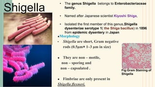
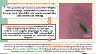
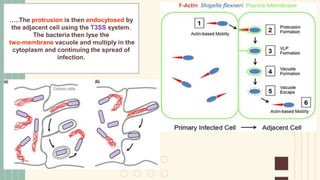
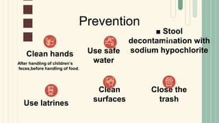
Shigella
Bacillary dysentery
Shigellosis
Irfana Azad
Sem:-M.sc 3rd sem
Enroll no:20072119008
CORD,Microbiology.
University of Kashmir
01
02
Symptoms
04
Abdominal Cramps,vomiting,fever
Clinical symptoms :
Bloodstained,mucopurulent stools
LaboratoryDiagnosis
05
Specimen collection,selective
media,transport media,microscopic
observations;etc
Treatment
Contents
Morphology,
culture characteristics,
Classification,etc
Shigella
Pathogenesis
Transmission..
virlunce factors:-virulence
plasmid,exotoxin,
endotoxin,etc ;pathogenesis.
06
03
Prevention
●Morphology
• Shigella are short, Gram negative
rods (0.5µm× 1–3 µm in size)
● They are non – motile,
non – sporing and
non – capsulated .
● Fimbriae are only present in
Shigella flexneri.
Fig:Gram Staining of
Shigella
• The genus Shigella belongs to Enterobacteriaceae
family.
• Named after Japanese scientist Kiyoshi Shiga,
• Isolated the first member of this genus,Shigella
dysenteriae serotype 1( the Shiga bacillus) in 1896
from epidemic dysentery in Japan
Shigella
Antigenic structure
of shigella:
• Posses Somatic “O“
antigens.
• Flagellar(H) antigen is
absent.
• Some strains posses
capsular “K“ antigens.
• Fimbrail antigens present
in some strains especially
Shigella flexneri.
Cultural characteristics:-
• Aerobes and facultative anaerobes. Optimum temperature is 37°C and optimum pH – 7.4.
• Non-lactose fermenters except Shigella sonnei.
• All shigella are mannitol fermenters except S.dysenteriae.
Media Colony Morphology
Xylose lysine deoxycholate
Agar(XLD)
Colonies are red__ no black centre
like salmonella
Deoxycholate citrate agar(DCA) Pale colonies,1-1.5mm
diameter,more translucent.
Salmonella shigella Agar Colorless colonies
Maconkey Agar Colorless colonies
Classification
Shigella are classified into 4
species(subgroups) and each
species further differentiated into
serotype:-
• Shigella dysenteriae (group A),
(16 serotypes);
• Shigella flexneri (group B), (6
serotypes);
• Shigella boydii (group C), (19
serotypes);
• Shigella sonnei (group D). (1
serotype).
• Transport medium: Carry Blair
medium;Sach’s buffered glycerol
saline
• Enrichment media:Salenite F broth
1:-Large virulence plasmid
214kb size
■ 31kb region(ipa-mxi –spa region) posses 50 to 60
virulence-associated genes.
■ This region encodes a type-III secretion system(T3SS)
and many effector proteins like invasion plasmid
antigens
( IpaA,IpaB,IpaC,IpaD) and IcsA
( Intracellular spread protein A).
■ Among Ipa, IpaB is an essential virulence factor》》
Control of T3SS secretion, phagsome escape and
macrophage apoptosis.
■IpaC 》Role in actin polymerization.
■ IpaD》Activation of T3SS
■IcsA》》secrete at the pole of Shigella daughter
cells,elicits polymerization of filamentous actin
2:Pathogenecity
islands(PAIs):-
■ Cluster of Genes
present on
plasmid and
chromosome.
■ Encode virulence
factors.
Virulence factors of shigella
a) Shigella enterotoxin 1
(ShET-1):-
● encoded by Genes on the
chromosome.
● found in S.flexneri.
b) Shigella enterotoxin 2
(ShET-2):-
• Plasmid encoded.
• Present in all four species of shigella.
ShET-1 and ShET-2 cause severe watery diarrhea(prodrome stage).
3:-Toxins
C) Shiga toxin(Stx):-
▪︎ Only produced by Shigella dysenteriae.
▪︎ AB5 toxin-one A subunit and 5 B subunits.
B subunit bind to GB3 (Globotriaosyceramide) receptor[GB3 receptor is present on
endothelial cells of the intestine, kidney
and the brain and thus these organs are susceptible to the toxicity of Shiga toxin.)
A subunit cleaves into A1-fragment and A2-fragment by furin.
A1-fragment has enzymatic activity,inhibits protein synthesis.
Infection dose
● 10-100 bacilli cells
● Acid tolerant
Transmission
• Transmitted by the faecal-oral route-primarily
via food,fingers,feces,flies and fomites (the
five “F'S")
Shigellosis
Or Bacillary
dysentry
Pathogenesis
Shigellae are Intracellular
parasites that multiply within the
cells of colonic epithelium..
• Infects intestinal
cells
Pathogenesis contd..
1:-The path through Microfold cells:when Shigella
reaches the large intestine,they are transcytosed
through the M (Microfold) cells into the subepithelial
space(membrane ruffling)
2:-The escape from immune cells:-In subepithelial space,
they are engulfed by the resident macrophages .
Inside the macrophage,the Shigella lyse the phagosome
and induce apoptosis.Shigella use T3SS to secrete IpaB in
order to escape from phagosome and induce macrophage
apoptosis.
The invasion process of Shigella is divided into 6 steps:-
Apoptotic or infected macrophage release cytokines,Interleukin:-IL-1B and IL-18 and these initiate
inflammatory and immune responses such as infiltration of PMN(neutrophils) for mediating bacterial
clearance.Transmigration of infiltrating PMNs through tight junctions of local epithelial cells into the
intestinal lumen facilitates invasion of Shigella from lumen into subepithelial space.
3:-Epithelial cell invasion:- The surviving
bacteria then invade colonic epithelial
cells basolaterally by assembling T3SS,
which secretes effector proteins into the
epithelial cells to induce bacterial
endocytosis.
4:-Intracellular multiplication:- The
endocytosed bacteria then lyse the endocytic
vacuoles by IpaB protein and replicate in the
cytoplasm of epithelial cell.
5:-Intra and intercellular spread:- Actin
polymerization occurs at the poles of daughter
cells and forms actin tail that help the bacteria to
move within the enterocyte cytoplasm and help
in cell to cell spread.Actin based motility.
In Intracellular spread, actin tails propel the
Shigella into protrusions impinging on adjacent
enterocytes.
…..The protrusion is then endocytosed by
the adjacent cell using the T3SS system.
The bacteria then lyse the
two-membrane vacuole and multiply in the
cytoplasm and continuing the spread of
infection.
6:- The destruction of host cells:- As a result of infection, colonic cells are
damaged and exhibit impaired absorption of water and nutrients, leading to
watery diarrhea accompanied by blood and mucus in stools.
Clinical symptoms:-
5 phases
1. Incubation period:-1-4days
2. Initial phase:- watery diarrhea with fever, malaise and vomiting.
3. Dysentery phase:- bloody mucopurulent stools,increased tenesmus and
abdominal cramps
4. Phase of complication:- commonly seen in children < 5 years age.
Intestinal complications:- toxic megacolon; perforations
Metabolic complications:- hypoglycemia;hyponatremia and dehydration.
Ekiri syndrome or toxic encephalopathy:- Metabolic complications+
neurological complications like abnormal posturing,delirium(confusion)
5. Postinfection phase:- patients expressing HLA-B27, develop an autoimmune
reaction months after infection……reactive arthritis; conjunctivitis and
urethritis.(Reiter syndrome)
163.4 million
cases per year worldwide due to shigellosis
5 to 11 lakh
deaths worldwide due to bacillary dysentery or shigellosis
Laboratory Diagnosis
Fresh Stool sample
Direct smear Shows large number of pus
cells, erythrocytes and macrophages
Culture analysis
XLD; DCA ; MacConkey agar
Gram staining
Treatment
■ Ciprofloxacin is the drug of choice.
■ Alternative drugs which are effective are
ceftriaxone,azithromycin,pivmecillinam
■ Duration of treatment is about 3days
except Shigella dysenteriae 1 infection-
5days.
■ ORS(Oral rehydration solution ) for
correction of dehydration.
■ Stool
decontamination with
sodium hypochlorite
Clean hands Use safe
water
Use latrines
Clean
surfaces
Close the
trash
Prevention
After handling of children’s
feces,before handling of food.
● Apurba S.Sastry(2021;third edition); Sandhya
Bhat.Essentials of medical microbiology.
● Prescott Microbiology.
● Molecular Pathogenesis of Shigella spp.:
Controlling Host Cell Signaling, Invasion, and
Death by Type III Secretion
Gunnar N. Schroeder and Hubert Hilbi
https://dx.doi.org/10.1128%2FCMR.00032-07
References
CREDITS: This presentation template was created by Slidesgo,
including icons by Flaticon, infographics & images by Freepik
Thanks!

shigellosis.pptx

  • 1.
    Shigella Bacillary dysentery Shigellosis Irfana Azad Sem:-M.sc3rd sem Enroll no:20072119008 CORD,Microbiology. University of Kashmir
  • 2.
    01 02 Symptoms 04 Abdominal Cramps,vomiting,fever Clinical symptoms: Bloodstained,mucopurulent stools LaboratoryDiagnosis 05 Specimen collection,selective media,transport media,microscopic observations;etc Treatment Contents Morphology, culture characteristics, Classification,etc Shigella Pathogenesis Transmission.. virlunce factors:-virulence plasmid,exotoxin, endotoxin,etc ;pathogenesis. 06 03 Prevention
  • 3.
    ●Morphology • Shigella areshort, Gram negative rods (0.5µm× 1–3 µm in size) ● They are non – motile, non – sporing and non – capsulated . ● Fimbriae are only present in Shigella flexneri. Fig:Gram Staining of Shigella • The genus Shigella belongs to Enterobacteriaceae family. • Named after Japanese scientist Kiyoshi Shiga, • Isolated the first member of this genus,Shigella dysenteriae serotype 1( the Shiga bacillus) in 1896 from epidemic dysentery in Japan Shigella
  • 4.
    Antigenic structure of shigella: •Posses Somatic “O“ antigens. • Flagellar(H) antigen is absent. • Some strains posses capsular “K“ antigens. • Fimbrail antigens present in some strains especially Shigella flexneri.
  • 5.
    Cultural characteristics:- • Aerobesand facultative anaerobes. Optimum temperature is 37°C and optimum pH – 7.4. • Non-lactose fermenters except Shigella sonnei. • All shigella are mannitol fermenters except S.dysenteriae. Media Colony Morphology Xylose lysine deoxycholate Agar(XLD) Colonies are red__ no black centre like salmonella Deoxycholate citrate agar(DCA) Pale colonies,1-1.5mm diameter,more translucent. Salmonella shigella Agar Colorless colonies Maconkey Agar Colorless colonies
  • 6.
    Classification Shigella are classifiedinto 4 species(subgroups) and each species further differentiated into serotype:- • Shigella dysenteriae (group A), (16 serotypes); • Shigella flexneri (group B), (6 serotypes); • Shigella boydii (group C), (19 serotypes); • Shigella sonnei (group D). (1 serotype). • Transport medium: Carry Blair medium;Sach’s buffered glycerol saline • Enrichment media:Salenite F broth
  • 7.
    1:-Large virulence plasmid 214kbsize ■ 31kb region(ipa-mxi –spa region) posses 50 to 60 virulence-associated genes. ■ This region encodes a type-III secretion system(T3SS) and many effector proteins like invasion plasmid antigens ( IpaA,IpaB,IpaC,IpaD) and IcsA ( Intracellular spread protein A). ■ Among Ipa, IpaB is an essential virulence factor》》 Control of T3SS secretion, phagsome escape and macrophage apoptosis. ■IpaC 》Role in actin polymerization. ■ IpaD》Activation of T3SS ■IcsA》》secrete at the pole of Shigella daughter cells,elicits polymerization of filamentous actin 2:Pathogenecity islands(PAIs):- ■ Cluster of Genes present on plasmid and chromosome. ■ Encode virulence factors. Virulence factors of shigella
  • 8.
    a) Shigella enterotoxin1 (ShET-1):- ● encoded by Genes on the chromosome. ● found in S.flexneri. b) Shigella enterotoxin 2 (ShET-2):- • Plasmid encoded. • Present in all four species of shigella. ShET-1 and ShET-2 cause severe watery diarrhea(prodrome stage). 3:-Toxins C) Shiga toxin(Stx):- ▪︎ Only produced by Shigella dysenteriae. ▪︎ AB5 toxin-one A subunit and 5 B subunits. B subunit bind to GB3 (Globotriaosyceramide) receptor[GB3 receptor is present on endothelial cells of the intestine, kidney and the brain and thus these organs are susceptible to the toxicity of Shiga toxin.) A subunit cleaves into A1-fragment and A2-fragment by furin. A1-fragment has enzymatic activity,inhibits protein synthesis.
  • 9.
    Infection dose ● 10-100bacilli cells ● Acid tolerant Transmission • Transmitted by the faecal-oral route-primarily via food,fingers,feces,flies and fomites (the five “F'S") Shigellosis Or Bacillary dysentry Pathogenesis
  • 10.
    Shigellae are Intracellular parasitesthat multiply within the cells of colonic epithelium.. • Infects intestinal cells Pathogenesis contd..
  • 11.
    1:-The path throughMicrofold cells:when Shigella reaches the large intestine,they are transcytosed through the M (Microfold) cells into the subepithelial space(membrane ruffling) 2:-The escape from immune cells:-In subepithelial space, they are engulfed by the resident macrophages . Inside the macrophage,the Shigella lyse the phagosome and induce apoptosis.Shigella use T3SS to secrete IpaB in order to escape from phagosome and induce macrophage apoptosis. The invasion process of Shigella is divided into 6 steps:- Apoptotic or infected macrophage release cytokines,Interleukin:-IL-1B and IL-18 and these initiate inflammatory and immune responses such as infiltration of PMN(neutrophils) for mediating bacterial clearance.Transmigration of infiltrating PMNs through tight junctions of local epithelial cells into the intestinal lumen facilitates invasion of Shigella from lumen into subepithelial space.
  • 12.
    3:-Epithelial cell invasion:-The surviving bacteria then invade colonic epithelial cells basolaterally by assembling T3SS, which secretes effector proteins into the epithelial cells to induce bacterial endocytosis. 4:-Intracellular multiplication:- The endocytosed bacteria then lyse the endocytic vacuoles by IpaB protein and replicate in the cytoplasm of epithelial cell. 5:-Intra and intercellular spread:- Actin polymerization occurs at the poles of daughter cells and forms actin tail that help the bacteria to move within the enterocyte cytoplasm and help in cell to cell spread.Actin based motility. In Intracellular spread, actin tails propel the Shigella into protrusions impinging on adjacent enterocytes.
  • 13.
    …..The protrusion isthen endocytosed by the adjacent cell using the T3SS system. The bacteria then lyse the two-membrane vacuole and multiply in the cytoplasm and continuing the spread of infection.
  • 14.
    6:- The destructionof host cells:- As a result of infection, colonic cells are damaged and exhibit impaired absorption of water and nutrients, leading to watery diarrhea accompanied by blood and mucus in stools. Clinical symptoms:- 5 phases 1. Incubation period:-1-4days 2. Initial phase:- watery diarrhea with fever, malaise and vomiting. 3. Dysentery phase:- bloody mucopurulent stools,increased tenesmus and abdominal cramps 4. Phase of complication:- commonly seen in children < 5 years age. Intestinal complications:- toxic megacolon; perforations Metabolic complications:- hypoglycemia;hyponatremia and dehydration. Ekiri syndrome or toxic encephalopathy:- Metabolic complications+ neurological complications like abnormal posturing,delirium(confusion) 5. Postinfection phase:- patients expressing HLA-B27, develop an autoimmune reaction months after infection……reactive arthritis; conjunctivitis and urethritis.(Reiter syndrome)
  • 15.
    163.4 million cases peryear worldwide due to shigellosis 5 to 11 lakh deaths worldwide due to bacillary dysentery or shigellosis
  • 16.
    Laboratory Diagnosis Fresh Stoolsample Direct smear Shows large number of pus cells, erythrocytes and macrophages Culture analysis XLD; DCA ; MacConkey agar Gram staining
  • 17.
    Treatment ■ Ciprofloxacin isthe drug of choice. ■ Alternative drugs which are effective are ceftriaxone,azithromycin,pivmecillinam ■ Duration of treatment is about 3days except Shigella dysenteriae 1 infection- 5days. ■ ORS(Oral rehydration solution ) for correction of dehydration.
  • 18.
    ■ Stool decontamination with sodiumhypochlorite Clean hands Use safe water Use latrines Clean surfaces Close the trash Prevention After handling of children’s feces,before handling of food.
  • 19.
    ● Apurba S.Sastry(2021;thirdedition); Sandhya Bhat.Essentials of medical microbiology. ● Prescott Microbiology. ● Molecular Pathogenesis of Shigella spp.: Controlling Host Cell Signaling, Invasion, and Death by Type III Secretion Gunnar N. Schroeder and Hubert Hilbi https://dx.doi.org/10.1128%2FCMR.00032-07 References
  • 20.
    CREDITS: This presentationtemplate was created by Slidesgo, including icons by Flaticon, infographics & images by Freepik Thanks!