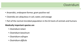
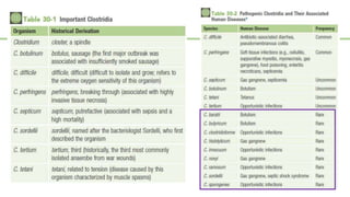
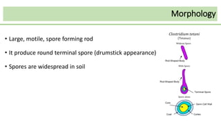
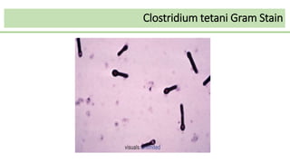
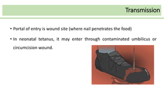
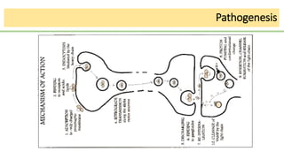
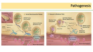
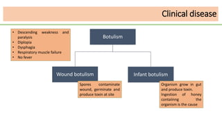
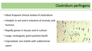
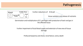
Spore forming Gram positive bacteria
Dr. Amirul Huda Bhuiyan
Lecturer, Department of Microbiology
Shaheed Syed Nazrul Islam Medical College, Kishorganj
classification
Spore forming GPB
Aerobic Bacillus
Anaerobic Clostridium
Clostridium
• Anaerobic, endospore former, gram positive rod
• Clostridia are ubiquitous in soil, water, and sewage
• Part of the normal microbial population in the GI tracts of animals and humans
Medically important species are
• Clostridium tetani
• Clostridium botulinum
• Clostridium erfingen
• Clostridium difficile
Clostridium tetani
Morphology
• Large, motile, spore forming rod
• It produce round terminal spore (drumstick appearance)
• Spores are widespread in soil
Clostridium tetani Gram Stain
Transmission
• Portal of entry is wound site (where nail penetrates the food)
• In neonatal tetanus, it may enter through contaminated umbilicus or
circumcision wound.
Toxin
Two toxins
• Oxygen-labile hemolysin (tetanolysin)
• Heat-labile neurotoxin (tetanospasmin), Plasmid encoded
Tetanolysin
• Clinical significance is unknown
• Inhibited by oxygen and serum cholesterol.
• serologically related to streptolysin O and the hemolysins.
Tetanospasmin
• Released during stationary phase of growth.
• Responsible for clinical manifestations of tetanus.
• Two part (A-B)
• Cleaved into a light (A-chain) subunit and a heavy
(B-chain) subunit by an endogenous protease.
• A chain has Zinc-endopeptidase activity.
Neuromuscular junction
Pathogenesis
Clinical disease
Incubation period
• varies from a few days to weeks.
• The duration of the incubation period is directly related to the distance of
the primary wound infection from the central nervous system.
Tetanus
Generalized
Tetanus
Neonatal tetanus Localized tetanus
Cephalic tetanus
Clinical disease
Generalized Tetanus
• Most common form.
• Involvement of the masseter muscles (trismus or lockjaw) is the presenting sign
in most patients.
• characteristic sardonic smile that results from the sustained contraction of the
facial muscles is known as risus sardonicus
• Other early signs are drooling, sweating, irritability, and persistent back spasms
(opisthotonos)
• The autonomic nervous system is involved in patients with more severe disease
Generalized Tetanus
Localized tetanus
• Disease remains confined to the musculature at the site of primary infection.
Cephalic tetanus
• A variant in which which the primary site of infection is the head
• The prognosis for patients with cephalic tetanus is very poor.
Neonatal tetanus
Neonatal tetanus (tetanus neonatorum)
• Typically associated with an initial infection of the umbilical stump
• progresses to become generalized.
• The mortality in infants exceeds 90%
• developmental defects are present in survivors.
• This is almost exclusively a disease in developing countries.
Neonatal tetanus
Diagnosis
• The diagnosis of tetanus is made on the basis of the clinical presentation.
• The microscopic detection of C. tetani or recovery in culture is useful but
frequently unsuccessful.
• Neither tetanus toxin nor antibodies to the toxin are detectable in the patient
because the toxin is rapidly bound to motor neurons and internalized
• If the organism is recovered in culture, production of toxin by the isolate can
be confirmed with the tetanus antitoxin neutralization test in mice
Treatment
• Debridement of the primary wound
• Use of penicillin or metronidazole to kill the bacteria and reduce toxin
production
• passive immunization with TIG to neutralize unbound toxin
• Vaccination with tetanus toxoid
• Toxic effects must be controlled symptomatically until the normal regulation
of synaptic transmission is restored.
Prevention
• Immunization with TT
• After trauma cleaning and debridement of wound and TT booster
• For grossly contaminated wound TT , TIG and penicillin administration
• Passive active immunization?
Prevention
Prevention
Prevention
Clostridium botulinum
• large, spore forming, anaerobic rods.
• Spores are widespread in soil, contaminate vegetables and meat.
• If foods are canned without sterilization, spore will survive and germinate
under anaerobic condition and bacteria will produce toxin
• Ingested preformed
Toxin
• Similar to tetanus toxin, C. botulinum toxin a protein (A-B toxin) consisting
of a small subunit (light, or A chain) with zinc-endopeptidase activity and
• A large, nontoxic subunit (B, or heavy chain).
• 08 immunologic type of toxin
• A,B and E are most common in human illness
Clinical use of toxin
Botox is a commercial preparation of exotoxin A used to remove wrinkles
on the face
Effective in minute amount in certain in muscle disorder such as
• Torticollis
• Writer’s cramp
• Blepharospasm
Pathogenesis
Clinical disease
Botulism
Wound botulism Infant botulism
Spores contaminate
wound, germinate and
produce toxin at site
Organism grow in gut
and produce toxin.
Ingestion of honey
contaiinng the
organism is the cause
• Descending weakness and
paralysis
• Diplopia
• Dysphagia
• Respiratory muscle failure
• No fever
Lab diagnosis
• Infant botulism is confirmed if toxin is detected in the infant’s feces or serum,
or the organism cultured from feces.
• Wound botulism is confirmed if toxin is detected in the patient’s serum or
wound, or if the organism is cultured from the wound
• Foodborne botulism is confirmed if toxin activity is demonstrated in the
implicated food or in the patient’s serum, feces, or gastric fluid.
• Isolation of C. botulinum from specimens contaminated with other
organisms (e.g., feces, wounds) can be improved by heating the specimen
for 10 minutes at 80° C to kill all non–spore-forming bacteria.
• Culture of the heated specimen on nutritionally enriched anaerobic media
allows the heat resistant C. botulinum spores to germinate.
• Demonstration of toxin production must be done with a mouse bioassay.
Lab diagnosis
Treatment
Patients with botulism require the following treatment measures:
• Adequate ventilatory support
• Elimination of the organism from the GI tract through the judicious use
of gastric lavage and metronidazole or penicillin therapy,
• Use of trivalent botulinum antitoxin versus toxins A, B, and E to
inactivate unbound toxin circulating in the bloodstream.
Prevention
Disease is prevented by
• Destroying the spores in food (virtually impossible for practical reasons)
• Preventing spore germination (by maintaining the food in an acid pH or storage at
4° C or colder)
• Destroying the preformed toxin (Toxins are inactivated by heating at 60° C to 100°
C for 10 minutes).
• Infant botulism has been associated with consumption of honey contaminated
with C. botulinum spores, so children younger than 1 year should not eat honey.
Clostridium perfingens
• Most frequent clinical isolate of clostridium
• Inhabits in soil and in intestine of animals and
humans.
• Rapidly grows in tissues and in culture
• Large, rectangular, gram positive bacilli
• Capsulated, non motile with subterminal
spore
Toxins and enzymes
Alpha toxin
• Phospholipase C (lecithinase);
• increase vascular permeability
• Haemolyti
• Produce necrotizing activity
Beta toxin
• Necrotizing activity in necrotizing enterocolitis
Epsilon toxin
• Increase vascular permeability of gastrointestinal wall
Iota toxin
• necrotizing activity
• increase vascular permeability
Entero toxin
• Produced primarily by type A strain
• Disrupts ion transport in ileum and jejunum by inserting into the cell membrane and
altering membrane permeability.
Toxins and enzymes
Clinical disease
Clostridial myonecrosis (gas gangrene)
• Military settings- Gutshot injuries
• Civilian cases - accidental injuries, surgical complication, injection of
medication such as epinephrine.
Reduced blood supply, tissue damage, blood clot and contamination of clostridial spores and
facultative organisms
Presence of foreign bodies eg. soil, clothing and metal fragments
Inflammatory reaction
Impaired tissue perfusion; stasis
Poor oxygenation, with reduction in local Eh
Impaired phagocytosis
Multiplication of facultative bacteria
Pyruvate of muscle is
incompletely oxidized and
lactic acid is accumulated
causing reducation of pH
Further reductions of Eh & pH
Pathogenesis
Germination and multiplication of Cl. perfingen with production of toxin and gas in
the affected tissue
Further reductions of Eh & pH
tissue autolysis and release of nutrients
Further impairment of local blood supply and extension of new area of tissue
damage
Profound toxaemia and shock; renal failure; coma; death
Pathogenesis
Lab diagnosis
Sample- faeces, food
Detection of toxin in stool by
• Reverse passive latex agglutination assay
• ELISA
• Tissue culture assay using vero cells with neutralizing antibody to
inhibit cytopathic effects.
Treatment
Simple wound contamination
• Care of wound
• removal of necrotic tissue
• cleansing
• Antibiotics - rarely
required
Anaerobic cellulitis:
• Opening the involved area
• removing all necrotic tissue
• cleansing thoroughly
• Antibiotics
Clostridial myonecrosis
• Care of wound : surgical removal of all infected and necrotic tissue
• Amputation- rapidly progressive infection involving limb
• Hysterectomy for uterine myonecrosis
• Antibiotics - Penicillin G/ Clindamycin or Metronidazole, Gentamicin or Tobramycin (for
facultative Gram negative organisms)
• Polyvalent gas gangrene antitoxin serum
• Hyperbaric oxygen - controversial
Treatment
Clostridium difficle
General criteria
• Part of normal microbial flora of GIT (3 – 5% adult, 40 – 50% healthy
neonates)
• Causes asymtomatic colonization, antibiotic associated diarrhea and
pseudomembranous colitis.
Morphology:
• large gram positive rod.
• having oval subterminal spore.
Virulence factors
Enterotoxin (Toxin A)
• produce chemotaxis; induce cytokine production, with hypersecretion of fluid;
• produces haemorrhagic necrosis.
Cytotoxin (Toxin B)
• induces depolymerization of actin with loss of cellular cytoskeleton.
Adhesin factor
• binding to colonic cells.
Transmission
• Exogenous - Person to person in hospital.
• Endogenous -Over growth of toxin producing strains after Rx of Antibiotics.
Antibiotics implicated in Cl.difficele Associated Diarrhoea and Colitis:
• Cephalosporin
• Ampicillin and amoxicillin
• Clindamycin.
• Other Penicillins, Macrolides, Tetracyclines,TMP-SMX.
• Disruption of normal colonic flora by Antibiotics.
• Colonization with Cl. difficile.
• Elaboration of toxin A and toxin B both of which mediate cytoskeletal
derangement in target cells
• mucosal injury and inflammation.
Pathogenesis
Lab diagnosis
Detection of C.difficile toxin in stool:
• Cytotoxicity assay (gold standard for diagnosis)
• ELISA
• Stool Culture (Anaerobic culture): Isolation of C.difficile is difficult.

Spore-forming gram Positive bacteria

  • 1.
    Spore forming Grampositive bacteria Dr. Amirul Huda Bhuiyan Lecturer, Department of Microbiology Shaheed Syed Nazrul Islam Medical College, Kishorganj
  • 2.
    classification Spore forming GPB AerobicBacillus Anaerobic Clostridium
  • 3.
    Clostridium • Anaerobic, endosporeformer, gram positive rod • Clostridia are ubiquitous in soil, water, and sewage • Part of the normal microbial population in the GI tracts of animals and humans Medically important species are • Clostridium tetani • Clostridium botulinum • Clostridium erfingen • Clostridium difficile
  • 5.
  • 6.
    Morphology • Large, motile,spore forming rod • It produce round terminal spore (drumstick appearance) • Spores are widespread in soil
  • 7.
  • 8.
    Transmission • Portal ofentry is wound site (where nail penetrates the food) • In neonatal tetanus, it may enter through contaminated umbilicus or circumcision wound.
  • 9.
    Toxin Two toxins • Oxygen-labilehemolysin (tetanolysin) • Heat-labile neurotoxin (tetanospasmin), Plasmid encoded
  • 10.
    Tetanolysin • Clinical significanceis unknown • Inhibited by oxygen and serum cholesterol. • serologically related to streptolysin O and the hemolysins.
  • 11.
    Tetanospasmin • Released duringstationary phase of growth. • Responsible for clinical manifestations of tetanus. • Two part (A-B) • Cleaved into a light (A-chain) subunit and a heavy (B-chain) subunit by an endogenous protease. • A chain has Zinc-endopeptidase activity.
  • 12.
  • 13.
  • 14.
    Clinical disease Incubation period •varies from a few days to weeks. • The duration of the incubation period is directly related to the distance of the primary wound infection from the central nervous system.
  • 15.
    Tetanus Generalized Tetanus Neonatal tetanus Localizedtetanus Cephalic tetanus Clinical disease
  • 16.
    Generalized Tetanus • Mostcommon form. • Involvement of the masseter muscles (trismus or lockjaw) is the presenting sign in most patients. • characteristic sardonic smile that results from the sustained contraction of the facial muscles is known as risus sardonicus • Other early signs are drooling, sweating, irritability, and persistent back spasms (opisthotonos) • The autonomic nervous system is involved in patients with more severe disease
  • 17.
  • 18.
    Localized tetanus • Diseaseremains confined to the musculature at the site of primary infection. Cephalic tetanus • A variant in which which the primary site of infection is the head • The prognosis for patients with cephalic tetanus is very poor.
  • 19.
    Neonatal tetanus Neonatal tetanus(tetanus neonatorum) • Typically associated with an initial infection of the umbilical stump • progresses to become generalized. • The mortality in infants exceeds 90% • developmental defects are present in survivors. • This is almost exclusively a disease in developing countries.
  • 20.
  • 21.
    Diagnosis • The diagnosisof tetanus is made on the basis of the clinical presentation. • The microscopic detection of C. tetani or recovery in culture is useful but frequently unsuccessful. • Neither tetanus toxin nor antibodies to the toxin are detectable in the patient because the toxin is rapidly bound to motor neurons and internalized • If the organism is recovered in culture, production of toxin by the isolate can be confirmed with the tetanus antitoxin neutralization test in mice
  • 22.
    Treatment • Debridement ofthe primary wound • Use of penicillin or metronidazole to kill the bacteria and reduce toxin production • passive immunization with TIG to neutralize unbound toxin • Vaccination with tetanus toxoid • Toxic effects must be controlled symptomatically until the normal regulation of synaptic transmission is restored.
  • 23.
    Prevention • Immunization withTT • After trauma cleaning and debridement of wound and TT booster • For grossly contaminated wound TT , TIG and penicillin administration • Passive active immunization?
  • 24.
  • 25.
  • 26.
  • 27.
  • 28.
    Clostridium botulinum • large,spore forming, anaerobic rods. • Spores are widespread in soil, contaminate vegetables and meat. • If foods are canned without sterilization, spore will survive and germinate under anaerobic condition and bacteria will produce toxin • Ingested preformed
  • 29.
    Toxin • Similar totetanus toxin, C. botulinum toxin a protein (A-B toxin) consisting of a small subunit (light, or A chain) with zinc-endopeptidase activity and • A large, nontoxic subunit (B, or heavy chain). • 08 immunologic type of toxin • A,B and E are most common in human illness
  • 30.
    Clinical use oftoxin Botox is a commercial preparation of exotoxin A used to remove wrinkles on the face Effective in minute amount in certain in muscle disorder such as • Torticollis • Writer’s cramp • Blepharospasm
  • 31.
  • 32.
    Clinical disease Botulism Wound botulismInfant botulism Spores contaminate wound, germinate and produce toxin at site Organism grow in gut and produce toxin. Ingestion of honey contaiinng the organism is the cause • Descending weakness and paralysis • Diplopia • Dysphagia • Respiratory muscle failure • No fever
  • 33.
    Lab diagnosis • Infantbotulism is confirmed if toxin is detected in the infant’s feces or serum, or the organism cultured from feces. • Wound botulism is confirmed if toxin is detected in the patient’s serum or wound, or if the organism is cultured from the wound • Foodborne botulism is confirmed if toxin activity is demonstrated in the implicated food or in the patient’s serum, feces, or gastric fluid.
  • 34.
    • Isolation ofC. botulinum from specimens contaminated with other organisms (e.g., feces, wounds) can be improved by heating the specimen for 10 minutes at 80° C to kill all non–spore-forming bacteria. • Culture of the heated specimen on nutritionally enriched anaerobic media allows the heat resistant C. botulinum spores to germinate. • Demonstration of toxin production must be done with a mouse bioassay. Lab diagnosis
  • 35.
    Treatment Patients with botulismrequire the following treatment measures: • Adequate ventilatory support • Elimination of the organism from the GI tract through the judicious use of gastric lavage and metronidazole or penicillin therapy, • Use of trivalent botulinum antitoxin versus toxins A, B, and E to inactivate unbound toxin circulating in the bloodstream.
  • 36.
    Prevention Disease is preventedby • Destroying the spores in food (virtually impossible for practical reasons) • Preventing spore germination (by maintaining the food in an acid pH or storage at 4° C or colder) • Destroying the preformed toxin (Toxins are inactivated by heating at 60° C to 100° C for 10 minutes). • Infant botulism has been associated with consumption of honey contaminated with C. botulinum spores, so children younger than 1 year should not eat honey.
  • 37.
  • 38.
    Clostridium perfingens • Mostfrequent clinical isolate of clostridium • Inhabits in soil and in intestine of animals and humans. • Rapidly grows in tissues and in culture • Large, rectangular, gram positive bacilli • Capsulated, non motile with subterminal spore
  • 39.
    Toxins and enzymes Alphatoxin • Phospholipase C (lecithinase); • increase vascular permeability • Haemolyti • Produce necrotizing activity Beta toxin • Necrotizing activity in necrotizing enterocolitis
  • 40.
    Epsilon toxin • Increasevascular permeability of gastrointestinal wall Iota toxin • necrotizing activity • increase vascular permeability Entero toxin • Produced primarily by type A strain • Disrupts ion transport in ileum and jejunum by inserting into the cell membrane and altering membrane permeability. Toxins and enzymes
  • 41.
    Clinical disease Clostridial myonecrosis(gas gangrene) • Military settings- Gutshot injuries • Civilian cases - accidental injuries, surgical complication, injection of medication such as epinephrine.
  • 42.
    Reduced blood supply,tissue damage, blood clot and contamination of clostridial spores and facultative organisms Presence of foreign bodies eg. soil, clothing and metal fragments Inflammatory reaction Impaired tissue perfusion; stasis Poor oxygenation, with reduction in local Eh Impaired phagocytosis Multiplication of facultative bacteria Pyruvate of muscle is incompletely oxidized and lactic acid is accumulated causing reducation of pH Further reductions of Eh & pH Pathogenesis
  • 43.
    Germination and multiplicationof Cl. perfingen with production of toxin and gas in the affected tissue Further reductions of Eh & pH tissue autolysis and release of nutrients Further impairment of local blood supply and extension of new area of tissue damage Profound toxaemia and shock; renal failure; coma; death Pathogenesis
  • 44.
    Lab diagnosis Sample- faeces,food Detection of toxin in stool by • Reverse passive latex agglutination assay • ELISA • Tissue culture assay using vero cells with neutralizing antibody to inhibit cytopathic effects.
  • 45.
    Treatment Simple wound contamination •Care of wound • removal of necrotic tissue • cleansing • Antibiotics - rarely required Anaerobic cellulitis: • Opening the involved area • removing all necrotic tissue • cleansing thoroughly • Antibiotics
  • 46.
    Clostridial myonecrosis • Careof wound : surgical removal of all infected and necrotic tissue • Amputation- rapidly progressive infection involving limb • Hysterectomy for uterine myonecrosis • Antibiotics - Penicillin G/ Clindamycin or Metronidazole, Gentamicin or Tobramycin (for facultative Gram negative organisms) • Polyvalent gas gangrene antitoxin serum • Hyperbaric oxygen - controversial Treatment
  • 47.
  • 48.
    General criteria • Partof normal microbial flora of GIT (3 – 5% adult, 40 – 50% healthy neonates) • Causes asymtomatic colonization, antibiotic associated diarrhea and pseudomembranous colitis. Morphology: • large gram positive rod. • having oval subterminal spore.
  • 49.
    Virulence factors Enterotoxin (ToxinA) • produce chemotaxis; induce cytokine production, with hypersecretion of fluid; • produces haemorrhagic necrosis. Cytotoxin (Toxin B) • induces depolymerization of actin with loss of cellular cytoskeleton. Adhesin factor • binding to colonic cells.
  • 50.
    Transmission • Exogenous -Person to person in hospital. • Endogenous -Over growth of toxin producing strains after Rx of Antibiotics. Antibiotics implicated in Cl.difficele Associated Diarrhoea and Colitis: • Cephalosporin • Ampicillin and amoxicillin • Clindamycin. • Other Penicillins, Macrolides, Tetracyclines,TMP-SMX.
  • 51.
    • Disruption ofnormal colonic flora by Antibiotics. • Colonization with Cl. difficile. • Elaboration of toxin A and toxin B both of which mediate cytoskeletal derangement in target cells • mucosal injury and inflammation. Pathogenesis
  • 52.
    Lab diagnosis Detection ofC.difficile toxin in stool: • Cytotoxicity assay (gold standard for diagnosis) • ELISA • Stool Culture (Anaerobic culture): Isolation of C.difficile is difficult.