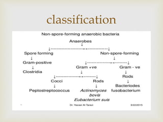
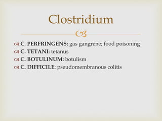
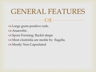
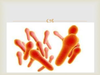
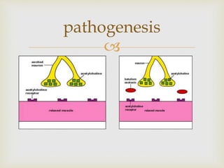
An anaerobe is an organism that does not require oxygen for growth. The document discusses various Clostridium species that are anaerobic bacteria. Clostridium perfringens can cause gas gangrene through secretion of exotoxins and enzymes. It is also a common cause of food poisoning. Clostridium botulinum produces a neurotoxin that causes botulism. Clostridium tetani secretes a toxin that causes the muscles spasms seen in tetanus. Clostridium difficile is associated with antibiotic-associated diarrhea and pseudomembranous colitis.
A presentation on food poisoning (bacterial and mycotoxins): Definition, causative agents, pathogenesis, clinical features and laboratory diagnosis of food poisoning.
my presentation on topic of yersinia species..... i think it will be enough for bachler student for knowledge ......
if there any question ... u guys may ask .......
hope all the best
thanks ....
The following presentation is only for quick reference. I would advise you to read the theoretical aspects of the respective topic and then use this presentation for your last minute revision. I hope it helps you..!!
Mayur D. Chauhan
These bacteria make spores, which act like protective coatings that help the bacteria survive. Under certain conditions, such as when food is kept at an unsafe temperature (between 40°F–140°F), C. perfringens can grow and multiply. After someone swallows the bacteria, it can produce a toxin (poison) that causes diarrhea.
Common sources of C. perfringens infection include meat, poultry, gravies, and other foods cooked in large batches and held at an unsafe temperature. Outbreaks tend to happen in places that serve large groups of people, such as hospitals, school cafeterias, prisons, and nursing homes, and at events with catered food. C. perfringens outbreaks occur most often in November and December. Many of these outbreaks have been linked to foods commonly served during the holidays, such as turkey and roast beef.
Anyone can get food poisoning from C. perfringens. Young children and older adults are at higher risk for severe illness.
Foodborne infection- ingestion of viable pathogens along with food e.g. typhoid
Foodborne intoxication (poisoning)- ingestion of foods containing preformed toxins e.g. botulism, staphylococcal poisoning
Toxoplasmosis is considered one of the neglected parasitic infections of the United States, a group of five parasitic diseases that have been targeted by CDC for public health action.Q fever is a disease caused by the bacteria Coxiella burnetii. This bacteria naturally infects some animals, such as goats, sheep, and cattle. C. burnetii bacteria are found in the birth products (i.e. placenta, amniotic fluid), urine, feces, and milk of infected animals.
MANAGEMENT OF ATRIOVENTRICULAR CONDUCTION BLOCK.pdfJim Jacob Roy
Cardiac conduction defects can occur due to various causes.
Atrioventricular conduction blocks ( AV blocks ) are classified into 3 types.
This document describes the acute management of AV block.
Report Back from SGO 2024: What’s the Latest in Cervical Cancer?bkling
Are you curious about what’s new in cervical cancer research or unsure what the findings mean? Join Dr. Emily Ko, a gynecologic oncologist at Penn Medicine, to learn about the latest updates from the Society of Gynecologic Oncology (SGO) 2024 Annual Meeting on Women’s Cancer. Dr. Ko will discuss what the research presented at the conference means for you and answer your questions about the new developments.
Title: Sense of Taste
Presenter: Dr. Faiza, Assistant Professor of Physiology
Qualifications:
MBBS (Best Graduate, AIMC Lahore)
FCPS Physiology
ICMT, CHPE, DHPE (STMU)
MPH (GC University, Faisalabad)
MBA (Virtual University of Pakistan)
Learning Objectives:
Describe the structure and function of taste buds.
Describe the relationship between the taste threshold and taste index of common substances.
Explain the chemical basis and signal transduction of taste perception for each type of primary taste sensation.
Recognize different abnormalities of taste perception and their causes.
Key Topics:
Significance of Taste Sensation:
Differentiation between pleasant and harmful food
Influence on behavior
Selection of food based on metabolic needs
Receptors of Taste:
Taste buds on the tongue
Influence of sense of smell, texture of food, and pain stimulation (e.g., by pepper)
Primary and Secondary Taste Sensations:
Primary taste sensations: Sweet, Sour, Salty, Bitter, Umami
Chemical basis and signal transduction mechanisms for each taste
Taste Threshold and Index:
Taste threshold values for Sweet (sucrose), Salty (NaCl), Sour (HCl), and Bitter (Quinine)
Taste index relationship: Inversely proportional to taste threshold
Taste Blindness:
Inability to taste certain substances, particularly thiourea compounds
Example: Phenylthiocarbamide
Structure and Function of Taste Buds:
Composition: Epithelial cells, Sustentacular/Supporting cells, Taste cells, Basal cells
Features: Taste pores, Taste hairs/microvilli, and Taste nerve fibers
Location of Taste Buds:
Found in papillae of the tongue (Fungiform, Circumvallate, Foliate)
Also present on the palate, tonsillar pillars, epiglottis, and proximal esophagus
Mechanism of Taste Stimulation:
Interaction of taste substances with receptors on microvilli
Signal transduction pathways for Umami, Sweet, Bitter, Sour, and Salty tastes
Taste Sensitivity and Adaptation:
Decrease in sensitivity with age
Rapid adaptation of taste sensation
Role of Saliva in Taste:
Dissolution of tastants to reach receptors
Washing away the stimulus
Taste Preferences and Aversions:
Mechanisms behind taste preference and aversion
Influence of receptors and neural pathways
Impact of Sensory Nerve Damage:
Degeneration of taste buds if the sensory nerve fiber is cut
Abnormalities of Taste Detection:
Conditions: Ageusia, Hypogeusia, Dysgeusia (parageusia)
Causes: Nerve damage, neurological disorders, infections, poor oral hygiene, adverse drug effects, deficiencies, aging, tobacco use, altered neurotransmitter levels
Neurotransmitters and Taste Threshold:
Effects of serotonin (5-HT) and norepinephrine (NE) on taste sensitivity
Supertasters:
25% of the population with heightened sensitivity to taste, especially bitterness
Increased number of fungiform papillae
Anti ulcer drugs and their Advance pharmacology ||
Anti-ulcer drugs are medications used to prevent and treat ulcers in the stomach and upper part of the small intestine (duodenal ulcers). These ulcers are often caused by an imbalance between stomach acid and the mucosal lining, which protects the stomach lining.
||Scope: Overview of various classes of anti-ulcer drugs, their mechanisms of action, indications, side effects, and clinical considerations.
These simplified slides by Dr. Sidra Arshad present an overview of the non-respiratory functions of the respiratory tract.
Learning objectives:
1. Enlist the non-respiratory functions of the respiratory tract
2. Briefly explain how these functions are carried out
3. Discuss the significance of dead space
4. Differentiate between minute ventilation and alveolar ventilation
5. Describe the cough and sneeze reflexes
Study Resources:
1. Chapter 39, Guyton and Hall Textbook of Medical Physiology, 14th edition
2. Chapter 34, Ganong’s Review of Medical Physiology, 26th edition
3. Chapter 17, Human Physiology by Lauralee Sherwood, 9th edition
4. Non-respiratory functions of the lungs https://academic.oup.com/bjaed/article/13/3/98/278874
ARTIFICIAL INTELLIGENCE IN HEALTHCARE.pdfAnujkumaranit
Artificial intelligence (AI) refers to the simulation of human intelligence processes by machines, especially computer systems. It encompasses tasks such as learning, reasoning, problem-solving, perception, and language understanding. AI technologies are revolutionizing various fields, from healthcare to finance, by enabling machines to perform tasks that typically require human intelligence.
2. Presented by
DR KHALID YOUSUF MEMON
LECTURER
LIAQUAT UNIVERSITY OF MEDICAL &
HEALTH SCIENCES JAMSHORO
3.
An anaerobic organism or anaerobe is any organism
that does not require oxygen for growth. It may react
negatively or even die if oxygen is present.
DEFINATION
9.
C. PERFRINGENS: gas gangrene; food poisoning
C. TETANI: tetanus
C. BOTULINUM: botulism
C. DIFFICILE: pseudomembranous colitis
Clostridium
10.
Large gram-positive rods.
Anaerobic.
Spore Forming: Rackit shape
Most clostridia are motile by flagella.
Mostly Non Capsulated
GENERAL FEATURES
12.
Clostridia, part of the intestinal flora They are found in
SOIL,
SEWAGE
when introduced into tissues (for example, by a break in
the skin resulting from surgery or trauma).
Endospore formation facilitates their persistence in the
environment.
Spores are resistant to chemical disinfectants and may
withstand ultraviolet irradiation or boiling temperatures
for some time, although not standard autoclaving
conditions (121°C for 15 minutes at increased pressure).
13.
C. perfringens is a large, non-motile, gram-positive,
encapsulated bacillus.
It is ubiquitous in nature
Its vegetative form as part of the normal flora of the
vagina and gastrointestinal (GI) tract.
Its spores are found in soil
Clostridium perfringens
14.
Can cause
ANAEROBIC CELLULITIS
MYONECROSIS (gas gangrene)
FOOD POISONING Some strains of C. perfringens
can cause food poisoning
C. perfringens
15.
C. perfringens secretes a variety of
EXOTOXINS,
ENTEROTOXINS
HYDROLYTIC ENZYMES (that facilitate the disease
process )
Pathogenesis:
16.
C. perfringens elaborates at least 12 exotoxins,
designated by Greek letters.
The most important of these
α TOXIN ( lesithenase C )
PERFRINGOLYSIN O,
THETA(Θ) TOXIN,
EXOTOXINS
18.
.C. perfringens strains are grouped A to E on the
basis of their spectrum of exotoxins.
TYPE A STRAINS, which produce both α toxin and
enterotoxin, are responsible for most human
clostridial infections.
STRAINS
19.
C. PERFRINGENS ENTEROTOXIN, a small, heat-
labile protein, acts in the lower portion of the small
intestine. The molecule binds to receptors on the
epithelial cell surface and alters the cell membrane,
disrupting ion transport (primarily in the ileum) and
leading to loss of fluid and intracellular proteins.
Interestingly, enterotoxin- They are heat resistant,
the spores remaining viable for longer than an hour
at 100oC, enhancing their threat as foodborne
pathogens.
Enterotoxin:
21. PROTEASES,
DNASES,
HYALURONIDASE,
COLLAGENASES,
These liquefy tissue and promote the spread of
infection. The resulting degradation products serve
as fermentation substrates for the rapid metabolism
of C. perfringens.
This organism has one of the fastest doubling times
recorded, at less than 10 minutes
Degradative enzymes:
23.
Clostridial spores are introduced into tissue, for
example, by
Contamination with infected soil,
Endogenous transfer from the intestinal tract.
Severe and open wounds, such as compound
fractures
Ischemia producing injuries (for example, crush
injuries), are a prime predisposing condition
1.MYONECROSIS (GAS GANGRENE)
25.
α Toxin and other exotoxins are secreted, and
extensive cell death ensues. Production of enzymes
that break down extracellular matrix facilitates the
spread of infection. Fermentation of tissue
carbohydrates, lipids, and amino acids yields gas,
and an accumulation of gas bubbles in the
subcutaneous spaces produces a crinkling sensation
on palpation (crepitation), hence, the name “gas
gangrene”
pathogenesis
26.
The exudates are copious and foul smelling. Leads
in systemic effects, such as shock, renal failure, and
intravascular hemolysis.
Untreated clostridial myonecrosis is uniformly fatal
within days of the initiation of gangrene
presentation
27.
This is a clostridial infection of connective tissue in
which the spread of bacterial growth along fascial
planes (fasciitis) does not involve invasion of muscle
tissue.
Necrotizing processes play a more limited role, but
surgical intervention is generally unsuccessful
(unless it is carried out very promptly and
aggressively) because of the rapid spread of infection
and compromise of blood supply due to swelling
beneath tight fascia.
2.Anaerobic cellulitis:
28.
C. perfringens is a common cause of foodborne infection
in the United States. Meats, meat products, and gravy are
the most commonly implicated foods in C. perfringens
foodborne illness.
Typically, the onset of
NAUSEA,
ABDOMINAL CRAMPS,
AND DIARRHEA occurs 8 to 18 hours after eating
contaminated food. Fever is absent and vomiting rare.
The attack is usually self-limited, with recovery within 1
to 2 days.
Foodborne infection
29.
Outbreaks of a necrotizing bowel disease with high
mortality (greater than 50 percent) caused by C.
perfringens have been sporadically reported.
Necrotic enteritis
30.
This condition is a grave complication of incomplete
abortion or the use of inadequately sterilized
instruments. Gangrenous infection of uterine tissue
is followed by illness due to toxins and bacteremia.
Clostridial endometritis
31.
Diagnosis of clostridial myonecrosis or cellulitis rests
largely on clinical presentation.
GRAM STAIN, however, specimens from diseased
tissue usually show vegetative clostridial forms
(large,gram-positive rods), accompanied by other
bacteria and cellular debris.
CULTURED ANAEROBICALLY on
blood agar,(double hemolysis) alpha toxin and
perfringolysin O
Lactose egg yolk medium
Laboratory identification:
32.
C perfringens produce an opacity in medium egg
yolk agar containing lecithin due to lecithenase c
activity (alpha toxin). This opacity is inhibited by
applying specific anti toxic serum to medium which
will inactivate lecithenase this technique is referred
as Nagler reaction
Nagler reaction
34. IMMEDIATE AND THOROUGH REMOVAL OF
FOREIGN MATERIAL and devitalized tissue and
exposure of the wound toO2.
HYPERBARIC OXYGEN CHAMBERS increase the
tissue O2 tension in the affected part and inhibit the
pathologic process.
If DEBRIDEMENT is unable to control the
progression of the gangrene,
AMPUTATION, when anatomically possible, is
mandatory in gangrene.
ADMINISTRATION OF ANTIBIOTICS
Penicillin
Treatment and prevention:
35.
C. botulinum causes botulism, which occurs in
several clinical forms.
Botulism is caused by the action of a neurotoxin that
is one of the most potent poisons known and causes
a flaccid paralysis. due to ingestion of toxin-
contaminated food.
CLOSTRIDIUM BOTULINUM
36.
C. botulinum is found worldwide in soil and aquatic
sediments, and the spores frequently contaminate
vegetables and meat or fish.
Under appropriate conditions,, outbreaks frequently
occur in families or other eating groups
Epidemiology:
37.
Types of botulinum toxin, A through G,
In human disease is almost always caused by types A, B,
or E.
Cause neurotoxicity arises from proteolytic cleavage of
specific synaptic vesicle peptides, causing subsequent
failure of neurotransmission.
Botulinum toxins affect peripheral cholinergic synapses
by blocking the neuromuscular junction and inhibiting
release of the neurotransmitter acetylcholine, preventing
contraction and causing flaccid paralysis (B).
Pathogenesis:
39.
Food poisoning in which a patient first begins to
experience difficulties in
FOCUSING VISION (12 to 36 hours).
A progressive paralysis of striated muscle groups
develops, and mortality rate is about 15 percent, with
the patient usually succumbing to respiratory
paralysis. Recovery, which involves regeneration of
the damaged nerves, is protracted, lasting several
weeks.
. Classic botulism
40.
Supplementation of infant foods (cereals or formula)
with raw honey, which is contaminated with C.
botulinum spores, may transmit the organism. The
condition is possibly a cause of sudden infant death
syndrome, but recovery is the usual outcome,
Following symptomatic treatment that may be
prolonged.
Infant botulism
41.
A rare form of botulism occurs when a wound
becomes contaminated with the organism, and toxin
is absorbed from that site. The molecular
pathogenesis of this infection is similar to that of
tetanus.
Wound botulism:
42.
The organism can be cultured and identified by
standard anaerobic methods . Toxin is also
identifiable in serum, stool, and food.
Laboratory identification:
43.
Antitoxin, which neutralizes unbound botulinum
toxin, should be administered as soon as possible in
suspected botulinal intoxication.
A trivalent (A, B, E) horse antiserum is available
from the Centers for Disease Control.
Supportive measures, including mechanical
ventilation, may be required. In wound and infant
botulism, the infection can be treated with penicillin
or other antibiotics to which the organism is
sensitive.
Treatment and prevention
44.
The introduction of C. tetani spores into even small
wounds via contaminated soil.
Growth of C. tetani is completely local, but it
produces a powerful neurotoxin that is transported
to the central nervous system, where it causes spastic
paralysis.
D. Clostridium tetani
45.
: C. tetani spores are common in barnyard,
garden,and other soils.
The most typical focus of infection in tetanus is a
puncture wound caused which tetanus spores can
germinate and grow.
Special circumstances may also lead to tetanus, for
example, after severe burns, surgery or ischemia.
Illicit drugs can contain spores that are introduced
by injection
Epidemiology
46.
Tetanus toxin, called tetanospasmin, is an extremely
potent toxin. It is transported from an infected locus
by retrograde neuronal flow or blood.
(A, or activity subunit) The A subunit blocks
neurotransmitter release at inhibitory synapses, there
by causing severe, prolonged muscle spasm small
synaptic vesicle protein (synaptobrevin) and
abolishing the flow of inhibitory neurotransmitters,
including glycine and γ-aminobutyric acid.
Pathogenesis:
48.
INCUBATION PERIOD---- 4 DAYS TO WEEKS
Tetanus presents as a spastic paralysis, in which muscle
spasms often first involve the site of infection.
In the early stages of the disease, the jaw muscles are
affected, so that the mouth cannot open (trismus, or
“lockjaw”).
Gradually, other voluntary muscles become involved and
any external stimulus (for example, noise or bright light)
precipitates a painful spasm and, sometimes, convulsions.
Death, which occurs in 50 to 60 percent of cases, is
usually the result of paralysis of chest muscles leading to
respiratory failure
Tetanus
54.
The diagnosis of tetanus is based largely on clinical
findings.
C. tetani has a characteristic morphology, with a
long, slender rod and round, terminal spore (racket-
shaped bacillus),
swarming growth on anaerobic blood agar.
Laboratory identification:
56.
ADMINISTRATION OF ANTITOXIN to neutralize any
toxin not yet bound to neurons is the first order of
treatment.
HUMAN HYPERIMMUNE GLOBULIN (tetanus
immune globulin) is preferred, but, in countries where it
is not available, horse antitoxin is used.
ANTIBIOTICS The organism is sensitive to penicillin,
DEBRIDEMENT OF NECROTIC TISSUE at the entry
wound.
SEDATIVES AND MUSCLE RELAXANTS to minimize
spasms and attention to maintenance of ventilation
Treatment:
57.
ACTIVE IMMUNIZATION WITH TETANUS TOXOID
(formalin inactivated toxin) prevents tetanus. It is usually
administered to children as a triple vaccine with diphtheria
toxoid and pertussis antigens (DTP).
Recent studies have confirmed that circulating antibody levels
gradually decline and that many older individuals lose
protection. Therefore, booster immunizations with a
preparation of diphtheria and tetanus toxoids given every 10
years throughout life are recommended.
TETANUS IMMUNOGLOBULIN can be used to give
immediate passive immunity to injury victims with no history
of immunization. Active immunization should also be started.
Antitoxin and toxoid, administered in different areas of the
body, can be given simultaneously
Prevention
58.
Diarrhea, a common complication of antimicrobial
drug treatment, can range from loose stools to life-
threatening pseudomembranous colitis (PMC)
C. difficile is estimated to be responsible for at least
one fourth of antibiotic-associated diarrheas
E. Clostridium difficile
59.
In hospitalized patients and almost all cases of PMC.
After its introduction to a site, the environment (that
is, dust, bedding, toilets, etc.) becomes persistently
contaminated with spores, and new residents are
easily colonized. They are then at higher risk for
developing the adverse intestinal effect of antibiotic
treatments.
60.
C. difficile is a minor component of the normal flora of
the large intestine.
When antimicrobial treatment suppresses more
predominant species in this community,
C. difficile proliferates Pathogenic strains produce two
toxic polypeptides, designated toxins A and B.
Toxin A is an enterotoxin that causes excessive fluid
secretion, but also stimulates an inflammatory response,
and has some cytopathic effect in tissue culture.
Toxin B is a cytotoxin. In tissue culture, it disrupts
protein synthesis and causes disorganization of the
cytoskeleton.
1. Pathogenesis
61.
The three drugs most commonly implicated are
CLINDAMYCIN,
AMPICILLIN
CEPHALOSPORINS.
The pseudo membranous exudate, composed of mucus,
fibrin, nflammatory cells, and cell debris overlying an
ulcerated epithelium, is best demonstrated by endoscopy.
PMC often begins some time after cessation of drug
treatment or may recur after what should be adequate
therapy. This is a consequence of the stability and
persistence of the spores formed by C. difficile
Clinical significance:
62.
CULTURED FROM STOOLS and identified by
routine anaerobic procedures, but the more rapid
and useful tests are directed at demonstrating toxin
production in stool extracts.
ENZYME IMMUNOASSAYS (ELISA, for exotoxins
A and B have replaced earlier immunologic or tissue
culture cytotoxicity assays.
POLYMERASE CHAIN REACTION (PCR)–based
detection strategies are also widely available.
3. Laboratory identification
63.
: Discontinuance of the predisposing drug and fluid
replacement usually lead to resolution of the
symptoms.
Relapses, however, are common. Oral
administration of metronidazole or vancomycin is
usually added Reconstitution of the host's normal
colonic flora may aid in the recover
Treatment
64.
Members of the genus Bacteroides are the predominant
anaerobes found in the human colon. They are part of the
normal flora and only cause disease when they gain access to
tissues or the blood during bowel penetration (for example,
during surgery or trauma).
They are, however, the most common cause of serious
infections by anaerobic organisms.
Bacteroides are slender rods or cocco - bacilli.
Their polysaccharide capsule is an important virulence factor ,
conveying resistance to phagocytosis.
Purified capsule alone is sufficient to induce abscess formation
in laboratory animals, indicating that this polysaccharide is key
to the pathology seen with Bacteroides infections.
A. Bacteroides
65.
Bacteroides are transmitted from the colon to the
blood or peritoneum following abdominal trauma.
Therefore, the source of infection is endogenous (it is
not transmitted from person to person
Epidemiology:
66.
The major disease-causing Bacteroides species is
Bacteroides fragilis.
When released from the colon into the blood, B.
fragilis multiplies rapidly, causing bacteremia.
If it is introduced into the abdominal cavity, B.
fragilis causes peritonitis, and/or abdominal
abscesse
Pathology and clinical
significance:
67.
: Exudates from mixed anaerobic lesions are often
copious and noticeably foul smelling.
A Gram stain of such exudates shows numerous
faint, slender, gram-negative rods, usually in mixed
flora. The organisms are easily obscured by debris
and polymorphonuclear leukocytes.
Laboratory identification
68.
Drug resistance is common among the Bacteroides.
Metronidazole is the antibiotic of choice for B. fragilis
infections.
Alternative choices include
Ampicillin-Sulbactam, Imipenem-Cilastatin,
Ticarcillin-clavulanate, Cefoxitin, or Clindamycin.
Surgical drainage of any abscess is essential to ensure
penetration of drugs. To prevent Bacteroides
contamination of a surgical wound,
Perioperative antibiotic, such as cefoxitin, can be
administered.
Treatment and prevention: