Downloaded 156 times
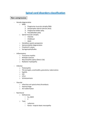
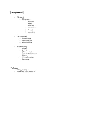
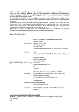

The document classifies spinal cord disorders into several categories: non-compressive (hereditodegenerative, inflammatory, infectious, vascular, and nutritional), and compressive (extradural, extramedullary, and intramedullary). It then provides examples of specific conditions that fall under each category. It describes a case of a 16-year old boy who developed sudden paraplegia and was found to have a focal demyelinating lesion of the spinal cord seen on MRI. His condition did not improve with IV steroids. The management of paraplegia involves symptomatic care of the bladder, bowels and limbs. It also requires supportive measures like skin care to prevent pressure sores, D


![APPROACH TO NON COMPRESSIVE MYELOPATHY [Autosaved].pptx](https://cdn.slidesharecdn.com/ss_thumbnails/approachtononcompressivemyelopathyautosaved-240930073343-8621cff7-thumbnail.jpg?width=640&height=640&fit=bounds)


