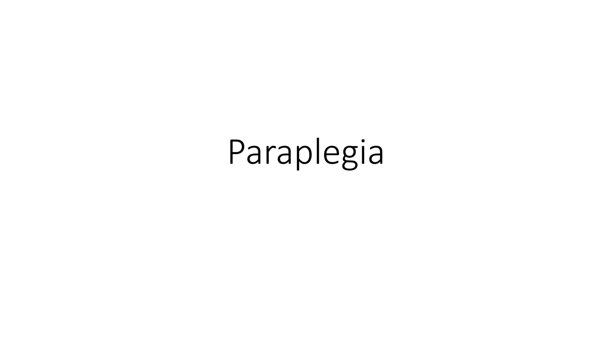
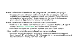
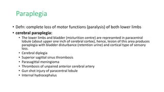
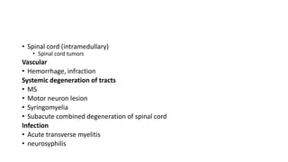
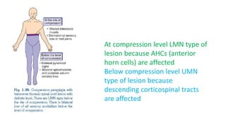
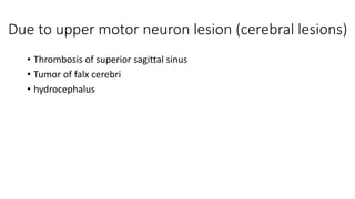
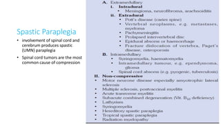
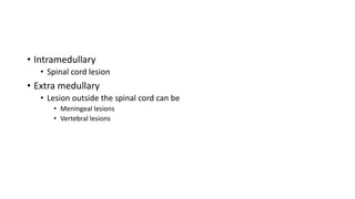
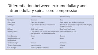
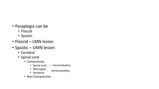
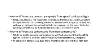
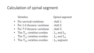
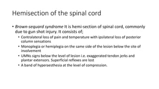
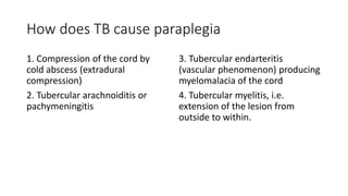
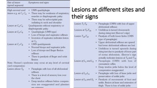
Paraplegia
• Paraplegia can be
• Flaccid
• Spastic
• Flaccid – LMN lesion
• Spastic – UMN lesion
• Cerebral
• Spinal cord
• Compressive
• Spinal cord
• Meningeal
• Vertebral
• Non-Compressive
extramedullary
intramedullary
• How to differentiate cerebral paraplegia from spinal cord paraplegia
• Headache, trauma, risk factors for thrombosis, frontal release signs, problem in
cognition (abstract thinking, memory), isolated cortical type of sensory loss with
preservation of sensation (can’t do stereognosis on the lower limbs but can do
graphesthesia or 2 point discrimination), retention of urine
• How to differentiate compressive from non-compressive?
• While we do the sensory examination we will find a segment that has LMN type of
lesion or a clear cut sensory level (with hyperesthesia, analgesia)
• In addition in compressive type there might be bone deformities, root pain
• How to differentiate intramedullary from extramedullary
• Root pain, vertebral tenderness, asymmetry, easily understandable sensory loss
(contralateral P & T with ipsilateral V & P if hemiparetic or monoparetic), bladder and
bowel involvement in early, prominent UMN sign
Paraplegia
• Defn: complete loss of motor functions (paralysis) of both lower limbs
• cerebral paraplegia:
• The lower limbs and bladder (micturition centre) are represented in paracentral
lobule (about upper one inch of cerebral cortex), hence, lesion of this area produces
paraplegia with bladder disturbance (retention urine) and cortical type of sensory
loss.
• Cerebral diplegia
• Superior sagittal sinus thrombosis
• Parasagittal meningioma
• Thrombosis of unpaired anterior cerebral artery
• Gun shot injury of paracentral lobule
• Internal hydrocephalus
Due to upper motor neuron lesion (spinal lesions)
Spinal cord compression
• Vertebral (extradural)
• Pott’s disease
• Mx carcinoma (from breast, lung, prostate)
• Multiple myeloma
• Intervertebral disc prolapse, fracture
• Cervical spondylosis
• Meninges (intradural)
• Tumors( meningioma, lymphoma, metastasis)
• Epidural abscess
• Spinal cord (intramedullary)
• Spinal cord tumors
Vascular
• Hemorrhage, infraction
Systemic degeneration of tracts
• MS
• Motor neuron lesion
• Syringomyelia
• Subacute combined degeneration of spinal cord
Infection
• Acute transverse myelitis
• neurosyphilis
At compression level LMN type of
lesion because AHCs (anterior
horn cells) are affected
Below compression level UMN
type of lesion because
descending corticospinal tracts
are affected
Due to upper motor neuron lesion (cerebral lesions)
• Thrombosis of superior sagittal sinus
• Tumor of falx cerebri
• hydrocephalus
Spastic Paraplegia
• involvement of spinal cord and
cerebrum produces spastic
(UMN) paraplegia
• Spinal cord tumors are the most
common cause of compression
Flaccid paraplegia (Due to lower motor
neuron lesion)
Flaccid paralysis resulting from
the diseases involving anterior
horns cells, radicals,
peripheral nerves and muscles
• Intramedullary
• Spinal cord lesion
• Extra medullary
• Lesion outside the spinal cord can be
• Meningeal lesions
• Vertebral lesions
Differentiation between extramedullary and
intramedullary spinal cord compression
• Paraplegia can be
• Flaccid
• Spastic
• Flaccid – LMN lesion
• Spastic – UMN lesion
• Cerebral
• Spinal cord
• Compressive
• Spinal cord
• Meningeal
• Vertebral
• Non-Compressive
extramedullary
intramedullary
• How to differentiate cerebral paraplegia from spinal cord paraplegia
• Headache, trauma, risk factors for thrombosis, frontal release signs, problem
in cognition (abstract thinking, memory), isolated cortical type of sensory loss
with preservation of sensation (can’t do stereognosis on the lower limbs but
can do graphesthesia or 2 point discrimination), retention of urine
• How to differentiate compressive from non-compressive?
• While we do the sensory examination we will find a segment that has LMN
type of lesion or a clear cut sensory level (with hyperesthesia, analgesia)
• In addition in compressive type there might be bone deformities, root pain
Calculation of spinal segment
Hemisection of the spinal cord
• Brown-sequard syndrome It is hemi-section of spinal cord, commonly
due to gun shot injury. It consists of;
• Contralateral loss of pain and temperature with ipsilateral loss of posterior
column sensations
• Monoplegia or hemiplegia on the same side of the lesion below the site of
involvement
• UMNs signs below the level of lesion i.e. exaggerated tendon jerks and
plantar extensors. Superficial reflexes are lost
• A band of hyperaesthesia at the level of compression.
Features of transverse myelitis
• Acute onset of fever with flaccid
paralysis. There may be neck or
back pain
• Cause is mostly viral
• Bladder involvement is early
• Girdle constriction (constriction
band) around the waist is common
indicating mid-thoracic region as
the common site of involvement
• Variable degree of sensory loss
(complete or incomplete) below
the level of the lesion. A zone of
hyperesthesia may be present
between the area of sensory loss
and area of normal sensation
• There is loss of all tendon reflexes
(areflexia) due to spinal shock.
Abdominal reflexes are absent.
Plantar flexors. As the spinal shock
passes off, hyper-reflexia returns
with plantar extensor response.
How does TB cause paraplegia
1. Compression of the cord by
cold abscess (extradural
compression)
2. Tubercular arachnoiditis or
pachymeningitis
3. Tubercular endarteritis
(vascular phenomenon) producing
myelomalacia of the cord
4. Tubercular myelitis, i.e.
extension of the lesion from
outside to within.
Albumino-cytologic dissociation
• It refers to increased protein content in CSF with no parallel rise in cell
count, hence, the word dissociation is used. The causes are:
1. GB syndrome
2. Froin’s syndrome (spinal block due to a spinal tumour)
3. Acoustic neurofibroma
4. Cauda equina syndrome.
Investigation
• Routine blood tests (TLC, DLC, ESR)
• Urine examination, urine for culture and sensitivity
• Blood biochemistry, e.g. urea, creatinine, electrolytes
• Chest X-ray for tuberculosis or malignancy lung or lymphoma
• Vertebral X-ray: TB spondylitis, herniated disc, # or dislocation of vertebrae, cervical spondylosis
• Lymph node biopsy – if lymph node enlarged
• CSF examination. Features of Froin’s syndrome below the level of compression will be evident if
spinal tumor is the cause of spinal block.
• Low CSF pressure
• Xanthochromia
• Increased protein
• Normal cellular count
• Positive Queckensted test
(i.e. no rise in CSF pressure following
compression of internal jugular vein)
Causes of xanthochrmoia in CSF
• Old SAH
• GB syndrome
• Froin’s syndrome
• Acoustic neuroma
• Deep jaundice.
• MRI to find out the cause of compression
• Before CT myelography used to be done
• Other tests depending on the cause or disease
Quadriplegia
• Cerebral palsy
• Bilateral brainstem lesion
• High cervical cord compression, e.g. craniovertebral anomaly (atlantoaxial
dislocation,…), high spinal cord injury (C1-C4) lesion, etc.
• Multiple sclerosis
• Motor neuron disease
• Acute anterior poliomyelitis
• Guillain-Barré syndrome
• Peripheral neuropathy
• Myopathy or polymyositis
• Periodic paralysis (transient quadriplegia)
Lesions at different sites and
their signs
Differentiate between conus medullaris and
cauda equina syndrome
Mgt
• Skin care
• Turned every 2-4 hours
• Dry and clean
• If pressure sores develop use pressure ring and antibiotics
• Bladder care
• Bowel care
• Rehabilitation
Causes of episodic weakness
• Hyperthyroidism
• Myasthenia Gravis
• GBS
• Periodic paralysis (hypokalemic and hyperkalemia)
• Conn’s syndrome (primary hyperaldosteronism)
• Botulinum poisoning

Paraplegia.pptx

  • 1.
  • 2.
    • Paraplegia canbe • Flaccid • Spastic • Flaccid – LMN lesion • Spastic – UMN lesion • Cerebral • Spinal cord • Compressive • Spinal cord • Meningeal • Vertebral • Non-Compressive extramedullary intramedullary
  • 3.
    • How todifferentiate cerebral paraplegia from spinal cord paraplegia • Headache, trauma, risk factors for thrombosis, frontal release signs, problem in cognition (abstract thinking, memory), isolated cortical type of sensory loss with preservation of sensation (can’t do stereognosis on the lower limbs but can do graphesthesia or 2 point discrimination), retention of urine • How to differentiate compressive from non-compressive? • While we do the sensory examination we will find a segment that has LMN type of lesion or a clear cut sensory level (with hyperesthesia, analgesia) • In addition in compressive type there might be bone deformities, root pain • How to differentiate intramedullary from extramedullary • Root pain, vertebral tenderness, asymmetry, easily understandable sensory loss (contralateral P & T with ipsilateral V & P if hemiparetic or monoparetic), bladder and bowel involvement in early, prominent UMN sign
  • 4.
    Paraplegia • Defn: completeloss of motor functions (paralysis) of both lower limbs • cerebral paraplegia: • The lower limbs and bladder (micturition centre) are represented in paracentral lobule (about upper one inch of cerebral cortex), hence, lesion of this area produces paraplegia with bladder disturbance (retention urine) and cortical type of sensory loss. • Cerebral diplegia • Superior sagittal sinus thrombosis • Parasagittal meningioma • Thrombosis of unpaired anterior cerebral artery • Gun shot injury of paracentral lobule • Internal hydrocephalus
  • 5.
    Due to uppermotor neuron lesion (spinal lesions) Spinal cord compression • Vertebral (extradural) • Pott’s disease • Mx carcinoma (from breast, lung, prostate) • Multiple myeloma • Intervertebral disc prolapse, fracture • Cervical spondylosis • Meninges (intradural) • Tumors( meningioma, lymphoma, metastasis) • Epidural abscess
  • 6.
    • Spinal cord(intramedullary) • Spinal cord tumors Vascular • Hemorrhage, infraction Systemic degeneration of tracts • MS • Motor neuron lesion • Syringomyelia • Subacute combined degeneration of spinal cord Infection • Acute transverse myelitis • neurosyphilis
  • 7.
    At compression levelLMN type of lesion because AHCs (anterior horn cells) are affected Below compression level UMN type of lesion because descending corticospinal tracts are affected
  • 8.
    Due to uppermotor neuron lesion (cerebral lesions) • Thrombosis of superior sagittal sinus • Tumor of falx cerebri • hydrocephalus
  • 9.
    Spastic Paraplegia • involvementof spinal cord and cerebrum produces spastic (UMN) paraplegia • Spinal cord tumors are the most common cause of compression
  • 10.
    Flaccid paraplegia (Dueto lower motor neuron lesion) Flaccid paralysis resulting from the diseases involving anterior horns cells, radicals, peripheral nerves and muscles
  • 13.
    • Intramedullary • Spinalcord lesion • Extra medullary • Lesion outside the spinal cord can be • Meningeal lesions • Vertebral lesions
  • 14.
    Differentiation between extramedullaryand intramedullary spinal cord compression
  • 15.
    • Paraplegia canbe • Flaccid • Spastic • Flaccid – LMN lesion • Spastic – UMN lesion • Cerebral • Spinal cord • Compressive • Spinal cord • Meningeal • Vertebral • Non-Compressive extramedullary intramedullary
  • 16.
    • How todifferentiate cerebral paraplegia from spinal cord paraplegia • Headache, trauma, risk factors for thrombosis, frontal release signs, problem in cognition (abstract thinking, memory), isolated cortical type of sensory loss with preservation of sensation (can’t do stereognosis on the lower limbs but can do graphesthesia or 2 point discrimination), retention of urine • How to differentiate compressive from non-compressive? • While we do the sensory examination we will find a segment that has LMN type of lesion or a clear cut sensory level (with hyperesthesia, analgesia) • In addition in compressive type there might be bone deformities, root pain
  • 17.
  • 18.
    Hemisection of thespinal cord • Brown-sequard syndrome It is hemi-section of spinal cord, commonly due to gun shot injury. It consists of; • Contralateral loss of pain and temperature with ipsilateral loss of posterior column sensations • Monoplegia or hemiplegia on the same side of the lesion below the site of involvement • UMNs signs below the level of lesion i.e. exaggerated tendon jerks and plantar extensors. Superficial reflexes are lost • A band of hyperaesthesia at the level of compression.
  • 20.
    Features of transversemyelitis • Acute onset of fever with flaccid paralysis. There may be neck or back pain • Cause is mostly viral • Bladder involvement is early • Girdle constriction (constriction band) around the waist is common indicating mid-thoracic region as the common site of involvement • Variable degree of sensory loss (complete or incomplete) below the level of the lesion. A zone of hyperesthesia may be present between the area of sensory loss and area of normal sensation • There is loss of all tendon reflexes (areflexia) due to spinal shock. Abdominal reflexes are absent. Plantar flexors. As the spinal shock passes off, hyper-reflexia returns with plantar extensor response.
  • 21.
    How does TBcause paraplegia 1. Compression of the cord by cold abscess (extradural compression) 2. Tubercular arachnoiditis or pachymeningitis 3. Tubercular endarteritis (vascular phenomenon) producing myelomalacia of the cord 4. Tubercular myelitis, i.e. extension of the lesion from outside to within.
  • 22.
    Albumino-cytologic dissociation • Itrefers to increased protein content in CSF with no parallel rise in cell count, hence, the word dissociation is used. The causes are: 1. GB syndrome 2. Froin’s syndrome (spinal block due to a spinal tumour) 3. Acoustic neurofibroma 4. Cauda equina syndrome.
  • 24.
    Investigation • Routine bloodtests (TLC, DLC, ESR) • Urine examination, urine for culture and sensitivity • Blood biochemistry, e.g. urea, creatinine, electrolytes • Chest X-ray for tuberculosis or malignancy lung or lymphoma • Vertebral X-ray: TB spondylitis, herniated disc, # or dislocation of vertebrae, cervical spondylosis • Lymph node biopsy – if lymph node enlarged • CSF examination. Features of Froin’s syndrome below the level of compression will be evident if spinal tumor is the cause of spinal block. • Low CSF pressure • Xanthochromia • Increased protein • Normal cellular count • Positive Queckensted test (i.e. no rise in CSF pressure following compression of internal jugular vein) Causes of xanthochrmoia in CSF • Old SAH • GB syndrome • Froin’s syndrome • Acoustic neuroma • Deep jaundice.
  • 25.
    • MRI tofind out the cause of compression • Before CT myelography used to be done • Other tests depending on the cause or disease
  • 26.
    Quadriplegia • Cerebral palsy •Bilateral brainstem lesion • High cervical cord compression, e.g. craniovertebral anomaly (atlantoaxial dislocation,…), high spinal cord injury (C1-C4) lesion, etc. • Multiple sclerosis • Motor neuron disease • Acute anterior poliomyelitis • Guillain-Barré syndrome • Peripheral neuropathy • Myopathy or polymyositis • Periodic paralysis (transient quadriplegia)
  • 27.
    Lesions at differentsites and their signs
  • 29.
    Differentiate between conusmedullaris and cauda equina syndrome
  • 30.
    Mgt • Skin care •Turned every 2-4 hours • Dry and clean • If pressure sores develop use pressure ring and antibiotics • Bladder care • Bowel care • Rehabilitation
  • 31.
    Causes of episodicweakness • Hyperthyroidism • Myasthenia Gravis • GBS • Periodic paralysis (hypokalemic and hyperkalemia) • Conn’s syndrome (primary hyperaldosteronism) • Botulinum poisoning

Editor's Notes

  • #25 Froin’s syndrome: an alteration in the cerebrospinal fluid, which is yellowish and coagulates spontaneously in a few seconds after withdrawal, owing to its greatly increased protein (albumin and globulin) content; noted in loculated portions of the subarachnoid space isolated from spinal fluid circulation by an inflammatory or neoplastic obstruction.
  • #26 CT preferred for vertebral lesion; MRI preferred for spinal cord lesions
  • #30 The conus medullaris is the terminal portion/ point at which spinal cord ends and cauda equina (a bunch of roots) starts.