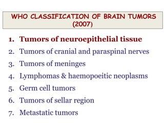
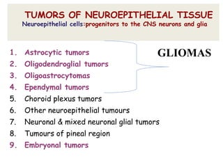
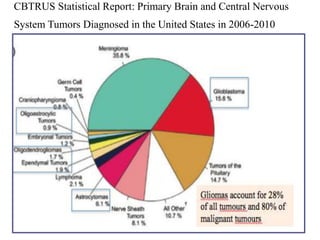
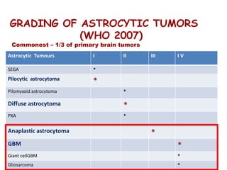
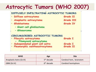
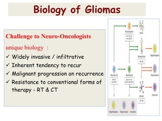
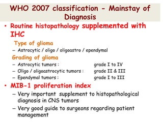
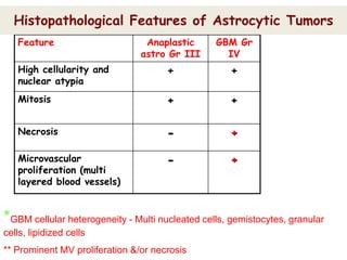
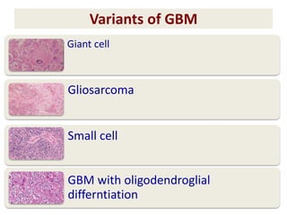
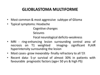
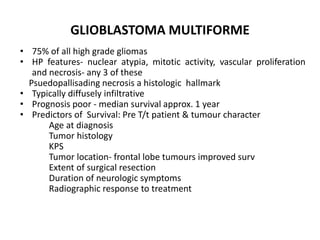
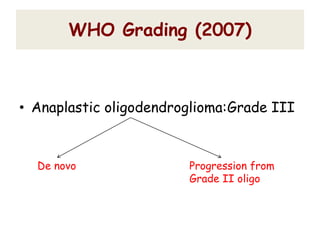
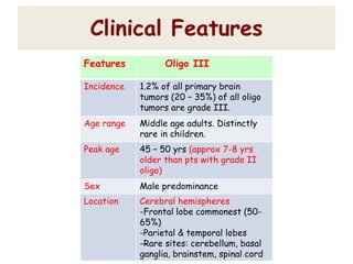
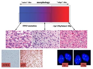
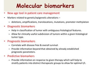
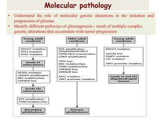
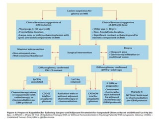
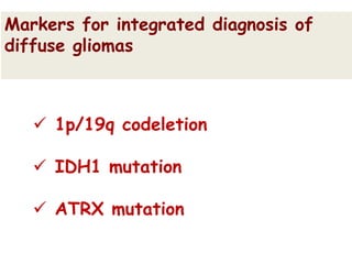
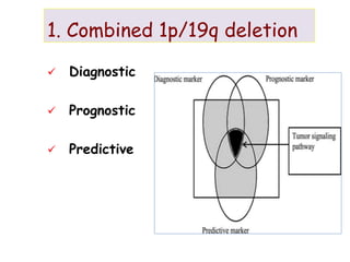
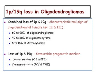
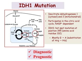
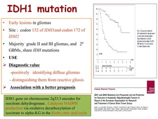
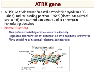
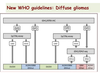
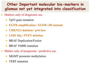
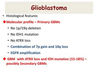
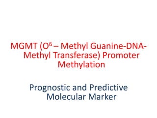
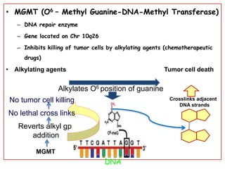
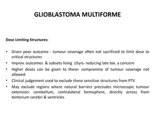
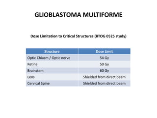
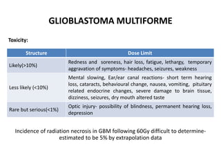
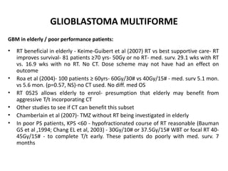
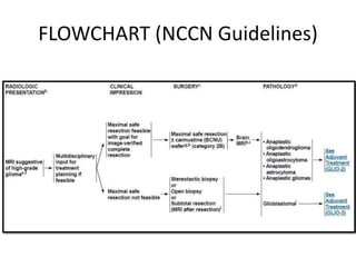
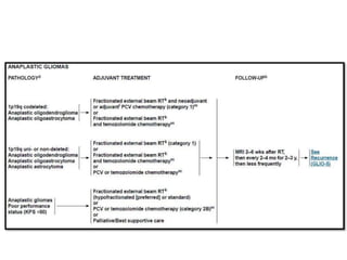
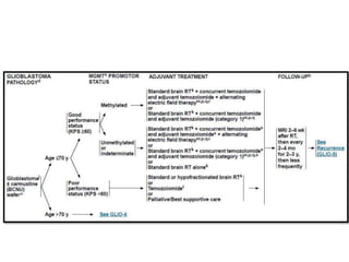
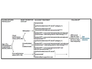
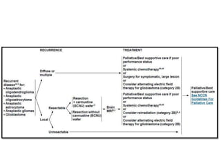
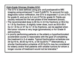
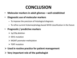
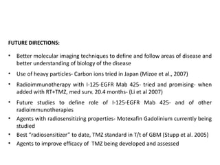
SlideShare a Scribd company logo
Dr. Ayush Garg
High Grade Glioma
WHO CLASSIFICATION OF BRAIN TUMORS
(2007)
1. Tumors of neuroepithelial tissue
2. Tumors of cranial and paraspinal nerves
3. Tumors of meninges
4. Lymphomas & haemopoeitic neoplasms
5. Germ cell tumors
6. Tumors of sellar region
7. Metastatic tumors
TUMORS OF NEUROEPITHELIAL TISSUE
Neuroepithelial cells:progenitors to the CNS neurons and glia
1. Astrocytic tumors
2. Oligodendroglial tumors
3. Oligoastrocytomas
4. Ependymal tumors
5. Choroid plexus tumors
6. Other neuroepithelial tumours
7. Neuronal & mixed neuronal glial tumors
8. Tumours of pineal region
9. Embryonal tumors
GLIOMAS
CBTRUS Statistical Report: Primary Brain and Central Nervous
System Tumors Diagnosed in the United States in 2006-2010
GRADING OF ASTROCYTIC TUMORS
(WHO 2007)
Commonest – 1/3 of primary brain tumors
Astrocytic Tumours I II III I V
SEGA *
Pilocytic astrocytoma
*
Pilomyxoid astrocytoma *
Diffuse astrocytoma *
PXA *
Anaplastic astrocytoma *
GBM *
Giant cellGBM *
Gliosarcoma *
Astrocytic Tumors (WHO 2007)
WHO Age Site
Anaplastic Astro (Gr.III) 5th decade Cerebral hem, brainstem
GBM (Gr.IV) 6th decade Cerebral hemisphere
Challenge to Neuro-Oncologists
unique biology :
 Widely invasive / infiltrative
 Inherent tendency to recur
 Malignant progression on recurrence
 Resistance to conventional forms of
therapy - RT & CT
Biology of Gliomas
WHO 2007 classification - Mainstay of
Diagnosis
• Routine histopathology supplemented with
IHC
Type of glioma
– Astrocytic / oligo / oligoastro / ependymal
Grading of glioma
– Astrocytic tumors : grade I to IV
– Oligo / oligoastrocytic tumors : grade II & III
– Ependymal tumors : grade I to III
• MIB-1 proliferation index
– Very important supplement to histopathological
diagnosis in CNS tumors
– Very good guide to surgeons regarding patient
management
Feature Anaplastic
astro Gr III
GBM Gr
IV
High cellularity and
nuclear atypia
+ +
Mitosis + +
Necrosis - +
Microvascular
proliferation (multi
layered blood vessels)
- +
*GBM cellular heterogeneity - Multi nucleated cells, gemistocytes, granular
cells, lipidized cells
** Prominent MV proliferation &/or necrosis
Histopathological Features of Astrocytic Tumors
Immunohistochemical Features
• GFAP : +ve (degree of cytoplasmic positivity highly
variable- related to grade)
• S-100 : +ve (less specific glial marker; but +ve even in
gliomas in which GFAP is –ve / equivocal)
• CK : –ve (false +ve with AE1/AE3 cocktail; not with CAM 5.2)
• EMA : –ve
Proliferation Index
 MIB-1 labeling index correlates with grade
– Grade I : 1-2%
– Grade II: <5%
– Grade III: 5-10%
– Grade IV: >10-20%
•Increased cellularity compared
to grade II
•Nuclear atypia, pleomorphism
Anaplastic Astrocytoma Grade III
Increased cellularity & pleomorphism
Classical GBM Gr.IV
Mitosis
Large ischemic necrosisPseudopallisading
Variants of GBM
Giant cell
Gliosarcoma
Small cell
GBM with oligodendroglial
differntiation
GLIOBLASTOMA MULTIFORME
• Most common & most aggressive subtype of Glioma
• Typical symptoms: Headache
Cognitive changes
Seizures
Focal neurological deficits-weakness
• MRI - ring-enhancing lesion surrounding central area of
necrosis on T1 weighted imaging- significant FLAIR
hyperintensity surrounding the lesion
• Most cases- grow inexorably- finally refractory to all T/t
• Recent data- 5-yr survival of almost 30% in patients with
favourable prognostic factors (age< 50 yrs & high PS)
GLIOBLASTOMA MULTIFORME
• 75% of all high grade gliomas
• HP features- nuclear atypia, mitotic activity, vascular proliferation
and necrosis- any 3 of these
Psuedopallisading necrosis a histologic hallmark
• Typically diffusely infiltrative
• Prognosis poor - median survival approx. 1 year
• Predictors of Survival: Pre T/t patient & tumour character
Age at diagnosis
Tumor histology
KPS
Tumor location- frontal lobe tumours improved surv
Extent of surgical resection
Duration of neurologic symptoms
Radiographic response to treatment
Oligodendroglial tumors
WHO Grading (2007)
• Anaplastic oligodendroglioma:Grade III
De novo Progression from
Grade II oligo
Clinical Features
Features Oligo III
Incidence 1.2% of all primary brain
tumors (20 – 35%) of all oligo
tumors are grade III.
Age range Middle age adults. Distinctly
rare in children.
Peak age 45 – 50 yrs (approx 7-8 yrs
older than pts with grade II
oligo)
Sex Male predominance
Location Cerebral hemispheres
-Frontal lobe commonest (50-
65%)
-Parietal & temporal lobes
-Rare sites: cerebellum, basal
ganglia, brainstem, spinal cord
Histopathological features of
Oligodendroglioma Grade III
Increased cellularity with endothelial proliferation
Endothelial proliferation with glomeruloid formation
High MIB
Frequent Mitosis
Necrosis
Oligoastrocytoma
Oligoastrocytoma
• Diffusely infiltrating gliomas.
• Admixture of tumor cells with oligodendroglial and
astrocytic differentiation
• Two variants:
– Biphasic or compact variant : Oligo and astro
components in geographically distinct zones.
– Intermingled or diffuse variant : both oligo and
astro components intimately intermixed.
• Clinical features & radiology :
– overlap with pure astro and pure oligo tumors
 Anaplastic oligoastrocytoma grade III
 ? Oligoastrocytoma grade IV / GBM with
oligodendroglioma component (GBMO).
WHO grading (2007)
Feature Grade III
Oligoastro
Grade IV
Oligoastro/GBMO
Cellularity &
cytological atypia
Moderate to severe Moderate to severe
Mitosis Frequent Frequent
Endothelial prolif. Present Present
Necrosis Absent Present
Mean survival time 2 – 4 yrs ~ 22 mths
WHO Grading
p53 1p 19qATRX
• New age tool in patient care management
• Markers related to genetic/epigenetic alterations –
– deletions, amplifications, translocations, mutations, promoter methylation
• Diagnostic biomarkers
– Help in classification of tumor with ambiguous histological features.
– Allow for clinically useful subdivision of tumors within a given histological
tumor type.
• Prognostic biomarkers
– Correlate with disease free & overall survival.
– Provide information beyond that obtained by already established
prognostic parameters.
• Predictive biomarkers
– Provide information on response to given therapy which will help to
stratify patients into distinct therapeutic groups to allow for optimal t/t.
Molecular biomarkers
Molecular pathology
• Understand the role of molecular genetic alterations in the initiation and
progression of gliomas
• Identify different pathways of gliomagenesis - result of multiple complex
genetic alterations that accumulate with tumor progression
 1p/19q codeletion
 IDH1 mutation
 ATRX mutation
Markers for integrated diagnosis of
diffuse gliomas
1. Combined 1p/19q deletion
 Diagnostic
 Prognostic
 Predictive
1p/19q loss in Oligodendrogliomas
 Combined loss of 1p & 19q – characteristic mol sign of
oligodendroglial tumors (Gr II & III)
 60 to 90% of oligodendrogliomas
 40 to 60% of oligoastrocytoma
 5 to 15% of Astrocytomas
 Loss of 1p & 19q - favourable prognostic marker
 Longer survival (OS & PFS)
 Chemosensitivity (PCV & TMZ)
IDH1 Mutation
 Diagnostic
 Prognostic
• Isocitrate dehydrogenase 1
(cytosol) and 2 (mitochondrial)
• Participates in the citric acid
cycle, NADP+ dependant
• IDH1: hot spot mutation at
position 395 (amino acid
residue 132)
– Mostly G  A (substitution
of Arg  His)
IDH1 mutation
• Early lesions in gliomas
• Site : codon 132 of IDH1and codon 172 of
IDH2
• Majority grade II and III gliomas, and 20
GBMs, share IDH mutations
• USE
 Diagnostic value
-positively identifying diffuse gliomas
- distinguishing them from reactive gliosis
 Association with a better prognosis
IDH1 gene on chromosome 2q33.3 encodes for
isocitrate dehydrogenase . Catalyzes NADPH
production via oxidative decarboxylation of
isocitrate to alpha-KG in the Krebs citric acid cycle
Alpha Thalassemia/Mental Retardation
Syndrome X-linked gene (ATRX)
Diagnostic
? Prognostic
ATRX gene
• ATRX (α thalassemia/mental retardation syndrome X-
linked) and its binding partner DAXX (death-associated
protein 6) are central components of a chromatin
remodeling complex
• Normal functions
– Chromatin remodelling and nucleosome assembly
– Regulates incorporation of histone H3.3 into telomeric chromatin
– Plays crucial role in normal telomere homeostasis
New WHO guidelines: Diffuse gliomas
Other Important molecular bio-markers in
gliomas not yet integrated into classification
 Markers only of diagnostic use
 Tp53 gene mutation
 EGFR amplification / EGFR vIII mutant
 CDKN2A deletion / p16 loss
 LOH 10q / PTEN deletion
 BRAF Duplication/Fusion
 BRAF V600E mutation
 Marker only of prognostic / predictive use
 MGMT promoter methylation
 TERT mutation
Glioblastoma
• Histological features
Molecular profile – Primary GBMs
• No 1p/19q deletion
• No IDH1 mutation
• No ATRX loss
• Combination of 7p gain and 10q loss
• EGFR amplification
 GBM with ATRX loss and IDH mutation (15-18%) –
possibly Secondary GBMs
MGMT (O6 – Methyl Guanine-DNA-
Methyl Transferase) Promoter
Methylation
Prognostic and Predictive
Molecular Marker
• MGMT (O6 – Methyl Guanine-DNA-Methyl Transferase)
– DNA repair enzyme
– Gene located on Chr 10q26
– Inhibits killing of tumor cells by alkylating agents (chemotherapeutic
drugs)
• Alkylating agents Tumor cell death
Alkylates O6 position of guanine
Crosslinks adjacent
DNA strands
MGMT
Reverts alkyl gp.
addition
No lethal cross links
No tumor cell killing
DNA
New Prognostic Marker
TERT Mutation
TERT mutations
• Recurrent mutations in promoter region of telomerase reverse
transcriptase (TERT)
• Gene encoding catalytic subunit of telomerase
• Two most common mutations - C228T, C250T
– Associated with marked upregulation of TERT expression
C228T mutationC250T mutation
Principles of Brain Imaging
Treatment
GLIOBLASTOMA MULTIFORME
Surgery
• A critical component of T/t
• Survival: extensive resection> partial resection>surgical
decompression
• Devaux et al (1993)- Resection & RT- med. surv.-50.6 wks
• Laws et al (2003) - Biopsy & RT- med. surv.-33.0 wks
• Lacroix et al (2001) - Resection of at least 98% tumour tissue
increased med. surv. (13 vs 8.8 months)
• Maximal surgical resection- currently accepted standard of care esp.
for patients <65 yrs
• Larger resection-increased diagnostic accuracy and tissue for
molecular profiling- may prognosticate and guide T/t
• Gliomas- “ïnfiltrating propensities” without clear demarcation from
normal tissues
• Include T/t with potential to target focal disease and microscopic
tumour cells throughout brain
GLIOBLASTOMA MULTIFORME
Radiotherapy
• Diffusely infiltrate brain beyond gross tumour & recur locally
• RT- a critical component - focus T/t to areas of highest risk
• In current form - GTV and margin of several cms
• Benefit clearly seen since 1970s. Use dates back to 1925
• Shapiro and Young (1976)- CT vs CT+RT. RT 45Gy+15Gy
RT+CT(BCNU+VCR)- med. surv.- 44.5 wks
CT-med. surv - 30 wks
• Coop. Gr. Trials: Improved surv. for RT ± nitrosurea - med surv. - 9-12
months vs. half of this when RT excluded
• Radiosurgery- interest in past - abandoned after negative trials
• Current standard - total of appr. 60Gy / 30#
• Different total dose, fractionation and delivery methods tried
• Ext. beam RT+Temozolamide & adjuvant Temozolamide
Chemotherapy
Agents
GLIOBLASTOMA MULTIFORME
• Chemotherapy
• Stupp trial randomized 573 patients with newly diagnosed glioblastoma to
either RT alone (total 60 Gy in 30 fractions; control arm) or RT + TMZ (total 60
Gy in 30 fractions; experimental arm). Patients on the experimental arm
received temozolomide daily during RT at a dose of 75 mg/m2, followed by
monthly temozolomide at a dose of 150 to 200 mg/m2 on a 5 of every 28 days
schedule for 6 cycles.
• Patients randomized to the experimental arm had a median survival of 14.6
months as compared to 12.1 months for the control arm. The 2-year survival
of patients treated with radiation therapy plus chemotherapy was 26% as
compared to 6% for radiation alone.
• The survival benefit from the addition of temozolomide has now been
demonstrated for at least 5 years out from initial treatment and in all clinical
prognostic subgroups, including patients aged 60 to 70 years and in RPA
classes III through V. Five-year overall survival was 9.8% for patients who
received combined temozolomide and radiotherapy as compared to 1.9% for
those who received radiotherapy alone.
• The RTOG recently completed a 1,100-patient, randomized, phase III trial
comparing standard adjuvant temozolomide with a dose-dense schedule
in newly diagnosed glioblastoma.
• A total of 833 patients were randomized to receive either standard
therapy (temozolomide plus radiotherapy followed by 6 to 12 cycles of
temozolomide at a dose of 150 to 200 mg/m2 on a 5/28 day schedule) or
dose-intense temozolomide (temozolomide plus radiotherapy followed by
6 to 12 cycles of temozolomide at a dose of 150 mg/m2 on a 21/28 day
schedule).
• There was no statistical difference between the experimental and
standard arms for overall survival (16.6 vs.14.9 months, p = .63) or
progression-free survival (5.5 vs. 6.7 months, p = .06), indicating no
additional benefit from dose-intense temozolomide.
• The trial prospectively stratified for MGMT methylation status, and no
survival benefit with dose-intense therapy was identified in any subgroup.
As expected, the dose-intense arm resulted in increased toxicity.
• Thus, at the present time, there is no role for dose-intense temozolomide
for newly diagnosed glioblastoma patients.
• Other chemotherapeutic regimens, such as the combination of CPT-11
and temozolomide, have shown promising results in a phase II trial with an
objective response rate of 25% and 6-month progression-free rate of 38%.
When tested prospectively in a single-arm RTOG trial, the regimen did not
show improved survival.
• Buckner et al. reported on a phase III trial of carmustine with or without
cisplatin before and concurrently with radiotherapy and observed
increased toxicity but no survival benefit with the addition of cisplatin.
• Two large phase III, randomized clinical trials investigating the addition of
bevacizumab to the EORTC/NCIC regimen have completed accrual, and
results are pending.
• Anaplastic Oligodendroglioma/Oligoastrocytoma
• Anaplastic oligodendroglioma and oligoastrocytoma are generally
chemosensitive primarily based on high response rates to PCV in
several studies.
• Two large randomized trials, described earlier, investigated the use
of sequential chemoradiotherapy compared to radiotherapy alone
with chemotherapy reserved for salvage in patients with anaplastic
oligodendroglioma and oligoastrocytoma.
• With 11-year follow-up, no difference in survival was found for the
entire cohort, but for the codeleted patients, there was a near-
doubling of survival, establishing chemoradiotherapy as a standard
for this subset.
• Because of the significant toxicity associated with PCV, many
clinicians now use temozolomide, which is much better tolerated.
GLIOBLASTOMA MULTIFORME
Early Brain Tumour Study Group Studies
Dose Response to Radiation based on 3 BTSG studies (Walker et al 1979)
Med. Surv.(weeks) P-value
BTSG 6901( Walker et al, 1978)
Best supportive care 14
BCNU (Carmustine) 18.5 0.119
Radiation 35 0.001
Radiation+BCNU 34.5 0.001
BTSG 7201(Walker et al, (1980)
MeCCNU (Semustine) 31
Radiation 37 0.003
Radiaiton+BCNU 49 <0.001
Radiaiton+MeCCNU 43 <0.001
No RT ≤45 Gy 50 Gy 55 Gy 60 Gy
Med. Surv (wks) 18 13.5 28 36 42
P-value 0.346 <0.001 <0.001 <0.001
GLIOBLASTOMA MULTIFORME
Brachytherapy for GBM
• Retrospective data- technique promising- I-125 improved med. surv. from 17.9
months in RTOG Class III patients to 28 months. Improvement also in Class IV & V
(Videtic et al. 1999)
• Prospective studies failed to support this
Med. Surv.(weeks) P-Value
Brain Tumour Cooperative Group (Selker 2002)
60.2 Gy 58.5
60.2 Gy+I-125 (60Gy) 68.1 0.101
Princess Margaret (Laperriere et al,1998)
50 Gy 57.2
50Gy+I-125 (60Gy) 59.8 0.49
UCSF (Sneed et al, 1998)
59.4 Gy+I-125 (60Gy) 76
59.4 Gy+ I-125(60Gy) + Hyperthermia 85 0.02
GLIOBLASTOMA MULTIFORME
Radiation Volumes:
• Historically margins to cover potential microscopic disease beyond visualised
area of disease-typically 2cm around gross tumour
• Better imaging and sophisticated radiation delivery- variation in margin
• Partial brain RT is standard - no benefit of WBT in terms of survival and
control (Shibamoto et al, 1990)
• 90% recurrence within 2cm of known primary tumour - typically 2-3cm margin
• Using oedema to delineate microscopic disease imperfect- imaging that is
more specific to tumour better
• UCSF- MRI spectroscopy to define volume (Park et al. 2007)
• Univ. Michigan- 11C-methionine PET (Lee et al, 2007)
GLIOBLASTOMA MULTIFORME
Radiation Volumes:
• Historically margins to cover potential microscopic disease beyond visualised
area of disease
• Typically 2cm around gross tumour
• Better imaging and sophisticated radiation delivery-variation in margin
RTOG (old) RTOG (new) EORTC NABTT
Total Dose 46 Gy 46 Gy 60 Gy 46 Gy
Initial Margin 2 cm block 2cm dosimetric to PTV 2-3 cm dosimet. to PTV 1 cm dosimetric to PTV
Initial Vol. Def. T2/FLAIR T2/FLAIR T1+ Contrast T2/FLAIR
Boost + + - +
Boost Dose 14Gy 14 Gy 14Gy
Boost Margin 2.5cm block 2.5 cm dosimet. to PTV 1cm dosimetric to PTV
Boost Vol. Def. T1 + Contrast T1+ Contrast T1+ Contrast
IMRT allowed No No No Yes
Final Dose 60Gy 60 Gy 60 Gy 60 Gy
GLIOBLASTOMA MULTIFORME
Radiation Volumes:
• IMRT as a means to hypofractionate / deliver more dose centrally in some
centre
• Preliminary studies- RT over 2-4 weeks without concurrent CT comparable to
full 6 weeks T/t (Floyd et al, 2004; Sultanem et al 2004)
• With this RT can be given safely and effectively in a shorter period of time
• IMRT using conventional fractionation- incorporated into current studies
including studies by NABTT- uses 5mm margin for CTV and PTV both for initial
and boost volume
GLIOBLASTOMA MULTIFORME
Simulation:
• CT based simulation typically used
• Thermoplastic mask and contrast usually given
• GBM may progress after postoperative images acquired- contrast used in
simulation may help identify progression following surgery
• After CT simulation, fusion of MRI image if available
• Critical structures typically included-lenses, eyes, optic nerve, optic chiasm,
pituitary, hypothalamus, cochleas, brainstem
GLIOBLASTOMA MULTIFORME
Dose Limiting Structures:
• Given poor outcome - tumour coverage often not sacrificed to limit dose to
critical structures
• Improv. outcomes & subsets living ≥5yrs- reducing late tox. a concern
• Higher doses can be given to these- compromise of tumour coverage not
allowed
• Clinical judgement used to exclude these sensitive structures from PTV
• May exclude regions where natural barriers precludes microscopic tumour
extension- cerebellum, contralateral hemisphere, directly across from
tentorium cerebri & ventricles
GLIOBLASTOMA MULTIFORME
Dose Limitation to Critical Structures (RTOG 0525 study)
Structure Dose Limit
Optic Chiasm / Optic nerve 54 Gy
Retina 50 Gy
Brainstem 60 Gy
Lens Shielded from direct beam
Cervical Spine Shielded from direct beam
GLIOBLASTOMA MULTIFORME
Toxicity:
Incidence of radiation necrosis in GBM following 60Gy difficult to determine-
estimated to be 5% by extrapolation data
Structure Dose Limit
Likely(>10%)
Redness and soreness, hair loss, fatigue, lethargy, temporary
aggravation of symptoms- headaches, seizures, weakness
Less likely (<10%)
Mental slowing, Ear/ear canal reactions- short term hearing
loss, cataracts, behavioural change, nausea, vomiting, pituitary
related endocrine changes, severe damage to brain tissue,
dizziness, seizures, dry mouth altered taste
Rare but serious(<1%)
Optic injury- possibility of blindness, permanent hearing loss,
depression
GLIOBLASTOMA MULTIFORME
GBM in elderly / poor performance patients:
• RT beneficial in elderly - Keime-Guibert et al (2007) RT vs best supportive care- RT
improves survival- 81 patients ≥70 yrs- 50Gy or no RT- med. surv. 29.1 wks with RT
vs. 16.9 wks with no RT. No CT. Dose scheme may not have had an effect on
outcome
• Roa et al (2004)- 100 patients ≥ 60yrs- 60Gy/30# vs 40Gy/15# - med. surv 5.1 mon.
vs 5.6 mon. (p=0.57, NS)-no CT used. No diff. med OS
• RT 0525 allows elderly to enrol- presumption that elderly may benefit from
aggressive T/t incorporating CT
• Other studies to see if CT can benefit this subset
• Chamberlain et al (2007)- TMZ without RT being investigated in elderly
• In poor PS patients, KPS <60 - hypofractionated course of RT reasonable (Bauman
GS et al ,1994; Chang EL et al, 2003) - 30Gy/10# or 37.5Gy/15# WBT or focal RT 40-
45Gy/15# - to complete T/t early. These patients do poorly with med. surv. 7
months
GLIOBLASTOMA MULTIFORME
Radiation sensitizers:
• Motexafin Gadolinium (Xcytrin) - previously known as Gadolinium Texaphyrin
or Gd-Tex- redox mediator selectively targets tumour cells- generation of
reactive oxygen species and fixation of damage by radiation
• Phase I study in GBM - max tolerated dose 5mg/kg/day daily for 2 wks, then 3
times per week till RT completion. TMZ not given (Ford et al 2007)
• Results from a single-arm phase II trial, RTOG 0513, of MGd and
conventional therapy in newly diagnosed GBM showed no
survival improvement.
GLIOBLASTOMA MULTIFORME
FOLLOW-UP:
• MRI scan 4 weeks after completion of CT+RT, 2-3 months thereafter
• Pseudoprogression- one area of controversy- worsening FLAIR or T1 contrast
soon after RT completion- may resolve if followed long enough rather than
changing planned T/t course
• Controversial how to image pseudoprogression and distinguish from tumour
progression
• Cause unknown- seen more frequently after using aggressive upfront T/t-
acute T/t related changes including blood-brain barrier disruption and oedema
• While FU of GBM, pseudoprogression a D/d
GLIOBLASTOMA MULTIFORME
RE-IRRADIATION
• Studied both for local and distant recurrence
• Often given stereotactically
Study Authors
Nos of
Pts.
Med. Dose in Gy Med. Surv
U.. Michigan Kim et al. 1997 20 36 (30.6-50.4) 9 mo
Germany
Vordermark et al..
2005
14
30. Hypo. Stereo.
Med 5Gy/#
7.9 mo
U. Heidelberg Combs et al. 2005 53
36. Med #-2Gy
1mm mar. stereo
8 mo
U. Wisconsin Tome et al. 2007 99
LDR radiation. 0.2
Gy pulses 3 min
apart
6 mo 31% surv
• Recurrence
• Single-agent bevacizumab was approved by the FDA in
2009 for the treatment of recurrent glioblastoma.
• In a study of 49 glioblastoma patients, Kreisl et al. reported
objective response rate of 35%, 6-month progression-free
survival of 29%, 3.7-month median progression-free
survival, and 7.2-month median overall survival.
• Similarly, Friedman et al. reported an objective response
rate of 28%, 6-month progression-free survival of 43%,
median progression-free survival of 4.2 months, and
median overall survival of 9.2 months in a total of 85
patients.
FLOWCHART (NCCN Guidelines)
• Molecular markers in adult gliomas – well established
• Diagnostic use of molecular markers
– To improve the precision of histological diagnosis
– To refine current histomorphology based WHO classification in the future
• Prognostic / predictive markers
 1p/19q deletion
 IDH1 mutation
 MGMT promoter methylation
 TERT mutation
 Used in routine practice for patient management
 Very important role of the pathologist
CONCLUSION
FUTURE DIRECTIONS:
• Better molecular imaging techniques to define and follow areas of disease and
better understanding of biology of the disease
• Use of heavy particles- Carbon ions tried in Japan (Mizoe et al., 2007)
• Radioimmunotherapy with I-125-EGFR Mab 425- tried and promising- when
added with RT+TMZ, med surv. 20.4 months- (Li et al 2007)
• Future studies to define role of I-125-EGFR Mab 425- and of other
radioimmunotherapies
• Agents with radiosensitizing properties- Motexafin Gadolinium currently being
studied
• Best “radiosensitizer” to date, TMZ standard in T/t of GBM (Stupp et al. 2005)
• Agents to improve efficacy of TMZ being developed and assessed
High-grade glioma: Where we are and where
are we going
• Systemic therapy-most often utilized treatment in recurrent HGG.
• Choice of therapy- varies and revolves around re-challenge with
temozolomide (TMZ), use of a nitrosourea (most often lomustine;
CCNU) or BEV (most frequently used angiogenic inhibitor)
• No clear recommendation regarding prefered agent or combination of
agents.
• Prognosis after progression of HGG remains poor, with unmet need to
improve therapy.
THANK YOU

More Related Content

What's hot

Medulloblastoma
MedulloblastomaMedulloblastoma
MEDULLOBLASTOMA
MEDULLOBLASTOMAMEDULLOBLASTOMA
MEDULLOBLASTOMA
suresh Bishokarma
 
Brain metastasis
Brain metastasis Brain metastasis
Brain metastasis
Kiran Ramakrishna
 
Primary CNS Lymphoma
Primary CNS Lymphoma Primary CNS Lymphoma
Primary CNS Lymphoma
Dr.Rashmi Yadav
 
Medulloblastoma n csi kiran
Medulloblastoma n csi kiranMedulloblastoma n csi kiran
Medulloblastoma n csi kiran
Kiran Ramakrishna
 
Medulloblastoma
MedulloblastomaMedulloblastoma
Medulloblastoma
DrAyush Garg
 
Glioblastoma multiforme (GBM) Radiotherapy planning and management principles
Glioblastoma multiforme (GBM) Radiotherapy planning and management principlesGlioblastoma multiforme (GBM) Radiotherapy planning and management principles
Glioblastoma multiforme (GBM) Radiotherapy planning and management principles
Gebrekirstos Hagos Gebrekirstos, MD
 
Management of brain metastases
Management of brain metastasesManagement of brain metastases
Management of brain metastases
Shreya Singh
 
Accelerated partial breast irradiation
Accelerated partial breast irradiationAccelerated partial breast irradiation
Accelerated partial breast irradiation
Bharti Devnani
 
High grade glioma, standard of care & new advances..
High grade glioma, standard of care & new advances.. High grade glioma, standard of care & new advances..
High grade glioma, standard of care & new advances..
Osama Elzaafarany, MD.
 
Diagnosis, Treatment & Management of Medulloblastoma
Diagnosis, Treatment & Management of Medulloblastoma Diagnosis, Treatment & Management of Medulloblastoma
Diagnosis, Treatment & Management of Medulloblastoma
Dr Vandana Singh Kushwaha
 
Glioma molecular markers
Glioma molecular markersGlioma molecular markers
Glioma molecular markers
kanwalpreet15
 
MANAGEMENT OF PITUITARY TUMORS.pptx
MANAGEMENT OF PITUITARY  TUMORS.pptxMANAGEMENT OF PITUITARY  TUMORS.pptx
MANAGEMENT OF PITUITARY TUMORS.pptx
Kiran Ramakrishna
 
Primary cns lymphoma ppt
Primary cns lymphoma pptPrimary cns lymphoma ppt
Primary cns lymphoma ppt
Shashank Bansal
 
HIGH GRADE GLIOMA MANAGEMENT
HIGH GRADE GLIOMA MANAGEMENTHIGH GRADE GLIOMA MANAGEMENT
HIGH GRADE GLIOMA MANAGEMENT
Nabeel Yahiya
 
MANAGEMENT OF LOW GARDE GLIOMA
MANAGEMENT OF LOW GARDE GLIOMAMANAGEMENT OF LOW GARDE GLIOMA
MANAGEMENT OF LOW GARDE GLIOMA
Kanhu Charan
 
EANO GUIDELINES FOR MANAGEMENT OF MENINGIOMA
EANO GUIDELINES FOR MANAGEMENT OF MENINGIOMAEANO GUIDELINES FOR MANAGEMENT OF MENINGIOMA
EANO GUIDELINES FOR MANAGEMENT OF MENINGIOMA
apollo seminar group
 
Low Grade Gliomas
Low  Grade  GliomasLow  Grade  Gliomas
Low Grade Gliomas
Arnab Bose
 

What's hot (20)

Medulloblastoma
MedulloblastomaMedulloblastoma
Medulloblastoma
 
Medulloblastomas
MedulloblastomasMedulloblastomas
Medulloblastomas
 
MEDULLOBLASTOMA
MEDULLOBLASTOMAMEDULLOBLASTOMA
MEDULLOBLASTOMA
 
Brain metastasis
Brain metastasis Brain metastasis
Brain metastasis
 
Primary CNS Lymphoma
Primary CNS Lymphoma Primary CNS Lymphoma
Primary CNS Lymphoma
 
Medulloblastoma n csi kiran
Medulloblastoma n csi kiranMedulloblastoma n csi kiran
Medulloblastoma n csi kiran
 
Medulloblastoma
MedulloblastomaMedulloblastoma
Medulloblastoma
 
Glioblastoma multiforme (GBM) Radiotherapy planning and management principles
Glioblastoma multiforme (GBM) Radiotherapy planning and management principlesGlioblastoma multiforme (GBM) Radiotherapy planning and management principles
Glioblastoma multiforme (GBM) Radiotherapy planning and management principles
 
Management of brain metastases
Management of brain metastasesManagement of brain metastases
Management of brain metastases
 
Accelerated partial breast irradiation
Accelerated partial breast irradiationAccelerated partial breast irradiation
Accelerated partial breast irradiation
 
High grade glioma, standard of care & new advances..
High grade glioma, standard of care & new advances.. High grade glioma, standard of care & new advances..
High grade glioma, standard of care & new advances..
 
Diagnosis, Treatment & Management of Medulloblastoma
Diagnosis, Treatment & Management of Medulloblastoma Diagnosis, Treatment & Management of Medulloblastoma
Diagnosis, Treatment & Management of Medulloblastoma
 
Glioma molecular markers
Glioma molecular markersGlioma molecular markers
Glioma molecular markers
 
MANAGEMENT OF PITUITARY TUMORS.pptx
MANAGEMENT OF PITUITARY  TUMORS.pptxMANAGEMENT OF PITUITARY  TUMORS.pptx
MANAGEMENT OF PITUITARY TUMORS.pptx
 
Primary cns lymphoma ppt
Primary cns lymphoma pptPrimary cns lymphoma ppt
Primary cns lymphoma ppt
 
HIGH GRADE GLIOMA MANAGEMENT
HIGH GRADE GLIOMA MANAGEMENTHIGH GRADE GLIOMA MANAGEMENT
HIGH GRADE GLIOMA MANAGEMENT
 
craniospinal irradiation
craniospinal irradiationcraniospinal irradiation
craniospinal irradiation
 
MANAGEMENT OF LOW GARDE GLIOMA
MANAGEMENT OF LOW GARDE GLIOMAMANAGEMENT OF LOW GARDE GLIOMA
MANAGEMENT OF LOW GARDE GLIOMA
 
EANO GUIDELINES FOR MANAGEMENT OF MENINGIOMA
EANO GUIDELINES FOR MANAGEMENT OF MENINGIOMAEANO GUIDELINES FOR MANAGEMENT OF MENINGIOMA
EANO GUIDELINES FOR MANAGEMENT OF MENINGIOMA
 
Low Grade Gliomas
Low  Grade  GliomasLow  Grade  Gliomas
Low Grade Gliomas
 

Viewers also liked

Glioblastoma vibhay
Glioblastoma vibhayGlioblastoma vibhay
Glioblastoma vibhay
Vibhay Pareek
 
Glioblastoma multiforme
Glioblastoma multiformeGlioblastoma multiforme
Glioblastoma multiforme
Anderson Burbano
 
Future direction in the management of high risk LOW GRADE GLIOMA
Future direction in the management of high risk LOW GRADE GLIOMAFuture direction in the management of high risk LOW GRADE GLIOMA
Future direction in the management of high risk LOW GRADE GLIOMA
apollo seminar group
 
Prezentacja2
Prezentacja2Prezentacja2
Prezentacja2
Noemie Fontenelle
 
Essentials of gliomas
Essentials of gliomas Essentials of gliomas
Essentials of gliomas NeurologyKota
 
Glioblastoma Multiforme.Dr NG NeuroEdu
Glioblastoma Multiforme.Dr NG NeuroEduGlioblastoma Multiforme.Dr NG NeuroEdu
Glioblastoma Multiforme.Dr NG NeuroEduslneurosurgery
 
Spinal cord disorders
Spinal cord disordersSpinal cord disorders
Spinal cord disorders
anoop k r
 
Glioblastoma Presentation
Glioblastoma PresentationGlioblastoma Presentation
Glioblastoma Presentation
Student Wilsonsbiologylab
 
CT Scan: Glioblastoma Multiforme
CT Scan: Glioblastoma MultiformeCT Scan: Glioblastoma Multiforme
CT Scan: Glioblastoma Multiformegueste19bfb0
 
Classification of spinal cord disorders
Classification of spinal cord disorders Classification of spinal cord disorders
Classification of spinal cord disorders
Yapa
 
Neuro-Oncology for the Radiation Oncologist - Gliomas
Neuro-Oncology for the Radiation Oncologist - GliomasNeuro-Oncology for the Radiation Oncologist - Gliomas
Neuro-Oncology for the Radiation Oncologist - Gliomas
Emil Lou, M.D., Ph.D, FACP
 
Diseases of the spinal cord
Diseases of the spinal cordDiseases of the spinal cord
Diseases of the spinal cordHiba Hassan
 
Functional analysis of proteomic biomarkers and targeting glioblastoma stem c...
Functional analysis of proteomic biomarkers and targeting glioblastoma stem c...Functional analysis of proteomic biomarkers and targeting glioblastoma stem c...
Functional analysis of proteomic biomarkers and targeting glioblastoma stem c...
Pasteur_Tunis
 
Anti-GBM Diseases
Anti-GBM DiseasesAnti-GBM Diseases
Anti-GBM Diseases
Amanda Valliant
 
Spinal cord tumor
Spinal cord tumorSpinal cord tumor
Classification of brain tumors AND MANAGEMENT OG LOW GRADE GLIOMA
Classification of brain tumors AND MANAGEMENT OG LOW GRADE GLIOMAClassification of brain tumors AND MANAGEMENT OG LOW GRADE GLIOMA
Classification of brain tumors AND MANAGEMENT OG LOW GRADE GLIOMA
radiation oncology
 

Viewers also liked (20)

Glioblastoma vibhay
Glioblastoma vibhayGlioblastoma vibhay
Glioblastoma vibhay
 
Glioblastoma multiforme
Glioblastoma multiformeGlioblastoma multiforme
Glioblastoma multiforme
 
Glioblastoma Multiforme
Glioblastoma MultiformeGlioblastoma Multiforme
Glioblastoma Multiforme
 
Glioblastoma Multiforme
Glioblastoma MultiformeGlioblastoma Multiforme
Glioblastoma Multiforme
 
Future direction in the management of high risk LOW GRADE GLIOMA
Future direction in the management of high risk LOW GRADE GLIOMAFuture direction in the management of high risk LOW GRADE GLIOMA
Future direction in the management of high risk LOW GRADE GLIOMA
 
Glioblastoma multiforme
Glioblastoma multiformeGlioblastoma multiforme
Glioblastoma multiforme
 
Snc Who 2007
Snc Who 2007Snc Who 2007
Snc Who 2007
 
Prezentacja2
Prezentacja2Prezentacja2
Prezentacja2
 
Essentials of gliomas
Essentials of gliomas Essentials of gliomas
Essentials of gliomas
 
Glioblastoma Multiforme.Dr NG NeuroEdu
Glioblastoma Multiforme.Dr NG NeuroEduGlioblastoma Multiforme.Dr NG NeuroEdu
Glioblastoma Multiforme.Dr NG NeuroEdu
 
Spinal cord disorders
Spinal cord disordersSpinal cord disorders
Spinal cord disorders
 
Glioblastoma Presentation
Glioblastoma PresentationGlioblastoma Presentation
Glioblastoma Presentation
 
CT Scan: Glioblastoma Multiforme
CT Scan: Glioblastoma MultiformeCT Scan: Glioblastoma Multiforme
CT Scan: Glioblastoma Multiforme
 
Classification of spinal cord disorders
Classification of spinal cord disorders Classification of spinal cord disorders
Classification of spinal cord disorders
 
Neuro-Oncology for the Radiation Oncologist - Gliomas
Neuro-Oncology for the Radiation Oncologist - GliomasNeuro-Oncology for the Radiation Oncologist - Gliomas
Neuro-Oncology for the Radiation Oncologist - Gliomas
 
Diseases of the spinal cord
Diseases of the spinal cordDiseases of the spinal cord
Diseases of the spinal cord
 
Functional analysis of proteomic biomarkers and targeting glioblastoma stem c...
Functional analysis of proteomic biomarkers and targeting glioblastoma stem c...Functional analysis of proteomic biomarkers and targeting glioblastoma stem c...
Functional analysis of proteomic biomarkers and targeting glioblastoma stem c...
 
Anti-GBM Diseases
Anti-GBM DiseasesAnti-GBM Diseases
Anti-GBM Diseases
 
Spinal cord tumor
Spinal cord tumorSpinal cord tumor
Spinal cord tumor
 
Classification of brain tumors AND MANAGEMENT OG LOW GRADE GLIOMA
Classification of brain tumors AND MANAGEMENT OG LOW GRADE GLIOMAClassification of brain tumors AND MANAGEMENT OG LOW GRADE GLIOMA
Classification of brain tumors AND MANAGEMENT OG LOW GRADE GLIOMA
 

Similar to High Grade Glioma

Molecular pathogenesis of CNS tumors
Molecular pathogenesis of  CNS tumorsMolecular pathogenesis of  CNS tumors
Molecular pathogenesis of CNS tumors
MD Patholgoy, AFMC
 
Glioma markers in neurosurgery
Glioma markers in neurosurgeryGlioma markers in neurosurgery
Glioma markers in neurosurgery
Dr. Shahnawaz Alam
 
Molecular studies in cns tumors
Molecular studies in cns tumorsMolecular studies in cns tumors
Molecular studies in cns tumors
Dr.Amrita Rakesh
 
LOW GRADE GLIOMA.pptx
LOW GRADE GLIOMA.pptxLOW GRADE GLIOMA.pptx
LOW GRADE GLIOMA.pptx
Brijesh Maheshwari
 
Pre-managment of Low Grade Glioma- low grade glioma
Pre-managment of Low Grade Glioma- low grade gliomaPre-managment of Low Grade Glioma- low grade glioma
Pre-managment of Low Grade Glioma- low grade glioma
7pwys74fgg
 
high grade glioma
high grade gliomahigh grade glioma
high grade glioma
HardikSharma590779
 
Evolution of treatment strategies of brain tumors
Evolution of treatment strategies of brain tumorsEvolution of treatment strategies of brain tumors
Evolution of treatment strategies of brain tumors
Anil Gupta
 
WHO CNS Classification of Tumors 2021-2.pptx
WHO CNS Classification of Tumors 2021-2.pptxWHO CNS Classification of Tumors 2021-2.pptx
WHO CNS Classification of Tumors 2021-2.pptx
RabiyyahBashir1
 
ThyroidIP.pptx
ThyroidIP.pptxThyroidIP.pptx
ThyroidIP.pptx
Ipsita Panda
 
Overview of brain tumors
Overview of brain tumorsOverview of brain tumors
Overview of brain tumors
DrAyush Garg
 
Updates in the Management of Primary CNS Malignancies
Updates in the Management of Primary CNS MalignanciesUpdates in the Management of Primary CNS Malignancies
Updates in the Management of Primary CNS Malignancies
Mary Ondinee Manalo Igot
 
Multiple Myeloma
Multiple MyelomaMultiple Myeloma
Multiple Myeloma
Sadia Sadiq
 
Gliomas - Brain Tumor
Gliomas - Brain TumorGliomas - Brain Tumor
Gliomas - Brain Tumor
suresh Bishokarma
 
Summary of 2021 WHO Classification of CNS Tumors
Summary of 2021 WHO Classification of CNS TumorsSummary of 2021 WHO Classification of CNS Tumors
Summary of 2021 WHO Classification of CNS Tumors
Dr Seena Tresa Samuel
 
THE gist OF GIST
THE gist OF GISTTHE gist OF GIST
THE gist OF GIST
Glee Thapa
 
Multiple myeloma - Etiopathogenesis, Clinical features, Advances in Management
Multiple myeloma - Etiopathogenesis, Clinical features, Advances in ManagementMultiple myeloma - Etiopathogenesis, Clinical features, Advances in Management
Multiple myeloma - Etiopathogenesis, Clinical features, Advances in Management
Chetan Ganteppanavar
 
Smm to mgus
Smm to mgusSmm to mgus
Smm to mgus
Faheema Hasan
 
neoplasia.pptx
neoplasia.pptxneoplasia.pptx
neoplasia.pptx
libin1986
 
Glioblastoma multiforme
Glioblastoma multiformeGlioblastoma multiforme
Glioblastoma multiforme
عبد الحافظ الحميري
 

Similar to High Grade Glioma (20)

Molecular pathogenesis of CNS tumors
Molecular pathogenesis of  CNS tumorsMolecular pathogenesis of  CNS tumors
Molecular pathogenesis of CNS tumors
 
Glioma markers in neurosurgery
Glioma markers in neurosurgeryGlioma markers in neurosurgery
Glioma markers in neurosurgery
 
Molecular studies in cns tumors
Molecular studies in cns tumorsMolecular studies in cns tumors
Molecular studies in cns tumors
 
LOW GRADE GLIOMA.pptx
LOW GRADE GLIOMA.pptxLOW GRADE GLIOMA.pptx
LOW GRADE GLIOMA.pptx
 
Pre-managment of Low Grade Glioma- low grade glioma
Pre-managment of Low Grade Glioma- low grade gliomaPre-managment of Low Grade Glioma- low grade glioma
Pre-managment of Low Grade Glioma- low grade glioma
 
high grade glioma
high grade gliomahigh grade glioma
high grade glioma
 
Evolution of treatment strategies of brain tumors
Evolution of treatment strategies of brain tumorsEvolution of treatment strategies of brain tumors
Evolution of treatment strategies of brain tumors
 
WHO CNS Classification of Tumors 2021-2.pptx
WHO CNS Classification of Tumors 2021-2.pptxWHO CNS Classification of Tumors 2021-2.pptx
WHO CNS Classification of Tumors 2021-2.pptx
 
ThyroidIP.pptx
ThyroidIP.pptxThyroidIP.pptx
ThyroidIP.pptx
 
Overview of brain tumors
Overview of brain tumorsOverview of brain tumors
Overview of brain tumors
 
Updates in the Management of Primary CNS Malignancies
Updates in the Management of Primary CNS MalignanciesUpdates in the Management of Primary CNS Malignancies
Updates in the Management of Primary CNS Malignancies
 
Multiple Myeloma
Multiple MyelomaMultiple Myeloma
Multiple Myeloma
 
Gliomas - Brain Tumor
Gliomas - Brain TumorGliomas - Brain Tumor
Gliomas - Brain Tumor
 
Summary of 2021 WHO Classification of CNS Tumors
Summary of 2021 WHO Classification of CNS TumorsSummary of 2021 WHO Classification of CNS Tumors
Summary of 2021 WHO Classification of CNS Tumors
 
THE gist OF GIST
THE gist OF GISTTHE gist OF GIST
THE gist OF GIST
 
Multiple myeloma - Etiopathogenesis, Clinical features, Advances in Management
Multiple myeloma - Etiopathogenesis, Clinical features, Advances in ManagementMultiple myeloma - Etiopathogenesis, Clinical features, Advances in Management
Multiple myeloma - Etiopathogenesis, Clinical features, Advances in Management
 
Glioblastoma
GlioblastomaGlioblastoma
Glioblastoma
 
Smm to mgus
Smm to mgusSmm to mgus
Smm to mgus
 
neoplasia.pptx
neoplasia.pptxneoplasia.pptx
neoplasia.pptx
 
Glioblastoma multiforme
Glioblastoma multiformeGlioblastoma multiforme
Glioblastoma multiforme
 

More from DrAyush Garg

Overview of Carcinoma Prostate and Genetics
Overview of Carcinoma Prostate and GeneticsOverview of Carcinoma Prostate and Genetics
Overview of Carcinoma Prostate and Genetics
DrAyush Garg
 
OSTEOSARCOMA
OSTEOSARCOMAOSTEOSARCOMA
OSTEOSARCOMA
DrAyush Garg
 
Hormonal therapy in breast cancer
Hormonal therapy in breast cancerHormonal therapy in breast cancer
Hormonal therapy in breast cancer
DrAyush Garg
 
Multiple myeloma
Multiple myelomaMultiple myeloma
Multiple myeloma
DrAyush Garg
 
Ear carcinoma
Ear carcinomaEar carcinoma
Ear carcinoma
DrAyush Garg
 
Role of palliative care in oncology
Role of palliative care in oncologyRole of palliative care in oncology
Role of palliative care in oncology
DrAyush Garg
 
Breast cancer screening, prevention and genetic counselling
Breast cancer screening, prevention and genetic counsellingBreast cancer screening, prevention and genetic counselling
Breast cancer screening, prevention and genetic counselling
DrAyush Garg
 
Role of SBRT in lung cancer
Role of SBRT in lung cancerRole of SBRT in lung cancer
Role of SBRT in lung cancer
DrAyush Garg
 
Role of hpv in head and neck tumors
Role of hpv in head and neck tumorsRole of hpv in head and neck tumors
Role of hpv in head and neck tumors
DrAyush Garg
 
Carcinoma esophagus
Carcinoma esophagusCarcinoma esophagus
Carcinoma esophagus
DrAyush Garg
 
Cancer vaccines final
Cancer vaccines finalCancer vaccines final
Cancer vaccines final
DrAyush Garg
 
EXTRANODAL EXTENSION
EXTRANODAL EXTENSIONEXTRANODAL EXTENSION
EXTRANODAL EXTENSION
DrAyush Garg
 
Management of ca prostate
Management of ca prostateManagement of ca prostate
Management of ca prostate
DrAyush Garg
 
Clinical Trials in Carcinoma Prostate
Clinical Trials in Carcinoma ProstateClinical Trials in Carcinoma Prostate
Clinical Trials in Carcinoma Prostate
DrAyush Garg
 
Prognostic index in brain metastasis
Prognostic index in brain metastasisPrognostic index in brain metastasis
Prognostic index in brain metastasis
DrAyush Garg
 
Supportive care and quality of life
Supportive care and quality of lifeSupportive care and quality of life
Supportive care and quality of life
DrAyush Garg
 
Palliation brain, spinal and bone mets
Palliation brain, spinal and bone metsPalliation brain, spinal and bone mets
Palliation brain, spinal and bone mets
DrAyush Garg
 
Retinoblastoma
RetinoblastomaRetinoblastoma
Retinoblastoma
DrAyush Garg
 
Small cell lung cancer
Small cell lung cancerSmall cell lung cancer
Small cell lung cancer
DrAyush Garg
 
LOCALLY ADVANCED BREAST CANCER
LOCALLY ADVANCED BREAST CANCERLOCALLY ADVANCED BREAST CANCER
LOCALLY ADVANCED BREAST CANCER
DrAyush Garg
 

More from DrAyush Garg (20)

Overview of Carcinoma Prostate and Genetics
Overview of Carcinoma Prostate and GeneticsOverview of Carcinoma Prostate and Genetics
Overview of Carcinoma Prostate and Genetics
 
OSTEOSARCOMA
OSTEOSARCOMAOSTEOSARCOMA
OSTEOSARCOMA
 
Hormonal therapy in breast cancer
Hormonal therapy in breast cancerHormonal therapy in breast cancer
Hormonal therapy in breast cancer
 
Multiple myeloma
Multiple myelomaMultiple myeloma
Multiple myeloma
 
Ear carcinoma
Ear carcinomaEar carcinoma
Ear carcinoma
 
Role of palliative care in oncology
Role of palliative care in oncologyRole of palliative care in oncology
Role of palliative care in oncology
 
Breast cancer screening, prevention and genetic counselling
Breast cancer screening, prevention and genetic counsellingBreast cancer screening, prevention and genetic counselling
Breast cancer screening, prevention and genetic counselling
 
Role of SBRT in lung cancer
Role of SBRT in lung cancerRole of SBRT in lung cancer
Role of SBRT in lung cancer
 
Role of hpv in head and neck tumors
Role of hpv in head and neck tumorsRole of hpv in head and neck tumors
Role of hpv in head and neck tumors
 
Carcinoma esophagus
Carcinoma esophagusCarcinoma esophagus
Carcinoma esophagus
 
Cancer vaccines final
Cancer vaccines finalCancer vaccines final
Cancer vaccines final
 
EXTRANODAL EXTENSION
EXTRANODAL EXTENSIONEXTRANODAL EXTENSION
EXTRANODAL EXTENSION
 
Management of ca prostate
Management of ca prostateManagement of ca prostate
Management of ca prostate
 
Clinical Trials in Carcinoma Prostate
Clinical Trials in Carcinoma ProstateClinical Trials in Carcinoma Prostate
Clinical Trials in Carcinoma Prostate
 
Prognostic index in brain metastasis
Prognostic index in brain metastasisPrognostic index in brain metastasis
Prognostic index in brain metastasis
 
Supportive care and quality of life
Supportive care and quality of lifeSupportive care and quality of life
Supportive care and quality of life
 
Palliation brain, spinal and bone mets
Palliation brain, spinal and bone metsPalliation brain, spinal and bone mets
Palliation brain, spinal and bone mets
 
Retinoblastoma
RetinoblastomaRetinoblastoma
Retinoblastoma
 
Small cell lung cancer
Small cell lung cancerSmall cell lung cancer
Small cell lung cancer
 
LOCALLY ADVANCED BREAST CANCER
LOCALLY ADVANCED BREAST CANCERLOCALLY ADVANCED BREAST CANCER
LOCALLY ADVANCED BREAST CANCER
 

Recently uploaded

Navigating Healthcare with Telemedicine
Navigating Healthcare with  TelemedicineNavigating Healthcare with  Telemedicine
Navigating Healthcare with Telemedicine
Iris Thiele Isip-Tan
 
The Importance of Community Nursing Care.pdf
The Importance of Community Nursing Care.pdfThe Importance of Community Nursing Care.pdf
The Importance of Community Nursing Care.pdf
AD Healthcare
 
The Docs PPG - 30.05.2024.pptx..........
The Docs PPG - 30.05.2024.pptx..........The Docs PPG - 30.05.2024.pptx..........
The Docs PPG - 30.05.2024.pptx..........
TheDocs
 
ICH Guidelines for Pharmacovigilance.pdf
ICH Guidelines for Pharmacovigilance.pdfICH Guidelines for Pharmacovigilance.pdf
ICH Guidelines for Pharmacovigilance.pdf
NEHA GUPTA
 
the IUA Administrative Board and General Assembly meeting
the IUA Administrative Board and General Assembly meetingthe IUA Administrative Board and General Assembly meeting
the IUA Administrative Board and General Assembly meeting
ssuser787e5c1
 
Medical Technology Tackles New Health Care Demand - Research Report - March 2...
Medical Technology Tackles New Health Care Demand - Research Report - March 2...Medical Technology Tackles New Health Care Demand - Research Report - March 2...
Medical Technology Tackles New Health Care Demand - Research Report - March 2...
pchutichetpong
 
GURGAON Call Girls ❤8901183002❤ #ℂALL# #gIRLS# In GURGAON ₹,2500 Cash Payment...
GURGAON Call Girls ❤8901183002❤ #ℂALL# #gIRLS# In GURGAON ₹,2500 Cash Payment...GURGAON Call Girls ❤8901183002❤ #ℂALL# #gIRLS# In GURGAON ₹,2500 Cash Payment...
GURGAON Call Girls ❤8901183002❤ #ℂALL# #gIRLS# In GURGAON ₹,2500 Cash Payment...
ranishasharma67
 
GLOBAL WARMING BY PRIYA BHOJWANI @..pptx
GLOBAL WARMING BY PRIYA BHOJWANI @..pptxGLOBAL WARMING BY PRIYA BHOJWANI @..pptx
GLOBAL WARMING BY PRIYA BHOJWANI @..pptx
priyabhojwani1200
 
Introduction to Forensic Pathology course
Introduction to Forensic Pathology courseIntroduction to Forensic Pathology course
Introduction to Forensic Pathology course
fprxsqvnz5
 
Antibiotic Stewardship by Anushri Srivastava.pptx
Antibiotic Stewardship by Anushri Srivastava.pptxAntibiotic Stewardship by Anushri Srivastava.pptx
Antibiotic Stewardship by Anushri Srivastava.pptx
AnushriSrivastav
 
💘Ludhiana ℂall Girls 📞]][89011★83002][[ 📱 ❤ESCORTS service in Ludhiana💃💦Ludhi...
💘Ludhiana ℂall Girls 📞]][89011★83002][[ 📱 ❤ESCORTS service in Ludhiana💃💦Ludhi...💘Ludhiana ℂall Girls 📞]][89011★83002][[ 📱 ❤ESCORTS service in Ludhiana💃💦Ludhi...
💘Ludhiana ℂall Girls 📞]][89011★83002][[ 📱 ❤ESCORTS service in Ludhiana💃💦Ludhi...
ranishasharma67
 
Jaipur ❤cALL gIRLS 89O1183002 ❤ℂall Girls IN JaiPuR ESCORT SERVICE
Jaipur ❤cALL gIRLS 89O1183002 ❤ℂall Girls IN JaiPuR ESCORT SERVICEJaipur ❤cALL gIRLS 89O1183002 ❤ℂall Girls IN JaiPuR ESCORT SERVICE
Jaipur ❤cALL gIRLS 89O1183002 ❤ℂall Girls IN JaiPuR ESCORT SERVICE
ranishasharma67
 
Global launch of the Healthy Ageing and Prevention Index 2nd wave – alongside...
Global launch of the Healthy Ageing and Prevention Index 2nd wave – alongside...Global launch of the Healthy Ageing and Prevention Index 2nd wave – alongside...
Global launch of the Healthy Ageing and Prevention Index 2nd wave – alongside...
ILC- UK
 
The Impact of Meeting: How It Can Change Your Life
The Impact of Meeting: How It Can Change Your LifeThe Impact of Meeting: How It Can Change Your Life
The Impact of Meeting: How It Can Change Your Life
ranishasharma67
 
Nursing Care of Client With Acute And Chronic Renal Failure.ppt
Nursing Care of Client With Acute And Chronic Renal Failure.pptNursing Care of Client With Acute And Chronic Renal Failure.ppt
Nursing Care of Client With Acute And Chronic Renal Failure.ppt
Rommel Luis III Israel
 
Empowering ACOs: Leveraging Quality Management Tools for MIPS and Beyond
Empowering ACOs: Leveraging Quality Management Tools for MIPS and BeyondEmpowering ACOs: Leveraging Quality Management Tools for MIPS and Beyond
Empowering ACOs: Leveraging Quality Management Tools for MIPS and Beyond
Health Catalyst
 
A Community health , health for prisoners
A Community health  , health for prisonersA Community health  , health for prisoners
A Community health , health for prisoners
Ahmed Elmi
 
Navigating Challenges: Mental Health, Legislation, and the Prison System in B...
Navigating Challenges: Mental Health, Legislation, and the Prison System in B...Navigating Challenges: Mental Health, Legislation, and the Prison System in B...
Navigating Challenges: Mental Health, Legislation, and the Prison System in B...
Guillermo Rivera
 
Contact ME {89011**83002} Haridwar ℂall Girls By Full Service Call Girl In Ha...
Contact ME {89011**83002} Haridwar ℂall Girls By Full Service Call Girl In Ha...Contact ME {89011**83002} Haridwar ℂall Girls By Full Service Call Girl In Ha...
Contact ME {89011**83002} Haridwar ℂall Girls By Full Service Call Girl In Ha...
ranishasharma67
 
CANCER CANCER CANCER CANCER CANCER CANCER
CANCER  CANCER  CANCER  CANCER  CANCER CANCERCANCER  CANCER  CANCER  CANCER  CANCER CANCER
CANCER CANCER CANCER CANCER CANCER CANCER
KRISTELLEGAMBOA2
 

Recently uploaded (20)

Navigating Healthcare with Telemedicine
Navigating Healthcare with  TelemedicineNavigating Healthcare with  Telemedicine
Navigating Healthcare with Telemedicine
 
The Importance of Community Nursing Care.pdf
The Importance of Community Nursing Care.pdfThe Importance of Community Nursing Care.pdf
The Importance of Community Nursing Care.pdf
 
The Docs PPG - 30.05.2024.pptx..........
The Docs PPG - 30.05.2024.pptx..........The Docs PPG - 30.05.2024.pptx..........
The Docs PPG - 30.05.2024.pptx..........
 
ICH Guidelines for Pharmacovigilance.pdf
ICH Guidelines for Pharmacovigilance.pdfICH Guidelines for Pharmacovigilance.pdf
ICH Guidelines for Pharmacovigilance.pdf
 
the IUA Administrative Board and General Assembly meeting
the IUA Administrative Board and General Assembly meetingthe IUA Administrative Board and General Assembly meeting
the IUA Administrative Board and General Assembly meeting
 
Medical Technology Tackles New Health Care Demand - Research Report - March 2...
Medical Technology Tackles New Health Care Demand - Research Report - March 2...Medical Technology Tackles New Health Care Demand - Research Report - March 2...
Medical Technology Tackles New Health Care Demand - Research Report - March 2...
 
GURGAON Call Girls ❤8901183002❤ #ℂALL# #gIRLS# In GURGAON ₹,2500 Cash Payment...
GURGAON Call Girls ❤8901183002❤ #ℂALL# #gIRLS# In GURGAON ₹,2500 Cash Payment...GURGAON Call Girls ❤8901183002❤ #ℂALL# #gIRLS# In GURGAON ₹,2500 Cash Payment...
GURGAON Call Girls ❤8901183002❤ #ℂALL# #gIRLS# In GURGAON ₹,2500 Cash Payment...
 
GLOBAL WARMING BY PRIYA BHOJWANI @..pptx
GLOBAL WARMING BY PRIYA BHOJWANI @..pptxGLOBAL WARMING BY PRIYA BHOJWANI @..pptx
GLOBAL WARMING BY PRIYA BHOJWANI @..pptx
 
Introduction to Forensic Pathology course
Introduction to Forensic Pathology courseIntroduction to Forensic Pathology course
Introduction to Forensic Pathology course
 
Antibiotic Stewardship by Anushri Srivastava.pptx
Antibiotic Stewardship by Anushri Srivastava.pptxAntibiotic Stewardship by Anushri Srivastava.pptx
Antibiotic Stewardship by Anushri Srivastava.pptx
 
💘Ludhiana ℂall Girls 📞]][89011★83002][[ 📱 ❤ESCORTS service in Ludhiana💃💦Ludhi...
💘Ludhiana ℂall Girls 📞]][89011★83002][[ 📱 ❤ESCORTS service in Ludhiana💃💦Ludhi...💘Ludhiana ℂall Girls 📞]][89011★83002][[ 📱 ❤ESCORTS service in Ludhiana💃💦Ludhi...
💘Ludhiana ℂall Girls 📞]][89011★83002][[ 📱 ❤ESCORTS service in Ludhiana💃💦Ludhi...
 
Jaipur ❤cALL gIRLS 89O1183002 ❤ℂall Girls IN JaiPuR ESCORT SERVICE
Jaipur ❤cALL gIRLS 89O1183002 ❤ℂall Girls IN JaiPuR ESCORT SERVICEJaipur ❤cALL gIRLS 89O1183002 ❤ℂall Girls IN JaiPuR ESCORT SERVICE
Jaipur ❤cALL gIRLS 89O1183002 ❤ℂall Girls IN JaiPuR ESCORT SERVICE
 
Global launch of the Healthy Ageing and Prevention Index 2nd wave – alongside...
Global launch of the Healthy Ageing and Prevention Index 2nd wave – alongside...Global launch of the Healthy Ageing and Prevention Index 2nd wave – alongside...
Global launch of the Healthy Ageing and Prevention Index 2nd wave – alongside...
 
The Impact of Meeting: How It Can Change Your Life
The Impact of Meeting: How It Can Change Your LifeThe Impact of Meeting: How It Can Change Your Life
The Impact of Meeting: How It Can Change Your Life
 
Nursing Care of Client With Acute And Chronic Renal Failure.ppt
Nursing Care of Client With Acute And Chronic Renal Failure.pptNursing Care of Client With Acute And Chronic Renal Failure.ppt
Nursing Care of Client With Acute And Chronic Renal Failure.ppt
 
Empowering ACOs: Leveraging Quality Management Tools for MIPS and Beyond
Empowering ACOs: Leveraging Quality Management Tools for MIPS and BeyondEmpowering ACOs: Leveraging Quality Management Tools for MIPS and Beyond
Empowering ACOs: Leveraging Quality Management Tools for MIPS and Beyond
 
A Community health , health for prisoners
A Community health  , health for prisonersA Community health  , health for prisoners
A Community health , health for prisoners
 
Navigating Challenges: Mental Health, Legislation, and the Prison System in B...
Navigating Challenges: Mental Health, Legislation, and the Prison System in B...Navigating Challenges: Mental Health, Legislation, and the Prison System in B...
Navigating Challenges: Mental Health, Legislation, and the Prison System in B...
 
Contact ME {89011**83002} Haridwar ℂall Girls By Full Service Call Girl In Ha...
Contact ME {89011**83002} Haridwar ℂall Girls By Full Service Call Girl In Ha...Contact ME {89011**83002} Haridwar ℂall Girls By Full Service Call Girl In Ha...
Contact ME {89011**83002} Haridwar ℂall Girls By Full Service Call Girl In Ha...
 
CANCER CANCER CANCER CANCER CANCER CANCER
CANCER  CANCER  CANCER  CANCER  CANCER CANCERCANCER  CANCER  CANCER  CANCER  CANCER CANCER
CANCER CANCER CANCER CANCER CANCER CANCER
 

High Grade Glioma

  • 1. Dr. Ayush Garg High Grade Glioma
  • 2. WHO CLASSIFICATION OF BRAIN TUMORS (2007) 1. Tumors of neuroepithelial tissue 2. Tumors of cranial and paraspinal nerves 3. Tumors of meninges 4. Lymphomas & haemopoeitic neoplasms 5. Germ cell tumors 6. Tumors of sellar region 7. Metastatic tumors
  • 3. TUMORS OF NEUROEPITHELIAL TISSUE Neuroepithelial cells:progenitors to the CNS neurons and glia 1. Astrocytic tumors 2. Oligodendroglial tumors 3. Oligoastrocytomas 4. Ependymal tumors 5. Choroid plexus tumors 6. Other neuroepithelial tumours 7. Neuronal & mixed neuronal glial tumors 8. Tumours of pineal region 9. Embryonal tumors GLIOMAS
  • 4. CBTRUS Statistical Report: Primary Brain and Central Nervous System Tumors Diagnosed in the United States in 2006-2010
  • 5. GRADING OF ASTROCYTIC TUMORS (WHO 2007) Commonest – 1/3 of primary brain tumors Astrocytic Tumours I II III I V SEGA * Pilocytic astrocytoma * Pilomyxoid astrocytoma * Diffuse astrocytoma * PXA * Anaplastic astrocytoma * GBM * Giant cellGBM * Gliosarcoma *
  • 6. Astrocytic Tumors (WHO 2007) WHO Age Site Anaplastic Astro (Gr.III) 5th decade Cerebral hem, brainstem GBM (Gr.IV) 6th decade Cerebral hemisphere
  • 7. Challenge to Neuro-Oncologists unique biology :  Widely invasive / infiltrative  Inherent tendency to recur  Malignant progression on recurrence  Resistance to conventional forms of therapy - RT & CT Biology of Gliomas
  • 8. WHO 2007 classification - Mainstay of Diagnosis • Routine histopathology supplemented with IHC Type of glioma – Astrocytic / oligo / oligoastro / ependymal Grading of glioma – Astrocytic tumors : grade I to IV – Oligo / oligoastrocytic tumors : grade II & III – Ependymal tumors : grade I to III • MIB-1 proliferation index – Very important supplement to histopathological diagnosis in CNS tumors – Very good guide to surgeons regarding patient management
  • 9. Feature Anaplastic astro Gr III GBM Gr IV High cellularity and nuclear atypia + + Mitosis + + Necrosis - + Microvascular proliferation (multi layered blood vessels) - + *GBM cellular heterogeneity - Multi nucleated cells, gemistocytes, granular cells, lipidized cells ** Prominent MV proliferation &/or necrosis Histopathological Features of Astrocytic Tumors
  • 10. Immunohistochemical Features • GFAP : +ve (degree of cytoplasmic positivity highly variable- related to grade) • S-100 : +ve (less specific glial marker; but +ve even in gliomas in which GFAP is –ve / equivocal) • CK : –ve (false +ve with AE1/AE3 cocktail; not with CAM 5.2) • EMA : –ve Proliferation Index  MIB-1 labeling index correlates with grade – Grade I : 1-2% – Grade II: <5% – Grade III: 5-10% – Grade IV: >10-20%
  • 11. •Increased cellularity compared to grade II •Nuclear atypia, pleomorphism Anaplastic Astrocytoma Grade III
  • 12. Increased cellularity & pleomorphism Classical GBM Gr.IV
  • 14. Variants of GBM Giant cell Gliosarcoma Small cell GBM with oligodendroglial differntiation
  • 15. GLIOBLASTOMA MULTIFORME • Most common & most aggressive subtype of Glioma • Typical symptoms: Headache Cognitive changes Seizures Focal neurological deficits-weakness • MRI - ring-enhancing lesion surrounding central area of necrosis on T1 weighted imaging- significant FLAIR hyperintensity surrounding the lesion • Most cases- grow inexorably- finally refractory to all T/t • Recent data- 5-yr survival of almost 30% in patients with favourable prognostic factors (age< 50 yrs & high PS)
  • 16. GLIOBLASTOMA MULTIFORME • 75% of all high grade gliomas • HP features- nuclear atypia, mitotic activity, vascular proliferation and necrosis- any 3 of these Psuedopallisading necrosis a histologic hallmark • Typically diffusely infiltrative • Prognosis poor - median survival approx. 1 year • Predictors of Survival: Pre T/t patient & tumour character Age at diagnosis Tumor histology KPS Tumor location- frontal lobe tumours improved surv Extent of surgical resection Duration of neurologic symptoms Radiographic response to treatment
  • 18. WHO Grading (2007) • Anaplastic oligodendroglioma:Grade III De novo Progression from Grade II oligo
  • 19. Clinical Features Features Oligo III Incidence 1.2% of all primary brain tumors (20 – 35%) of all oligo tumors are grade III. Age range Middle age adults. Distinctly rare in children. Peak age 45 – 50 yrs (approx 7-8 yrs older than pts with grade II oligo) Sex Male predominance Location Cerebral hemispheres -Frontal lobe commonest (50- 65%) -Parietal & temporal lobes -Rare sites: cerebellum, basal ganglia, brainstem, spinal cord
  • 21. Increased cellularity with endothelial proliferation Endothelial proliferation with glomeruloid formation
  • 24. Oligoastrocytoma • Diffusely infiltrating gliomas. • Admixture of tumor cells with oligodendroglial and astrocytic differentiation • Two variants: – Biphasic or compact variant : Oligo and astro components in geographically distinct zones. – Intermingled or diffuse variant : both oligo and astro components intimately intermixed. • Clinical features & radiology : – overlap with pure astro and pure oligo tumors
  • 25.  Anaplastic oligoastrocytoma grade III  ? Oligoastrocytoma grade IV / GBM with oligodendroglioma component (GBMO). WHO grading (2007)
  • 26. Feature Grade III Oligoastro Grade IV Oligoastro/GBMO Cellularity & cytological atypia Moderate to severe Moderate to severe Mitosis Frequent Frequent Endothelial prolif. Present Present Necrosis Absent Present Mean survival time 2 – 4 yrs ~ 22 mths WHO Grading
  • 28. • New age tool in patient care management • Markers related to genetic/epigenetic alterations – – deletions, amplifications, translocations, mutations, promoter methylation • Diagnostic biomarkers – Help in classification of tumor with ambiguous histological features. – Allow for clinically useful subdivision of tumors within a given histological tumor type. • Prognostic biomarkers – Correlate with disease free & overall survival. – Provide information beyond that obtained by already established prognostic parameters. • Predictive biomarkers – Provide information on response to given therapy which will help to stratify patients into distinct therapeutic groups to allow for optimal t/t. Molecular biomarkers
  • 29. Molecular pathology • Understand the role of molecular genetic alterations in the initiation and progression of gliomas • Identify different pathways of gliomagenesis - result of multiple complex genetic alterations that accumulate with tumor progression
  • 30.
  • 31.  1p/19q codeletion  IDH1 mutation  ATRX mutation Markers for integrated diagnosis of diffuse gliomas
  • 32. 1. Combined 1p/19q deletion  Diagnostic  Prognostic  Predictive
  • 33. 1p/19q loss in Oligodendrogliomas  Combined loss of 1p & 19q – characteristic mol sign of oligodendroglial tumors (Gr II & III)  60 to 90% of oligodendrogliomas  40 to 60% of oligoastrocytoma  5 to 15% of Astrocytomas  Loss of 1p & 19q - favourable prognostic marker  Longer survival (OS & PFS)  Chemosensitivity (PCV & TMZ)
  • 34. IDH1 Mutation  Diagnostic  Prognostic • Isocitrate dehydrogenase 1 (cytosol) and 2 (mitochondrial) • Participates in the citric acid cycle, NADP+ dependant • IDH1: hot spot mutation at position 395 (amino acid residue 132) – Mostly G  A (substitution of Arg  His)
  • 35. IDH1 mutation • Early lesions in gliomas • Site : codon 132 of IDH1and codon 172 of IDH2 • Majority grade II and III gliomas, and 20 GBMs, share IDH mutations • USE  Diagnostic value -positively identifying diffuse gliomas - distinguishing them from reactive gliosis  Association with a better prognosis IDH1 gene on chromosome 2q33.3 encodes for isocitrate dehydrogenase . Catalyzes NADPH production via oxidative decarboxylation of isocitrate to alpha-KG in the Krebs citric acid cycle
  • 36. Alpha Thalassemia/Mental Retardation Syndrome X-linked gene (ATRX) Diagnostic ? Prognostic
  • 37. ATRX gene • ATRX (α thalassemia/mental retardation syndrome X- linked) and its binding partner DAXX (death-associated protein 6) are central components of a chromatin remodeling complex • Normal functions – Chromatin remodelling and nucleosome assembly – Regulates incorporation of histone H3.3 into telomeric chromatin – Plays crucial role in normal telomere homeostasis
  • 38. New WHO guidelines: Diffuse gliomas
  • 39. Other Important molecular bio-markers in gliomas not yet integrated into classification  Markers only of diagnostic use  Tp53 gene mutation  EGFR amplification / EGFR vIII mutant  CDKN2A deletion / p16 loss  LOH 10q / PTEN deletion  BRAF Duplication/Fusion  BRAF V600E mutation  Marker only of prognostic / predictive use  MGMT promoter methylation  TERT mutation
  • 40. Glioblastoma • Histological features Molecular profile – Primary GBMs • No 1p/19q deletion • No IDH1 mutation • No ATRX loss • Combination of 7p gain and 10q loss • EGFR amplification  GBM with ATRX loss and IDH mutation (15-18%) – possibly Secondary GBMs
  • 41. MGMT (O6 – Methyl Guanine-DNA- Methyl Transferase) Promoter Methylation Prognostic and Predictive Molecular Marker
  • 42. • MGMT (O6 – Methyl Guanine-DNA-Methyl Transferase) – DNA repair enzyme – Gene located on Chr 10q26 – Inhibits killing of tumor cells by alkylating agents (chemotherapeutic drugs) • Alkylating agents Tumor cell death Alkylates O6 position of guanine Crosslinks adjacent DNA strands MGMT Reverts alkyl gp. addition No lethal cross links No tumor cell killing DNA
  • 44. TERT mutations • Recurrent mutations in promoter region of telomerase reverse transcriptase (TERT) • Gene encoding catalytic subunit of telomerase • Two most common mutations - C228T, C250T – Associated with marked upregulation of TERT expression C228T mutationC250T mutation
  • 47. GLIOBLASTOMA MULTIFORME Surgery • A critical component of T/t • Survival: extensive resection> partial resection>surgical decompression • Devaux et al (1993)- Resection & RT- med. surv.-50.6 wks • Laws et al (2003) - Biopsy & RT- med. surv.-33.0 wks • Lacroix et al (2001) - Resection of at least 98% tumour tissue increased med. surv. (13 vs 8.8 months) • Maximal surgical resection- currently accepted standard of care esp. for patients <65 yrs • Larger resection-increased diagnostic accuracy and tissue for molecular profiling- may prognosticate and guide T/t • Gliomas- “ïnfiltrating propensities” without clear demarcation from normal tissues • Include T/t with potential to target focal disease and microscopic tumour cells throughout brain
  • 48. GLIOBLASTOMA MULTIFORME Radiotherapy • Diffusely infiltrate brain beyond gross tumour & recur locally • RT- a critical component - focus T/t to areas of highest risk • In current form - GTV and margin of several cms • Benefit clearly seen since 1970s. Use dates back to 1925 • Shapiro and Young (1976)- CT vs CT+RT. RT 45Gy+15Gy RT+CT(BCNU+VCR)- med. surv.- 44.5 wks CT-med. surv - 30 wks • Coop. Gr. Trials: Improved surv. for RT ± nitrosurea - med surv. - 9-12 months vs. half of this when RT excluded • Radiosurgery- interest in past - abandoned after negative trials • Current standard - total of appr. 60Gy / 30# • Different total dose, fractionation and delivery methods tried • Ext. beam RT+Temozolamide & adjuvant Temozolamide
  • 50. GLIOBLASTOMA MULTIFORME • Chemotherapy • Stupp trial randomized 573 patients with newly diagnosed glioblastoma to either RT alone (total 60 Gy in 30 fractions; control arm) or RT + TMZ (total 60 Gy in 30 fractions; experimental arm). Patients on the experimental arm received temozolomide daily during RT at a dose of 75 mg/m2, followed by monthly temozolomide at a dose of 150 to 200 mg/m2 on a 5 of every 28 days schedule for 6 cycles. • Patients randomized to the experimental arm had a median survival of 14.6 months as compared to 12.1 months for the control arm. The 2-year survival of patients treated with radiation therapy plus chemotherapy was 26% as compared to 6% for radiation alone. • The survival benefit from the addition of temozolomide has now been demonstrated for at least 5 years out from initial treatment and in all clinical prognostic subgroups, including patients aged 60 to 70 years and in RPA classes III through V. Five-year overall survival was 9.8% for patients who received combined temozolomide and radiotherapy as compared to 1.9% for those who received radiotherapy alone.
  • 51. • The RTOG recently completed a 1,100-patient, randomized, phase III trial comparing standard adjuvant temozolomide with a dose-dense schedule in newly diagnosed glioblastoma. • A total of 833 patients were randomized to receive either standard therapy (temozolomide plus radiotherapy followed by 6 to 12 cycles of temozolomide at a dose of 150 to 200 mg/m2 on a 5/28 day schedule) or dose-intense temozolomide (temozolomide plus radiotherapy followed by 6 to 12 cycles of temozolomide at a dose of 150 mg/m2 on a 21/28 day schedule). • There was no statistical difference between the experimental and standard arms for overall survival (16.6 vs.14.9 months, p = .63) or progression-free survival (5.5 vs. 6.7 months, p = .06), indicating no additional benefit from dose-intense temozolomide. • The trial prospectively stratified for MGMT methylation status, and no survival benefit with dose-intense therapy was identified in any subgroup. As expected, the dose-intense arm resulted in increased toxicity. • Thus, at the present time, there is no role for dose-intense temozolomide for newly diagnosed glioblastoma patients.
  • 52. • Other chemotherapeutic regimens, such as the combination of CPT-11 and temozolomide, have shown promising results in a phase II trial with an objective response rate of 25% and 6-month progression-free rate of 38%. When tested prospectively in a single-arm RTOG trial, the regimen did not show improved survival. • Buckner et al. reported on a phase III trial of carmustine with or without cisplatin before and concurrently with radiotherapy and observed increased toxicity but no survival benefit with the addition of cisplatin. • Two large phase III, randomized clinical trials investigating the addition of bevacizumab to the EORTC/NCIC regimen have completed accrual, and results are pending.
  • 53. • Anaplastic Oligodendroglioma/Oligoastrocytoma • Anaplastic oligodendroglioma and oligoastrocytoma are generally chemosensitive primarily based on high response rates to PCV in several studies. • Two large randomized trials, described earlier, investigated the use of sequential chemoradiotherapy compared to radiotherapy alone with chemotherapy reserved for salvage in patients with anaplastic oligodendroglioma and oligoastrocytoma. • With 11-year follow-up, no difference in survival was found for the entire cohort, but for the codeleted patients, there was a near- doubling of survival, establishing chemoradiotherapy as a standard for this subset. • Because of the significant toxicity associated with PCV, many clinicians now use temozolomide, which is much better tolerated.
  • 54. GLIOBLASTOMA MULTIFORME Early Brain Tumour Study Group Studies Dose Response to Radiation based on 3 BTSG studies (Walker et al 1979) Med. Surv.(weeks) P-value BTSG 6901( Walker et al, 1978) Best supportive care 14 BCNU (Carmustine) 18.5 0.119 Radiation 35 0.001 Radiation+BCNU 34.5 0.001 BTSG 7201(Walker et al, (1980) MeCCNU (Semustine) 31 Radiation 37 0.003 Radiaiton+BCNU 49 <0.001 Radiaiton+MeCCNU 43 <0.001 No RT ≤45 Gy 50 Gy 55 Gy 60 Gy Med. Surv (wks) 18 13.5 28 36 42 P-value 0.346 <0.001 <0.001 <0.001
  • 55. GLIOBLASTOMA MULTIFORME Brachytherapy for GBM • Retrospective data- technique promising- I-125 improved med. surv. from 17.9 months in RTOG Class III patients to 28 months. Improvement also in Class IV & V (Videtic et al. 1999) • Prospective studies failed to support this Med. Surv.(weeks) P-Value Brain Tumour Cooperative Group (Selker 2002) 60.2 Gy 58.5 60.2 Gy+I-125 (60Gy) 68.1 0.101 Princess Margaret (Laperriere et al,1998) 50 Gy 57.2 50Gy+I-125 (60Gy) 59.8 0.49 UCSF (Sneed et al, 1998) 59.4 Gy+I-125 (60Gy) 76 59.4 Gy+ I-125(60Gy) + Hyperthermia 85 0.02
  • 56. GLIOBLASTOMA MULTIFORME Radiation Volumes: • Historically margins to cover potential microscopic disease beyond visualised area of disease-typically 2cm around gross tumour • Better imaging and sophisticated radiation delivery- variation in margin • Partial brain RT is standard - no benefit of WBT in terms of survival and control (Shibamoto et al, 1990) • 90% recurrence within 2cm of known primary tumour - typically 2-3cm margin • Using oedema to delineate microscopic disease imperfect- imaging that is more specific to tumour better • UCSF- MRI spectroscopy to define volume (Park et al. 2007) • Univ. Michigan- 11C-methionine PET (Lee et al, 2007)
  • 57. GLIOBLASTOMA MULTIFORME Radiation Volumes: • Historically margins to cover potential microscopic disease beyond visualised area of disease • Typically 2cm around gross tumour • Better imaging and sophisticated radiation delivery-variation in margin RTOG (old) RTOG (new) EORTC NABTT Total Dose 46 Gy 46 Gy 60 Gy 46 Gy Initial Margin 2 cm block 2cm dosimetric to PTV 2-3 cm dosimet. to PTV 1 cm dosimetric to PTV Initial Vol. Def. T2/FLAIR T2/FLAIR T1+ Contrast T2/FLAIR Boost + + - + Boost Dose 14Gy 14 Gy 14Gy Boost Margin 2.5cm block 2.5 cm dosimet. to PTV 1cm dosimetric to PTV Boost Vol. Def. T1 + Contrast T1+ Contrast T1+ Contrast IMRT allowed No No No Yes Final Dose 60Gy 60 Gy 60 Gy 60 Gy
  • 58. GLIOBLASTOMA MULTIFORME Radiation Volumes: • IMRT as a means to hypofractionate / deliver more dose centrally in some centre • Preliminary studies- RT over 2-4 weeks without concurrent CT comparable to full 6 weeks T/t (Floyd et al, 2004; Sultanem et al 2004) • With this RT can be given safely and effectively in a shorter period of time • IMRT using conventional fractionation- incorporated into current studies including studies by NABTT- uses 5mm margin for CTV and PTV both for initial and boost volume
  • 59. GLIOBLASTOMA MULTIFORME Simulation: • CT based simulation typically used • Thermoplastic mask and contrast usually given • GBM may progress after postoperative images acquired- contrast used in simulation may help identify progression following surgery • After CT simulation, fusion of MRI image if available • Critical structures typically included-lenses, eyes, optic nerve, optic chiasm, pituitary, hypothalamus, cochleas, brainstem
  • 60. GLIOBLASTOMA MULTIFORME Dose Limiting Structures: • Given poor outcome - tumour coverage often not sacrificed to limit dose to critical structures • Improv. outcomes & subsets living ≥5yrs- reducing late tox. a concern • Higher doses can be given to these- compromise of tumour coverage not allowed • Clinical judgement used to exclude these sensitive structures from PTV • May exclude regions where natural barriers precludes microscopic tumour extension- cerebellum, contralateral hemisphere, directly across from tentorium cerebri & ventricles
  • 61. GLIOBLASTOMA MULTIFORME Dose Limitation to Critical Structures (RTOG 0525 study) Structure Dose Limit Optic Chiasm / Optic nerve 54 Gy Retina 50 Gy Brainstem 60 Gy Lens Shielded from direct beam Cervical Spine Shielded from direct beam
  • 62. GLIOBLASTOMA MULTIFORME Toxicity: Incidence of radiation necrosis in GBM following 60Gy difficult to determine- estimated to be 5% by extrapolation data Structure Dose Limit Likely(>10%) Redness and soreness, hair loss, fatigue, lethargy, temporary aggravation of symptoms- headaches, seizures, weakness Less likely (<10%) Mental slowing, Ear/ear canal reactions- short term hearing loss, cataracts, behavioural change, nausea, vomiting, pituitary related endocrine changes, severe damage to brain tissue, dizziness, seizures, dry mouth altered taste Rare but serious(<1%) Optic injury- possibility of blindness, permanent hearing loss, depression
  • 63. GLIOBLASTOMA MULTIFORME GBM in elderly / poor performance patients: • RT beneficial in elderly - Keime-Guibert et al (2007) RT vs best supportive care- RT improves survival- 81 patients ≥70 yrs- 50Gy or no RT- med. surv. 29.1 wks with RT vs. 16.9 wks with no RT. No CT. Dose scheme may not have had an effect on outcome • Roa et al (2004)- 100 patients ≥ 60yrs- 60Gy/30# vs 40Gy/15# - med. surv 5.1 mon. vs 5.6 mon. (p=0.57, NS)-no CT used. No diff. med OS • RT 0525 allows elderly to enrol- presumption that elderly may benefit from aggressive T/t incorporating CT • Other studies to see if CT can benefit this subset • Chamberlain et al (2007)- TMZ without RT being investigated in elderly • In poor PS patients, KPS <60 - hypofractionated course of RT reasonable (Bauman GS et al ,1994; Chang EL et al, 2003) - 30Gy/10# or 37.5Gy/15# WBT or focal RT 40- 45Gy/15# - to complete T/t early. These patients do poorly with med. surv. 7 months
  • 64. GLIOBLASTOMA MULTIFORME Radiation sensitizers: • Motexafin Gadolinium (Xcytrin) - previously known as Gadolinium Texaphyrin or Gd-Tex- redox mediator selectively targets tumour cells- generation of reactive oxygen species and fixation of damage by radiation • Phase I study in GBM - max tolerated dose 5mg/kg/day daily for 2 wks, then 3 times per week till RT completion. TMZ not given (Ford et al 2007) • Results from a single-arm phase II trial, RTOG 0513, of MGd and conventional therapy in newly diagnosed GBM showed no survival improvement.
  • 65. GLIOBLASTOMA MULTIFORME FOLLOW-UP: • MRI scan 4 weeks after completion of CT+RT, 2-3 months thereafter • Pseudoprogression- one area of controversy- worsening FLAIR or T1 contrast soon after RT completion- may resolve if followed long enough rather than changing planned T/t course • Controversial how to image pseudoprogression and distinguish from tumour progression • Cause unknown- seen more frequently after using aggressive upfront T/t- acute T/t related changes including blood-brain barrier disruption and oedema • While FU of GBM, pseudoprogression a D/d
  • 66. GLIOBLASTOMA MULTIFORME RE-IRRADIATION • Studied both for local and distant recurrence • Often given stereotactically Study Authors Nos of Pts. Med. Dose in Gy Med. Surv U.. Michigan Kim et al. 1997 20 36 (30.6-50.4) 9 mo Germany Vordermark et al.. 2005 14 30. Hypo. Stereo. Med 5Gy/# 7.9 mo U. Heidelberg Combs et al. 2005 53 36. Med #-2Gy 1mm mar. stereo 8 mo U. Wisconsin Tome et al. 2007 99 LDR radiation. 0.2 Gy pulses 3 min apart 6 mo 31% surv
  • 67. • Recurrence • Single-agent bevacizumab was approved by the FDA in 2009 for the treatment of recurrent glioblastoma. • In a study of 49 glioblastoma patients, Kreisl et al. reported objective response rate of 35%, 6-month progression-free survival of 29%, 3.7-month median progression-free survival, and 7.2-month median overall survival. • Similarly, Friedman et al. reported an objective response rate of 28%, 6-month progression-free survival of 43%, median progression-free survival of 4.2 months, and median overall survival of 9.2 months in a total of 85 patients.
  • 69.
  • 70.
  • 71.
  • 72.
  • 73.
  • 74. • Molecular markers in adult gliomas – well established • Diagnostic use of molecular markers – To improve the precision of histological diagnosis – To refine current histomorphology based WHO classification in the future • Prognostic / predictive markers  1p/19q deletion  IDH1 mutation  MGMT promoter methylation  TERT mutation  Used in routine practice for patient management  Very important role of the pathologist CONCLUSION
  • 75. FUTURE DIRECTIONS: • Better molecular imaging techniques to define and follow areas of disease and better understanding of biology of the disease • Use of heavy particles- Carbon ions tried in Japan (Mizoe et al., 2007) • Radioimmunotherapy with I-125-EGFR Mab 425- tried and promising- when added with RT+TMZ, med surv. 20.4 months- (Li et al 2007) • Future studies to define role of I-125-EGFR Mab 425- and of other radioimmunotherapies • Agents with radiosensitizing properties- Motexafin Gadolinium currently being studied • Best “radiosensitizer” to date, TMZ standard in T/t of GBM (Stupp et al. 2005) • Agents to improve efficacy of TMZ being developed and assessed
  • 76. High-grade glioma: Where we are and where are we going • Systemic therapy-most often utilized treatment in recurrent HGG. • Choice of therapy- varies and revolves around re-challenge with temozolomide (TMZ), use of a nitrosourea (most often lomustine; CCNU) or BEV (most frequently used angiogenic inhibitor) • No clear recommendation regarding prefered agent or combination of agents. • Prognosis after progression of HGG remains poor, with unmet need to improve therapy.