SlideShare a Scribd company logo
1 of 39
Spinal cord disorders
Mohamed rizk khodair
Lecturer of neurology
October 6 university
Mohamedrizk.med@o6u.edu.eg
Myelopathy: It is an injury to the spinal cord due to severe
compression that may result from trauma, congenital stenosis,
degenerative disease, or disc herniation.
Myelitis: is an inflammation of the spinal cord. This inflammation
can cause the myelin and axon to be damaged resulting in
symptoms such as paralysis and sensory loss.
Radiculopathy: is an injury or damage to nerve roots in the area
where they leave the spine. This condition can affect anyone and
can be the result disc degeneration, disc herniation or other trauma.
Myelopathy vs Radiculopathy
Myelopathy refers to a condition known as spinal cord
compression, while radiculopathy occurs when a nerve root (but
not the cord itself) becomes compressed. The latter is more
commonly known as a pinched nerve root.
Internal Divisions of the Spinal Cord
A cross section of the spinal cord shows an H shaped central grey matter surrounded by a peripheral white matter.
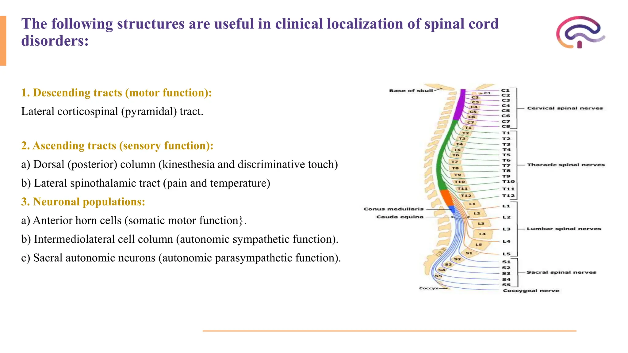
The following structures are useful in clinical localization of spinal cord
disorders:
1. Descending tracts (motor function):
Lateral corticospinal (pyramidal) tract.
2. Ascending tracts (sensory function):
a) Dorsal (posterior) column (kinesthesia and discriminative touch)
b) Lateral spinothalamic tract (pain and temperature)
3. Neuronal populations:
a) Anterior horn cells (somatic motor function}.
b) Intermediolateral cell column (autonomic sympathetic function).
c) Sacral autonomic neurons (autonomic parasympathetic function).
Spinal Cord Tracts
Dorsal (Posterior) columns
Ascending tracts (sensory:
fine touch,
proprioception)
Fasciculus gracilis Lower extremity
Fasciculus cuneatus Upper extremity
Lateral columns
Descending tracts Lateral corticospinal Motor: skeletal muscle
Rubrospinal Motor: cerebellum to skeletal muscle
Ascending tracts
Spinothalamic (anterolateral) Sensory: pain and temperature
Posterior (dorsal) spinocerebellar Sensory: proprioception to cerebellum
Anterior (ventral) spinocerebellar Sensory: proprioception to cerebellum
Ventral (Anterior) columns
Descending tracts
Anterior corticospinal Motor: skeletal muscle
Vestibulospinal Sensory: vestibular, balance to motor
neurons
Reticulospinal Sensory: pain, autonomic to motor
neurons
Tectospinal Sensory: visual to cervical motor neurons
Localization of spinal cord disorders
Spinal cord level Corresponding vertebral body
Cervical Same as cord level (C1 to C4)
Lower cervical 1 Level higher (C5-T6)
Upper thoracic 2 Level higher (T3 to T6)
Lower thoracic 2 to 3 level higher (T7 to T9)
Lumbar T10-T12
Sacral T12-L1
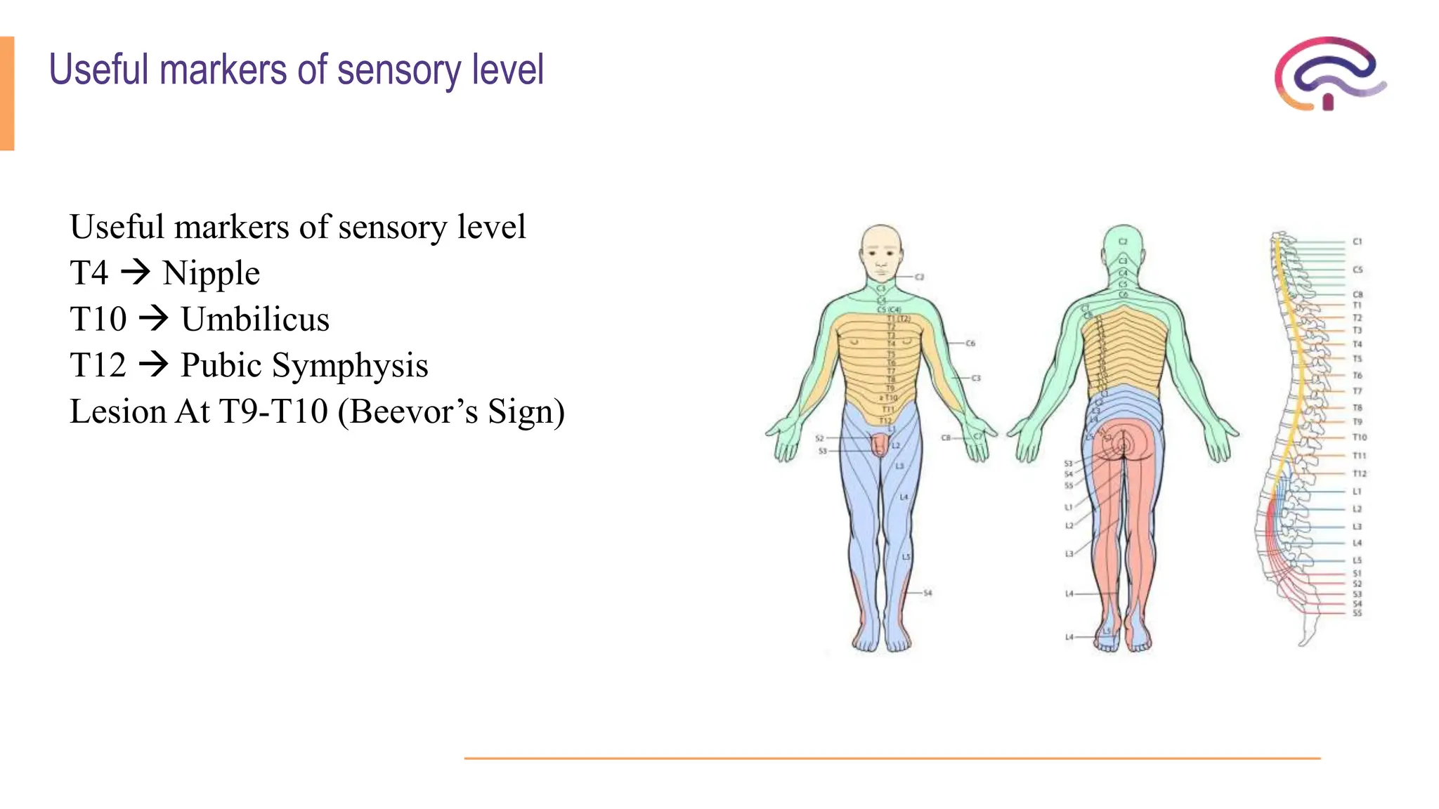
Useful markers of sensory level
T4  Nipple
T10  Umbilicus
T12  Pubic Symphysis
Lesion At T9-T10 (Beevor’s Sign)
Paraplegia
Paraplegia is paralysis or weakness (paraparesis) of both lower limbs.
- Pyramidal (U.M.N) lesion resulting in spastic paraplegia.
- Lower motor neuron (L.M.N.) lesion resulting in flaccid paraplegia.
Spastic paraplegia
It is paralysis or weakness of both lower limbs due to bilateral pyramidal tract lesion, most commonly in the
spinal cord (spinal paraplegia and less commonly in the in the brain stem or the cerebral parasagittal region
(cerebral paraplegia).
Spinal paraplegia may be:
- Focal: paraplegia with level
- Systemic: paraplegia without level
- Disseminated: paraplegia without level
Causes of spinal paraplegia
1) Focal causes:
a. Compression
1. Vertebral:
- Traumatic: fracture of vertebra
- Degenerative: spondylosis, disc prolapse, kyphoscoliosis
- Pott’s disease.
- Neoplastic diseases: primary as osteosarcoma & hemangioma or Metastatic from 1ry carcinoma of thyroid, breast, kidney, stomach & prostate.
2.Meningeal (extramedullary):
- Extradural: leukemic deposits
- Dural: meningioma
- Intradural: neurofibroma, cystic arachnoiditis
3. Cord (intramedullary):
Syringomyelia, Glioma or Ependymoma of cord
b. Inflammatory:
Transverse myelitis – Myelomeningitis – Myeloradiculitis
C, Vascular:
Anterior spinal artery occlusion.
• 2) Systemic causes:
• A systemic disease in neurology is a disease which affects one or more systems
selectively and is usual bilateral and symmetrical. When a systemic disease affects the
pyramidal tracts either alone or with other tracts, paraplegia will result.
• Heridofamilial: Hereditary spastic paraplegia, Hereditary ataxia: Friedreich’s ataxia.
• Symptomatic: pellagra lateral sclerosis, Subacute combined degeneration.
• Idiopathic: motor neuron disease.
•
• 3) Disseminated causes:
• A disseminated disease in neurology is a multifocal disease of the same nature:
• Multiple sclerosis (MS) & Acute disseminated encephalomyelitis (ADEM).
Causes of cerebral paraplegia
A. Causes in the parasagittal region: (area of cortical presentation of LL)
1.Traumatic: depressed fracture of the vault of the skull &Subdural hematoma.
2.Vascular: superior sagittal sinus thrombosis.
3.Inflammatory: encephalitis, meningoencephalitis.
4.Neoplastic: parasagittal meningioma.
5.Degenerative: cerebral palsy.
B. Causes in the Brain stem:
Syringobulbia & Midline brainstem tumors.
 these lesions arise in the midline and involve the innermost pyramidal fibers
which are those of the lower limbs.
Clinical picture of focal paraplegia
A. At the level of the lesion:
1. Vertebral manifestations: only if the cause is vertebral
- Localized pain or tenderness.
- Localized deformity or swelling.
2. Radicular manifestation: only present in extramedullary cause.
a) Posterior root affection:
- Early pain in the back referred to distribution of affected root and described as girdle pain; it is exaggerated by
coughing, sneezing & straining.
-Later there is hypoesthesia or anesthesia in dermatome supplied the affected root.
b) Anterior root affection:
- Localized L.M.N weakness in the muscles supplied by the affected root.
N.B. As the lesion in paraplegia is below cervical segments (which supply the muscle of the arm), the L.M.N affection at
the level of the lesion is not clinically evident; this is due to difficulty in eliciting wasting and difficulty of testing for tone
and deep reflexes in the trunk and abdominal muscles. If the lesion involves the cervical segments there is quadriplegia
with evident of LMNL in the upper limb.
B. Below the level of the lesion (Cord manifestation):
1.Motor manifestations: they depend on whether the cause of lesions is acute or gradual.
a) If the cause is acute:
(Inflammatory, Vascular, Or Traumatic).
The paraplegia passes by 2 stages:
 Stage of flaccidity due to neuronal shock:
Immediately following the lesion there is sudden paralysis of lower limbs associated with complete loss of
tone and absence of reflexes (flaccid paralysis), this stage lasts from 2 to 6 weeks.
 Stage of spasticity due to recovery from the neuronal shock:
On recovery from the shock stage, the picture of U.M.N.L. will be established including hypertonia,
hyperreflexia, positive Babinski sign & may be clonus.
b) If the cause is gradual (e.g., neoplastic):
 The shock stage is absent and there will be gradual progressive weakness of the lower limbs with hypertonia
and hyperreflexia:
1.The weakness affects the distal group of muscles more than the proximal group
and the flexor muscles more than the extensors.
2.The hypertonia and hyperreflexia affect the extensor group of muscles
(antigravity) more than the flexor group (progravity), the paraplegia in this stage
is described as (paraplegia in extension).
3.With further progression of the lesion the extrapyramidal fibers in the cord will be
affected, the hypertonia and hyperreflexia will be more in flexor group of muscles
than in the extensors. In this stage the paraplegia is described as (paraplegia in
flexion).
4.This last stage might be associated with the mass reflex where there is
spontaneous urination, defecation and sweating on scratching the skin over medial
side of the thigh associated with reflex erection and ejaculation on squeezing the
glans penis.
N.B.: Pierre Marie Foix test is done by firm passive planter flexing of the toes and foot. This will result in spontaneous “withdrawal reflex” i.e.,
spontaneous flexion of the hip, knee, and dorsiflexion of the ankle if the paraplegia is passing from extension to flexion.
Paraplegia In Extension Paraplegia In Flexion
Cause Pyramidal lesion Pyramidal and extrapyramidal
Hypertonia More in extensors More in flexors
Position Of L.L. Extended Flexed
Deep Reflexes Exaggerated Less exaggerated
Clonus Present Absent
Mass Reflex Absent May be present
Bladder Precipitancy Automatic bladder
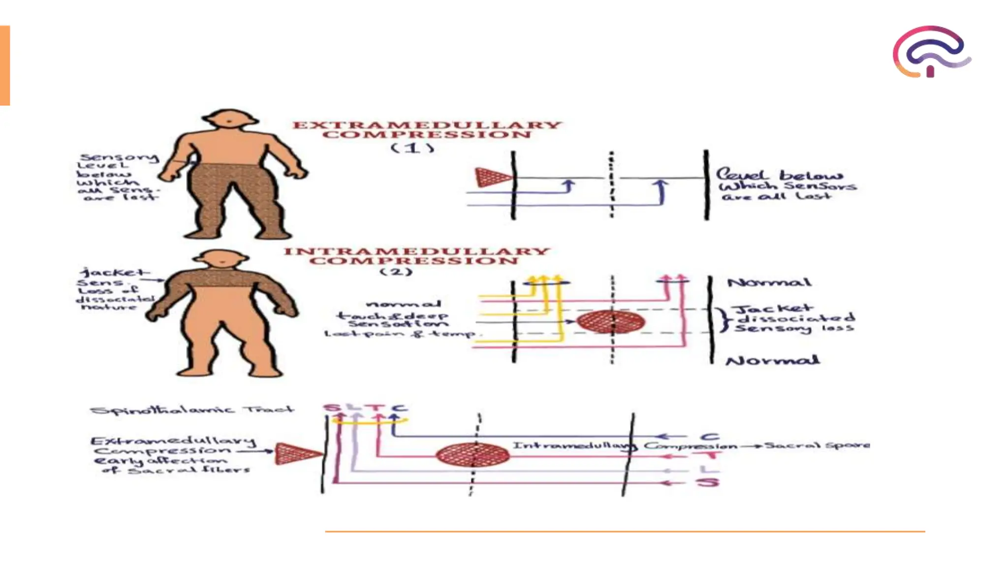
2. Sensory manifestations:
A) If the cause of the lesion is extramedullary, encroachment on the ascending tracts at the site of lesion results in a
sensory level below which all types of sensations are diminished. There is early loss of sensation in the saddle area (S
3,4,5), as the sacral fibers lie in the outermost part of spinothalamic tracts in the cord.
B) If the cause of the lesion is intramedullary, there will be jacket sensory loss (hyposthetic area with normal sensations
above and below it). The sensory loss of a dissociated nature i.e., pain and temperature sensations are lost but touch and
deep sensations are preserved; this is due to the interruption of crossing fibers carrying pain and temperature by the midline
lesion, while touch and deep sensation fibers ascend in the posterior column without decussation.
The sensations over the saddle area are preserved (sacral spare), as the sacral fibers lie far from the midline lesion.
3. sphincter manifestations:
a) In acute lesions retention of urine in
the shock stage followed by precipitancy
of micturition
b) In gradual lesions: precipitancy of
micturition which may terminate in
automatic bladder when complete
transection of the cord occurs.
These changes start late in the
extramedullary lesions and early in
intramedullary lesions as the pyramidal
fibers controlling the bladder center lie
medially in the cord.
Features Extramedullary Intramedullary
Radicular pain Early and common Rare
Sensory deficit No dissociation of
sensation
Dissociation of sensation
common
Sacral sensation Lost early Sacral spare
UMN involvement Early and prominent Less pronounced
LMN Segmental Marked with widespread
atrophy, fasciculations seen
Reflexes Brisk early feature Less brisk late feature
Autonomic
involvement
Late early
Trophic changes Usually not marked common
Vertebral tenderness May be sensitive to local
pressure
No bony tenderness
Changes in CSF Frequent rare
Clinical Localization
 Higher Cervical Cord Lesion:
- Sensory loss over occipital (C2).
- Involvement of spinal accessory nerve.
- Wasting of small muscles of hands.
- Quadriplegia with diaphragm weakness.
- Sensory loss of upper portion of face along with loss of corneal reflex due to involvement of spinal nucleus of
5th cranial nerve.
 Lower Cervical Cord:
- C5-C6  weakness in deltoid, supraspinatus, loss of power and reflexes in the biceps.
- C7  weakness is found in triceps, finger, and wrist extensors (radial nerve).
- C8  in finger and wrist flexion are impaired (ulnar nerve).
- Horner’s syndrome occurs lesion at any level.
 Thoracic Cord Lesion:
- Sensory level on trunk
- Site of midline back pain
- Paraparesis / paraplegia of lower limb
- Beevor’s sign positive
- Bladder bowel involvement
Lumbar cord lesions:
L2 - L4:
- Weakness of flexion and adduction of thigh
- Weakness leg extension at knee loss patellar reflex
- Exaggerated ankle jerk
L5 - S1:
- Paralyze movement of foot and ankle
- Weakness of flexion at knee
- Weakness of extension of the thigh
- Loss of ankle jerks (L5, S1)
Segmental Syndromes
Pathologies that affect all functions of the spinal cord at one or more levels produce a segmental syndrome.
Loss of function may be total or incomplete.
Total Cord Transection Syndrome
Results from the cessation of function in all ascending and descending spinal cord pathways and results in the
loss of all types of sensation and loss of movement below the level of the lesion
Acute transection can cause spinal shock, with a flaccid paralysis, urinary retention, and diminished tendon
reflexes.
this is usually temporary, and increased tone, spasticity, and hyperreflexia will usually supervene in days or
weeks after the event.
Causes of a cord segmental syndrome include:
1. Acute myelopathies, such as traumatic injury and spinal cord hemorrhage.
2. Epidural or intramedullary abscess, tumors, and transverse myelitis may have a more subacute presentation
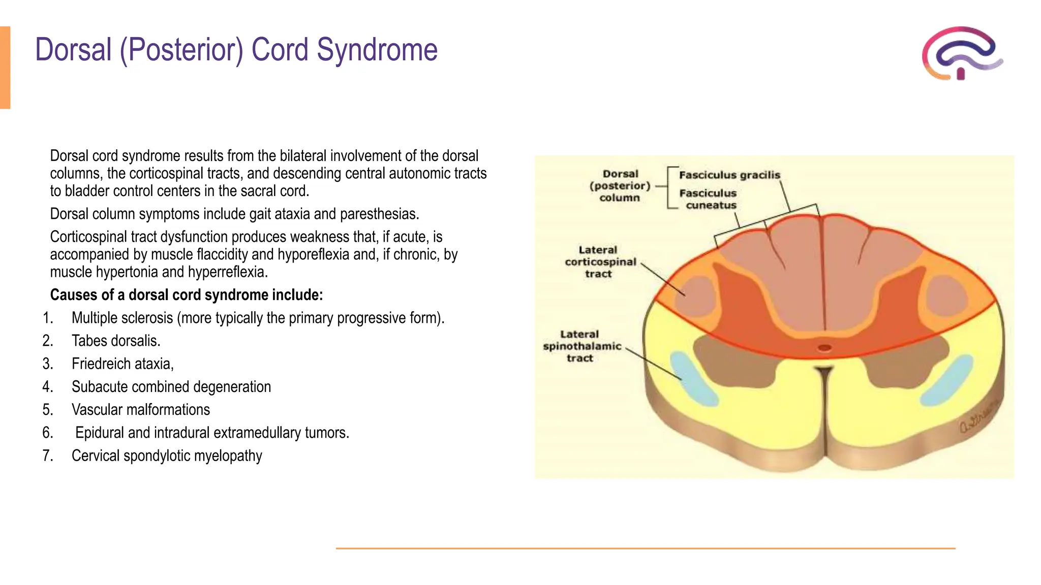
Dorsal (Posterior) Cord Syndrome
Dorsal cord syndrome results from the bilateral involvement of the dorsal
columns, the corticospinal tracts, and descending central autonomic tracts
to bladder control centers in the sacral cord.
Dorsal column symptoms include gait ataxia and paresthesias.
Corticospinal tract dysfunction produces weakness that, if acute, is
accompanied by muscle flaccidity and hyporeflexia and, if chronic, by
muscle hypertonia and hyperreflexia.
Causes of a dorsal cord syndrome include:
1. Multiple sclerosis (more typically the primary progressive form).
2. Tabes dorsalis.
3. Friedreich ataxia,
4. Subacute combined degeneration
5. Vascular malformations
6. Epidural and intradural extramedullary tumors.
7. Cervical spondylotic myelopathy
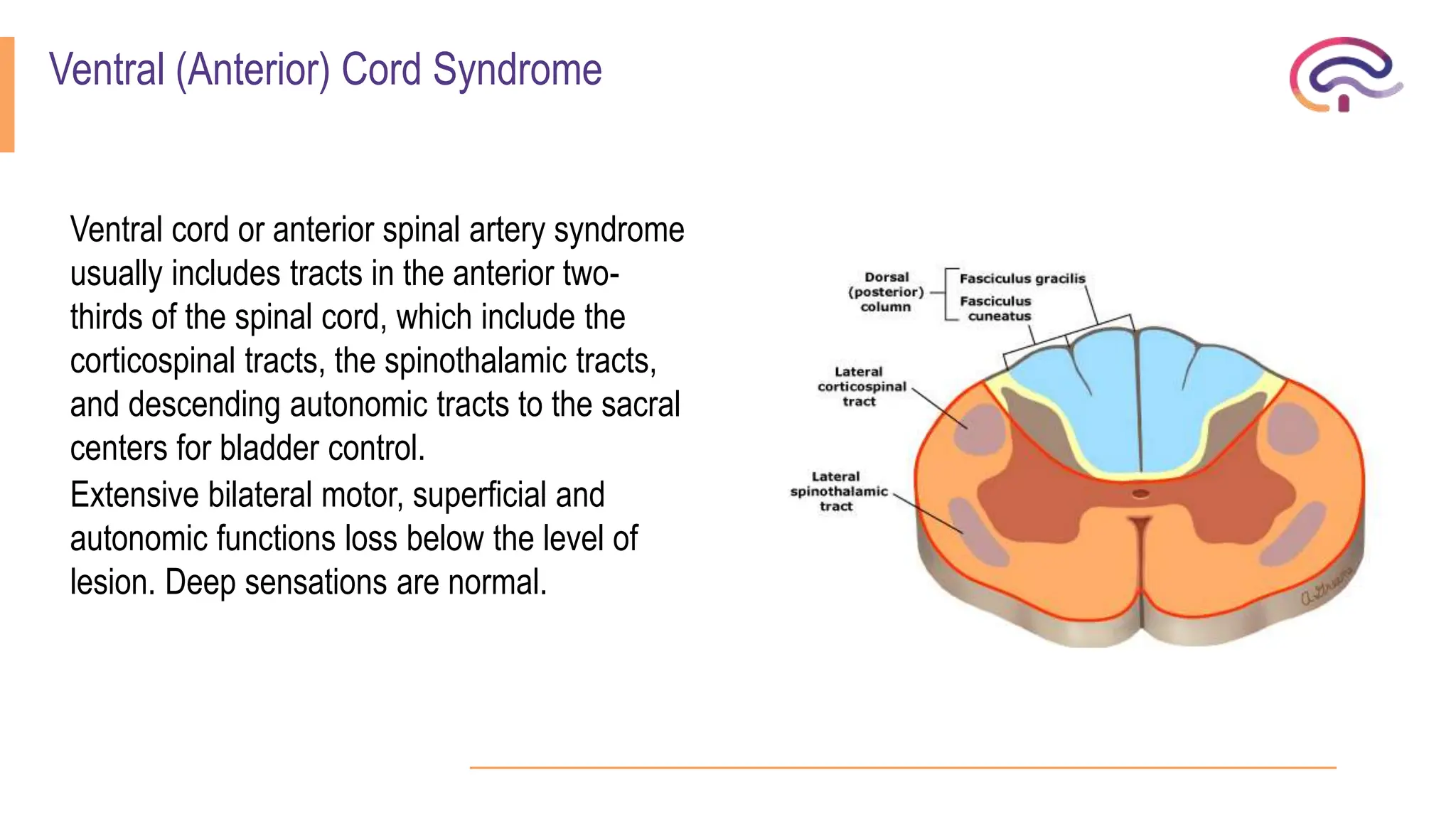
Ventral (Anterior) Cord Syndrome
Ventral cord or anterior spinal artery syndrome
usually includes tracts in the anterior two-
thirds of the spinal cord, which include the
corticospinal tracts, the spinothalamic tracts,
and descending autonomic tracts to the sacral
centers for bladder control.
Extensive bilateral motor, superficial and
autonomic functions loss below the level of
lesion. Deep sensations are normal.
Brown-Sequard (Hemi-Cord) Syndrome
A lateral hemi section syndrome, also known as the Brown–
Sequard syndrome, involves the dorsal column, corticospinal
tract, and spinothalamic tract unilaterally.
At the level of the lesion:
 Motor: Ipsilateral LMN
 Sensory: Ipsilateral loss of deep and superficial sensation.
Below level of lesion:
 Motor: Ipsilateral UMN
 Sensory: Ipsilateral loss of deep sensation and contralateral
loss of superficial sensation.
While there are many causes of this syndrome, knife or bullet
injuries and demyelination are the most common.
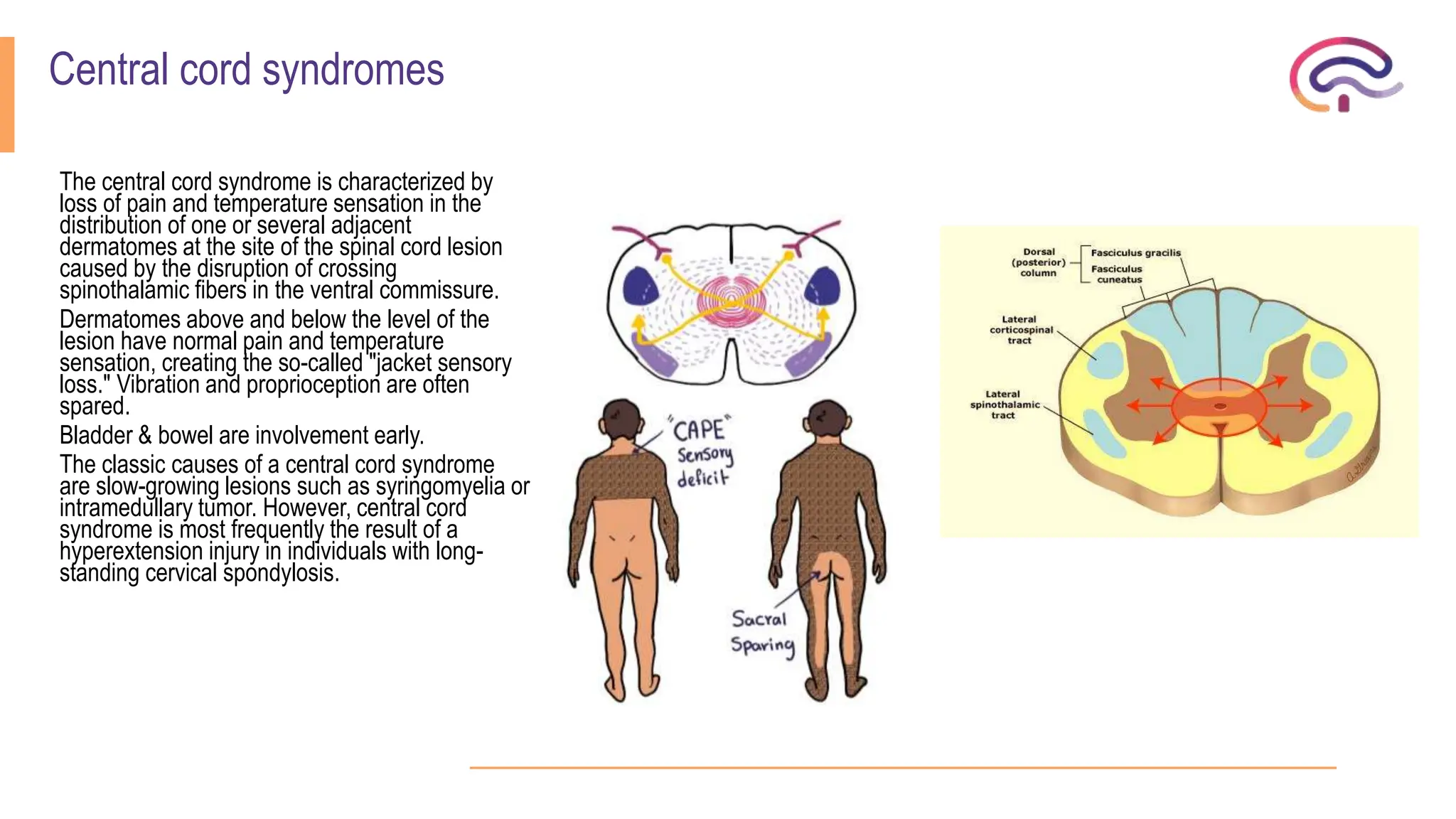
Central cord syndromes
The central cord syndrome is characterized by
loss of pain and temperature sensation in the
distribution of one or several adjacent
dermatomes at the site of the spinal cord lesion
caused by the disruption of crossing
spinothalamic fibers in the ventral commissure.
Dermatomes above and below the level of the
lesion have normal pain and temperature
sensation, creating the so-called "jacket sensory
loss." Vibration and proprioception are often
spared.
Bladder & bowel are involvement early.
The classic causes of a central cord syndrome
are slow-growing lesions such as syringomyelia or
intramedullary tumor. However, central cord
syndrome is most frequently the result of a
hyperextension injury in individuals with long-
standing cervical spondylosis.
Conus Medullaris Syndrome
• Lesions at vertebral level L2 often affect the conus medullaris.
• There is early and prominent sphincter dysfunction with flaccid paralysis of the bladder and rectum, impotence, and
saddle (S3-S5) anesthesia.
• Preserved motor function of lower limbs including ankle jerk.
• Causes include disc herniation, spinal fracture, and tumors.
Cauda Equina Syndrome
Deficits usually affect both legs but are often asymmetric.
Symptoms include:
 Low back pain accompanied by pain radiating into one or both legs.
 Radicular pain reflects involvement of dorsal nerve roots and may have localizing value.
 Weakness of plantar flexion of the feet with loss of ankle jerks occurs with mid cauda equina lesions, involving S1, S2
roots.
 Relative sparing of Bladder and rectal sphincter function.
 Sensory loss of all sensory modalities occurs in the dermatomal distribution of the affected nerve roots.
Many etiologies can cause a cauda equina syndrome including:
1. Intervertebral disc herniation.
2. Epidural abscess & tumor.
3. Intradural extramedullary tumor.
4. Lumbar spine spondylosis.
5. Spinal arachnoiditis.
6. Chronic inflammatory demyelinating polyneuropathy.
7. Sarcoidosis.
Conus medullaris syndrome Cauda equina syndrome
Vertebral level L1-L2 L2-sacrum
Spinal level Sacral cord segment and roots Lumbosacral nerve roots
Presentation Sudden and bilateral Gradual and unilateral
Radicular pain Less severe More severe
Low back pain More Less
Motor strength Symmetrical, less marked hyperreflexia distal paresis of
LL, fasciculation
More marked asymmetric areflexic paraplegia, atrophy
more common
Reflexes Ankle jerks affected Both knee and ankle jerks affected
Sensory Localized numbness to perianal area, symmetrical and
bilateral
Localized numbness at saddle area, asymmetrical and
unilateral
Sphincter dysfunction Early urinary and fecal incontinence Tend to present late
Impotence Frequent Less frequent
Pattern of sensory loss
spinal cord disorders  and  paraplegia .

More Related Content

What's hot

Neuropathy complete2
Neuropathy complete2Neuropathy complete2
Neuropathy complete2
udom
 
Pathology of Epilepsy
Pathology of EpilepsyPathology of Epilepsy
Pathology of Epilepsy
ML Cohen
 

What's hot (20)

Neurological history taking (2024) .
Neurological  history  taking  (2024)  .Neurological  history  taking  (2024)  .
Neurological history taking (2024) .
 
relfexes
relfexes relfexes
relfexes
 
Myasthenia gravis
Myasthenia gravisMyasthenia gravis
Myasthenia gravis
 
cranial nerve examination & theoritical
cranial nerve examination & theoritical cranial nerve examination & theoritical
cranial nerve examination & theoritical
 
approach to comatose patient
approach to comatose patient approach to comatose patient
approach to comatose patient
 
movement disorder for physiotherapy .pptx
movement disorder for physiotherapy .pptxmovement disorder for physiotherapy .pptx
movement disorder for physiotherapy .pptx
 
Spinal Cord Disease - Clinical Presentations and Management
Spinal Cord Disease - Clinical Presentations and ManagementSpinal Cord Disease - Clinical Presentations and Management
Spinal Cord Disease - Clinical Presentations and Management
 
spinal cord injury
spinal cord injuryspinal cord injury
spinal cord injury
 
ISAAC’S Syndrome.pptx
ISAAC’S Syndrome.pptxISAAC’S Syndrome.pptx
ISAAC’S Syndrome.pptx
 
Neuropathy complete2
Neuropathy complete2Neuropathy complete2
Neuropathy complete2
 
Pathology of Epilepsy
Pathology of EpilepsyPathology of Epilepsy
Pathology of Epilepsy
 
Multiple sclerosis
Multiple sclerosisMultiple sclerosis
Multiple sclerosis
 
approach to Dystonia and myoclonus movement disorders
approach to Dystonia and myoclonus movement disordersapproach to Dystonia and myoclonus movement disorders
approach to Dystonia and myoclonus movement disorders
 
cranial nerve examination
cranial nerve examinationcranial nerve examination
cranial nerve examination
 
Neuropathy ..paras
Neuropathy ..parasNeuropathy ..paras
Neuropathy ..paras
 
Cranial nerves
Cranial nervesCranial nerves
Cranial nerves
 
Reflex and Voluntary Control of Movement
Reflex and Voluntary Control of MovementReflex and Voluntary Control of Movement
Reflex and Voluntary Control of Movement
 
Motor Neuron Disease and PT Mx
Motor Neuron Disease and PT MxMotor Neuron Disease and PT Mx
Motor Neuron Disease and PT Mx
 
Neurology 4th epilepsy(1)
Neurology 4th epilepsy(1)Neurology 4th epilepsy(1)
Neurology 4th epilepsy(1)
 
Lecture section...Myasthenia Gravis
Lecture section...Myasthenia GravisLecture section...Myasthenia Gravis
Lecture section...Myasthenia Gravis
 

Similar to spinal cord disorders and paraplegia .

Spinal shock Physiology
Spinal shock PhysiologySpinal shock Physiology
Spinal shock Physiology
Raghu Veer
 
Diseases of the spinal cord
Diseases of the spinal cordDiseases of the spinal cord
Diseases of the spinal cord
Hiba Hassan
 

Similar to spinal cord disorders and paraplegia . (20)

Paraparesis biplave nams
Paraparesis biplave namsParaparesis biplave nams
Paraparesis biplave nams
 
paraparesisbiplavenams-190401002135.pdf
paraparesisbiplavenams-190401002135.pdfparaparesisbiplavenams-190401002135.pdf
paraparesisbiplavenams-190401002135.pdf
 
Paraplegia
ParaplegiaParaplegia
Paraplegia
 
SPINAL INJURY.pdf
SPINAL INJURY.pdfSPINAL INJURY.pdf
SPINAL INJURY.pdf
 
Limb Weakness Part I
Limb Weakness Part ILimb Weakness Part I
Limb Weakness Part I
 
Spinal cord injury
Spinal cord injurySpinal cord injury
Spinal cord injury
 
Spinal cord disorders
Spinal cord disordersSpinal cord disorders
Spinal cord disorders
 
Localization
LocalizationLocalization
Localization
 
Spinal shock Physiology
Spinal shock PhysiologySpinal shock Physiology
Spinal shock Physiology
 
Spinal cord injuries..
Spinal cord injuries..Spinal cord injuries..
Spinal cord injuries..
 
Spinal cord injuries
Spinal cord injuriesSpinal cord injuries
Spinal cord injuries
 
Diseases of the spinal cord
Diseases of the spinal cordDiseases of the spinal cord
Diseases of the spinal cord
 
Clinical problems related to the spinal cord
Clinical problems related to the spinal cordClinical problems related to the spinal cord
Clinical problems related to the spinal cord
 
Neuroscience board review.pptx
Neuroscience board review.pptxNeuroscience board review.pptx
Neuroscience board review.pptx
 
spinal cord anushka.pptx - BY ANUSHKA - MBBS 1ST YR
spinal cord anushka.pptx - BY ANUSHKA  - MBBS 1ST YRspinal cord anushka.pptx - BY ANUSHKA  - MBBS 1ST YR
spinal cord anushka.pptx - BY ANUSHKA - MBBS 1ST YR
 
Limb Weakness II Osama S. M. Amin
Limb Weakness II Osama S. M. AminLimb Weakness II Osama S. M. Amin
Limb Weakness II Osama S. M. Amin
 
Plexopathy
PlexopathyPlexopathy
Plexopathy
 
Spinal cord injury
Spinal cord injurySpinal cord injury
Spinal cord injury
 
Approach to myelopathy
Approach to myelopathyApproach to myelopathy
Approach to myelopathy
 
Brainstem leisons
Brainstem leisonsBrainstem leisons
Brainstem leisons
 

More from Mohamed Rizk Khodair (7)

motor system examination .pptx
motor system examination .pptxmotor system examination .pptx
motor system examination .pptx
 
SAH
SAHSAH
SAH
 
ataxia
ataxia ataxia
ataxia
 
sensory examination, speech , neurogenic bladder
sensory examination, speech , neurogenic bladdersensory examination, speech , neurogenic bladder
sensory examination, speech , neurogenic bladder
 
myopthies.pptx
myopthies.pptxmyopthies.pptx
myopthies.pptx
 
motor system examination.pptx
motor system examination.pptxmotor system examination.pptx
motor system examination.pptx
 
sensory system examination
sensory system examinationsensory system examination
sensory system examination
 

Recently uploaded

Best medicine 100% Effective&Safe Mifepristion ௵+918133066128௹Abortion pills ...
Best medicine 100% Effective&Safe Mifepristion ௵+918133066128௹Abortion pills ...Best medicine 100% Effective&Safe Mifepristion ௵+918133066128௹Abortion pills ...
Best medicine 100% Effective&Safe Mifepristion ௵+918133066128௹Abortion pills ...
Abortion pills in Kuwait Cytotec pills in Kuwait
 

Recently uploaded (20)

Is Rheumatoid Arthritis a Metabolic Disorder.pptx
Is Rheumatoid Arthritis a Metabolic Disorder.pptxIs Rheumatoid Arthritis a Metabolic Disorder.pptx
Is Rheumatoid Arthritis a Metabolic Disorder.pptx
 
ESC HF 2024 Spotlights Day-2.pptx heart failure
ESC HF 2024 Spotlights Day-2.pptx heart failureESC HF 2024 Spotlights Day-2.pptx heart failure
ESC HF 2024 Spotlights Day-2.pptx heart failure
 
DR. Neha Mehta Best Psychologist.in India
DR. Neha Mehta Best Psychologist.in IndiaDR. Neha Mehta Best Psychologist.in India
DR. Neha Mehta Best Psychologist.in India
 
Results For Love Spell Is Guaranteed In 1 Day +27834335081 [BACK LOST LOVE SP...
Results For Love Spell Is Guaranteed In 1 Day +27834335081 [BACK LOST LOVE SP...Results For Love Spell Is Guaranteed In 1 Day +27834335081 [BACK LOST LOVE SP...
Results For Love Spell Is Guaranteed In 1 Day +27834335081 [BACK LOST LOVE SP...
 
Quality control tests of suppository ...
Quality control tests  of suppository ...Quality control tests  of suppository ...
Quality control tests of suppository ...
 
TEST BANK For Lewis's Medical Surgical Nursing in Canada, 4th Edition by Jane...
TEST BANK For Lewis's Medical Surgical Nursing in Canada, 4th Edition by Jane...TEST BANK For Lewis's Medical Surgical Nursing in Canada, 4th Edition by Jane...
TEST BANK For Lewis's Medical Surgical Nursing in Canada, 4th Edition by Jane...
 
Denture base resins materials and its mechanism of action
Denture base resins materials and its mechanism of actionDenture base resins materials and its mechanism of action
Denture base resins materials and its mechanism of action
 
Case presentation on Antibody screening- how to solve 3 cell and 11 cell panel?
Case presentation on Antibody screening- how to solve 3 cell and 11 cell panel?Case presentation on Antibody screening- how to solve 3 cell and 11 cell panel?
Case presentation on Antibody screening- how to solve 3 cell and 11 cell panel?
 
The Clean Living Project Episode 24 - Subconscious
The Clean Living Project Episode 24 - SubconsciousThe Clean Living Project Episode 24 - Subconscious
The Clean Living Project Episode 24 - Subconscious
 
Dermatome and myotome test & pathology.pdf
Dermatome and myotome test & pathology.pdfDermatome and myotome test & pathology.pdf
Dermatome and myotome test & pathology.pdf
 
Let's Talk About It: Ovarian Cancer (The Emotional Toll of Treatment Decision...
Let's Talk About It: Ovarian Cancer (The Emotional Toll of Treatment Decision...Let's Talk About It: Ovarian Cancer (The Emotional Toll of Treatment Decision...
Let's Talk About It: Ovarian Cancer (The Emotional Toll of Treatment Decision...
 
Mgr university bsc nursing adult health previous question paper with answers
Mgr university  bsc nursing adult health previous question paper with answersMgr university  bsc nursing adult health previous question paper with answers
Mgr university bsc nursing adult health previous question paper with answers
 
CONGENITAL HYPERTROPHIC PYLORIC STENOSIS by Dr M.KARTHIK EMMANUEL
CONGENITAL HYPERTROPHIC PYLORIC STENOSIS  by Dr M.KARTHIK EMMANUELCONGENITAL HYPERTROPHIC PYLORIC STENOSIS  by Dr M.KARTHIK EMMANUEL
CONGENITAL HYPERTROPHIC PYLORIC STENOSIS by Dr M.KARTHIK EMMANUEL
 
Treatment Choices for Slip Disc at Gokuldas Hospital
Treatment Choices for Slip Disc at Gokuldas HospitalTreatment Choices for Slip Disc at Gokuldas Hospital
Treatment Choices for Slip Disc at Gokuldas Hospital
 
Tissue Banking and Umbilical Cord Blood Banking
Tissue Banking and Umbilical Cord Blood BankingTissue Banking and Umbilical Cord Blood Banking
Tissue Banking and Umbilical Cord Blood Banking
 
ANAPHYLAXIS BY DR.SOHAN BISWAS,MBBS,DNB(INTERNAL MEDICINE) RESIDENT.pptx
ANAPHYLAXIS BY DR.SOHAN BISWAS,MBBS,DNB(INTERNAL MEDICINE) RESIDENT.pptxANAPHYLAXIS BY DR.SOHAN BISWAS,MBBS,DNB(INTERNAL MEDICINE) RESIDENT.pptx
ANAPHYLAXIS BY DR.SOHAN BISWAS,MBBS,DNB(INTERNAL MEDICINE) RESIDENT.pptx
 
Varicose Veins Treatment Aftercare Tips by Gokuldas Hospital
Varicose Veins Treatment Aftercare Tips by Gokuldas HospitalVaricose Veins Treatment Aftercare Tips by Gokuldas Hospital
Varicose Veins Treatment Aftercare Tips by Gokuldas Hospital
 
Making Patient-Centric Immunotherapy a Reality in Lung Cancer: Best Practices...
Making Patient-Centric Immunotherapy a Reality in Lung Cancer: Best Practices...Making Patient-Centric Immunotherapy a Reality in Lung Cancer: Best Practices...
Making Patient-Centric Immunotherapy a Reality in Lung Cancer: Best Practices...
 
Best medicine 100% Effective&Safe Mifepristion ௵+918133066128௹Abortion pills ...
Best medicine 100% Effective&Safe Mifepristion ௵+918133066128௹Abortion pills ...Best medicine 100% Effective&Safe Mifepristion ௵+918133066128௹Abortion pills ...
Best medicine 100% Effective&Safe Mifepristion ௵+918133066128௹Abortion pills ...
 
NDCT Rules, 2019: An Overview | New Drugs and Clinical Trial Rules 2019
NDCT Rules, 2019: An Overview | New Drugs and Clinical Trial Rules 2019NDCT Rules, 2019: An Overview | New Drugs and Clinical Trial Rules 2019
NDCT Rules, 2019: An Overview | New Drugs and Clinical Trial Rules 2019
 

spinal cord disorders and paraplegia .

  • 1. Spinal cord disorders Mohamed rizk khodair Lecturer of neurology October 6 university Mohamedrizk.med@o6u.edu.eg
  • 2. Myelopathy: It is an injury to the spinal cord due to severe compression that may result from trauma, congenital stenosis, degenerative disease, or disc herniation. Myelitis: is an inflammation of the spinal cord. This inflammation can cause the myelin and axon to be damaged resulting in symptoms such as paralysis and sensory loss. Radiculopathy: is an injury or damage to nerve roots in the area where they leave the spine. This condition can affect anyone and can be the result disc degeneration, disc herniation or other trauma. Myelopathy vs Radiculopathy Myelopathy refers to a condition known as spinal cord compression, while radiculopathy occurs when a nerve root (but not the cord itself) becomes compressed. The latter is more commonly known as a pinched nerve root.
  • 3.
  • 4.
  • 5. Internal Divisions of the Spinal Cord A cross section of the spinal cord shows an H shaped central grey matter surrounded by a peripheral white matter.
  • 6.
  • 7. The following structures are useful in clinical localization of spinal cord disorders: 1. Descending tracts (motor function): Lateral corticospinal (pyramidal) tract. 2. Ascending tracts (sensory function): a) Dorsal (posterior) column (kinesthesia and discriminative touch) b) Lateral spinothalamic tract (pain and temperature) 3. Neuronal populations: a) Anterior horn cells (somatic motor function}. b) Intermediolateral cell column (autonomic sympathetic function). c) Sacral autonomic neurons (autonomic parasympathetic function).
  • 8. Spinal Cord Tracts Dorsal (Posterior) columns Ascending tracts (sensory: fine touch, proprioception) Fasciculus gracilis Lower extremity Fasciculus cuneatus Upper extremity Lateral columns Descending tracts Lateral corticospinal Motor: skeletal muscle Rubrospinal Motor: cerebellum to skeletal muscle Ascending tracts Spinothalamic (anterolateral) Sensory: pain and temperature Posterior (dorsal) spinocerebellar Sensory: proprioception to cerebellum Anterior (ventral) spinocerebellar Sensory: proprioception to cerebellum Ventral (Anterior) columns Descending tracts Anterior corticospinal Motor: skeletal muscle Vestibulospinal Sensory: vestibular, balance to motor neurons Reticulospinal Sensory: pain, autonomic to motor neurons Tectospinal Sensory: visual to cervical motor neurons
  • 9. Localization of spinal cord disorders Spinal cord level Corresponding vertebral body Cervical Same as cord level (C1 to C4) Lower cervical 1 Level higher (C5-T6) Upper thoracic 2 Level higher (T3 to T6) Lower thoracic 2 to 3 level higher (T7 to T9) Lumbar T10-T12 Sacral T12-L1
  • 10. Useful markers of sensory level Useful markers of sensory level T4  Nipple T10  Umbilicus T12  Pubic Symphysis Lesion At T9-T10 (Beevor’s Sign)
  • 11.
  • 12. Paraplegia Paraplegia is paralysis or weakness (paraparesis) of both lower limbs. - Pyramidal (U.M.N) lesion resulting in spastic paraplegia. - Lower motor neuron (L.M.N.) lesion resulting in flaccid paraplegia. Spastic paraplegia It is paralysis or weakness of both lower limbs due to bilateral pyramidal tract lesion, most commonly in the spinal cord (spinal paraplegia and less commonly in the in the brain stem or the cerebral parasagittal region (cerebral paraplegia). Spinal paraplegia may be: - Focal: paraplegia with level - Systemic: paraplegia without level - Disseminated: paraplegia without level
  • 13. Causes of spinal paraplegia 1) Focal causes: a. Compression 1. Vertebral: - Traumatic: fracture of vertebra - Degenerative: spondylosis, disc prolapse, kyphoscoliosis - Pott’s disease. - Neoplastic diseases: primary as osteosarcoma & hemangioma or Metastatic from 1ry carcinoma of thyroid, breast, kidney, stomach & prostate. 2.Meningeal (extramedullary): - Extradural: leukemic deposits - Dural: meningioma - Intradural: neurofibroma, cystic arachnoiditis 3. Cord (intramedullary): Syringomyelia, Glioma or Ependymoma of cord b. Inflammatory: Transverse myelitis – Myelomeningitis – Myeloradiculitis C, Vascular: Anterior spinal artery occlusion.
  • 14. • 2) Systemic causes: • A systemic disease in neurology is a disease which affects one or more systems selectively and is usual bilateral and symmetrical. When a systemic disease affects the pyramidal tracts either alone or with other tracts, paraplegia will result. • Heridofamilial: Hereditary spastic paraplegia, Hereditary ataxia: Friedreich’s ataxia. • Symptomatic: pellagra lateral sclerosis, Subacute combined degeneration. • Idiopathic: motor neuron disease. • • 3) Disseminated causes: • A disseminated disease in neurology is a multifocal disease of the same nature: • Multiple sclerosis (MS) & Acute disseminated encephalomyelitis (ADEM).
  • 15. Causes of cerebral paraplegia A. Causes in the parasagittal region: (area of cortical presentation of LL) 1.Traumatic: depressed fracture of the vault of the skull &Subdural hematoma. 2.Vascular: superior sagittal sinus thrombosis. 3.Inflammatory: encephalitis, meningoencephalitis. 4.Neoplastic: parasagittal meningioma. 5.Degenerative: cerebral palsy. B. Causes in the Brain stem: Syringobulbia & Midline brainstem tumors.  these lesions arise in the midline and involve the innermost pyramidal fibers which are those of the lower limbs.
  • 16. Clinical picture of focal paraplegia A. At the level of the lesion: 1. Vertebral manifestations: only if the cause is vertebral - Localized pain or tenderness. - Localized deformity or swelling. 2. Radicular manifestation: only present in extramedullary cause. a) Posterior root affection: - Early pain in the back referred to distribution of affected root and described as girdle pain; it is exaggerated by coughing, sneezing & straining. -Later there is hypoesthesia or anesthesia in dermatome supplied the affected root. b) Anterior root affection: - Localized L.M.N weakness in the muscles supplied by the affected root. N.B. As the lesion in paraplegia is below cervical segments (which supply the muscle of the arm), the L.M.N affection at the level of the lesion is not clinically evident; this is due to difficulty in eliciting wasting and difficulty of testing for tone and deep reflexes in the trunk and abdominal muscles. If the lesion involves the cervical segments there is quadriplegia with evident of LMNL in the upper limb.
  • 17. B. Below the level of the lesion (Cord manifestation): 1.Motor manifestations: they depend on whether the cause of lesions is acute or gradual. a) If the cause is acute: (Inflammatory, Vascular, Or Traumatic). The paraplegia passes by 2 stages:  Stage of flaccidity due to neuronal shock: Immediately following the lesion there is sudden paralysis of lower limbs associated with complete loss of tone and absence of reflexes (flaccid paralysis), this stage lasts from 2 to 6 weeks.  Stage of spasticity due to recovery from the neuronal shock: On recovery from the shock stage, the picture of U.M.N.L. will be established including hypertonia, hyperreflexia, positive Babinski sign & may be clonus. b) If the cause is gradual (e.g., neoplastic):
  • 18.  The shock stage is absent and there will be gradual progressive weakness of the lower limbs with hypertonia and hyperreflexia: 1.The weakness affects the distal group of muscles more than the proximal group and the flexor muscles more than the extensors. 2.The hypertonia and hyperreflexia affect the extensor group of muscles (antigravity) more than the flexor group (progravity), the paraplegia in this stage is described as (paraplegia in extension). 3.With further progression of the lesion the extrapyramidal fibers in the cord will be affected, the hypertonia and hyperreflexia will be more in flexor group of muscles than in the extensors. In this stage the paraplegia is described as (paraplegia in flexion). 4.This last stage might be associated with the mass reflex where there is spontaneous urination, defecation and sweating on scratching the skin over medial side of the thigh associated with reflex erection and ejaculation on squeezing the glans penis.
  • 19. N.B.: Pierre Marie Foix test is done by firm passive planter flexing of the toes and foot. This will result in spontaneous “withdrawal reflex” i.e., spontaneous flexion of the hip, knee, and dorsiflexion of the ankle if the paraplegia is passing from extension to flexion. Paraplegia In Extension Paraplegia In Flexion Cause Pyramidal lesion Pyramidal and extrapyramidal Hypertonia More in extensors More in flexors Position Of L.L. Extended Flexed Deep Reflexes Exaggerated Less exaggerated Clonus Present Absent Mass Reflex Absent May be present Bladder Precipitancy Automatic bladder
  • 20. 2. Sensory manifestations: A) If the cause of the lesion is extramedullary, encroachment on the ascending tracts at the site of lesion results in a sensory level below which all types of sensations are diminished. There is early loss of sensation in the saddle area (S 3,4,5), as the sacral fibers lie in the outermost part of spinothalamic tracts in the cord. B) If the cause of the lesion is intramedullary, there will be jacket sensory loss (hyposthetic area with normal sensations above and below it). The sensory loss of a dissociated nature i.e., pain and temperature sensations are lost but touch and deep sensations are preserved; this is due to the interruption of crossing fibers carrying pain and temperature by the midline lesion, while touch and deep sensation fibers ascend in the posterior column without decussation. The sensations over the saddle area are preserved (sacral spare), as the sacral fibers lie far from the midline lesion.
  • 21.
  • 22. 3. sphincter manifestations: a) In acute lesions retention of urine in the shock stage followed by precipitancy of micturition b) In gradual lesions: precipitancy of micturition which may terminate in automatic bladder when complete transection of the cord occurs. These changes start late in the extramedullary lesions and early in intramedullary lesions as the pyramidal fibers controlling the bladder center lie medially in the cord.
  • 23. Features Extramedullary Intramedullary Radicular pain Early and common Rare Sensory deficit No dissociation of sensation Dissociation of sensation common Sacral sensation Lost early Sacral spare UMN involvement Early and prominent Less pronounced LMN Segmental Marked with widespread atrophy, fasciculations seen Reflexes Brisk early feature Less brisk late feature Autonomic involvement Late early Trophic changes Usually not marked common Vertebral tenderness May be sensitive to local pressure No bony tenderness Changes in CSF Frequent rare
  • 24. Clinical Localization  Higher Cervical Cord Lesion: - Sensory loss over occipital (C2). - Involvement of spinal accessory nerve. - Wasting of small muscles of hands. - Quadriplegia with diaphragm weakness. - Sensory loss of upper portion of face along with loss of corneal reflex due to involvement of spinal nucleus of 5th cranial nerve.  Lower Cervical Cord: - C5-C6  weakness in deltoid, supraspinatus, loss of power and reflexes in the biceps. - C7  weakness is found in triceps, finger, and wrist extensors (radial nerve). - C8  in finger and wrist flexion are impaired (ulnar nerve). - Horner’s syndrome occurs lesion at any level.
  • 25.  Thoracic Cord Lesion: - Sensory level on trunk - Site of midline back pain - Paraparesis / paraplegia of lower limb - Beevor’s sign positive - Bladder bowel involvement Lumbar cord lesions: L2 - L4: - Weakness of flexion and adduction of thigh - Weakness leg extension at knee loss patellar reflex - Exaggerated ankle jerk L5 - S1: - Paralyze movement of foot and ankle - Weakness of flexion at knee - Weakness of extension of the thigh - Loss of ankle jerks (L5, S1)
  • 26. Segmental Syndromes Pathologies that affect all functions of the spinal cord at one or more levels produce a segmental syndrome. Loss of function may be total or incomplete.
  • 27. Total Cord Transection Syndrome Results from the cessation of function in all ascending and descending spinal cord pathways and results in the loss of all types of sensation and loss of movement below the level of the lesion Acute transection can cause spinal shock, with a flaccid paralysis, urinary retention, and diminished tendon reflexes. this is usually temporary, and increased tone, spasticity, and hyperreflexia will usually supervene in days or weeks after the event. Causes of a cord segmental syndrome include: 1. Acute myelopathies, such as traumatic injury and spinal cord hemorrhage. 2. Epidural or intramedullary abscess, tumors, and transverse myelitis may have a more subacute presentation
  • 28. Dorsal (Posterior) Cord Syndrome Dorsal cord syndrome results from the bilateral involvement of the dorsal columns, the corticospinal tracts, and descending central autonomic tracts to bladder control centers in the sacral cord. Dorsal column symptoms include gait ataxia and paresthesias. Corticospinal tract dysfunction produces weakness that, if acute, is accompanied by muscle flaccidity and hyporeflexia and, if chronic, by muscle hypertonia and hyperreflexia. Causes of a dorsal cord syndrome include: 1. Multiple sclerosis (more typically the primary progressive form). 2. Tabes dorsalis. 3. Friedreich ataxia, 4. Subacute combined degeneration 5. Vascular malformations 6. Epidural and intradural extramedullary tumors. 7. Cervical spondylotic myelopathy
  • 29. Ventral (Anterior) Cord Syndrome Ventral cord or anterior spinal artery syndrome usually includes tracts in the anterior two- thirds of the spinal cord, which include the corticospinal tracts, the spinothalamic tracts, and descending autonomic tracts to the sacral centers for bladder control. Extensive bilateral motor, superficial and autonomic functions loss below the level of lesion. Deep sensations are normal.
  • 30. Brown-Sequard (Hemi-Cord) Syndrome A lateral hemi section syndrome, also known as the Brown– Sequard syndrome, involves the dorsal column, corticospinal tract, and spinothalamic tract unilaterally. At the level of the lesion:  Motor: Ipsilateral LMN  Sensory: Ipsilateral loss of deep and superficial sensation. Below level of lesion:  Motor: Ipsilateral UMN  Sensory: Ipsilateral loss of deep sensation and contralateral loss of superficial sensation. While there are many causes of this syndrome, knife or bullet injuries and demyelination are the most common.
  • 31.
  • 32. Central cord syndromes The central cord syndrome is characterized by loss of pain and temperature sensation in the distribution of one or several adjacent dermatomes at the site of the spinal cord lesion caused by the disruption of crossing spinothalamic fibers in the ventral commissure. Dermatomes above and below the level of the lesion have normal pain and temperature sensation, creating the so-called "jacket sensory loss." Vibration and proprioception are often spared. Bladder & bowel are involvement early. The classic causes of a central cord syndrome are slow-growing lesions such as syringomyelia or intramedullary tumor. However, central cord syndrome is most frequently the result of a hyperextension injury in individuals with long- standing cervical spondylosis.
  • 33. Conus Medullaris Syndrome • Lesions at vertebral level L2 often affect the conus medullaris. • There is early and prominent sphincter dysfunction with flaccid paralysis of the bladder and rectum, impotence, and saddle (S3-S5) anesthesia. • Preserved motor function of lower limbs including ankle jerk. • Causes include disc herniation, spinal fracture, and tumors.
  • 34. Cauda Equina Syndrome Deficits usually affect both legs but are often asymmetric. Symptoms include:  Low back pain accompanied by pain radiating into one or both legs.  Radicular pain reflects involvement of dorsal nerve roots and may have localizing value.  Weakness of plantar flexion of the feet with loss of ankle jerks occurs with mid cauda equina lesions, involving S1, S2 roots.  Relative sparing of Bladder and rectal sphincter function.  Sensory loss of all sensory modalities occurs in the dermatomal distribution of the affected nerve roots. Many etiologies can cause a cauda equina syndrome including: 1. Intervertebral disc herniation. 2. Epidural abscess & tumor. 3. Intradural extramedullary tumor. 4. Lumbar spine spondylosis. 5. Spinal arachnoiditis. 6. Chronic inflammatory demyelinating polyneuropathy. 7. Sarcoidosis.
  • 35. Conus medullaris syndrome Cauda equina syndrome Vertebral level L1-L2 L2-sacrum Spinal level Sacral cord segment and roots Lumbosacral nerve roots Presentation Sudden and bilateral Gradual and unilateral Radicular pain Less severe More severe Low back pain More Less Motor strength Symmetrical, less marked hyperreflexia distal paresis of LL, fasciculation More marked asymmetric areflexic paraplegia, atrophy more common Reflexes Ankle jerks affected Both knee and ankle jerks affected Sensory Localized numbness to perianal area, symmetrical and bilateral Localized numbness at saddle area, asymmetrical and unilateral Sphincter dysfunction Early urinary and fecal incontinence Tend to present late Impotence Frequent Less frequent
  • 36.