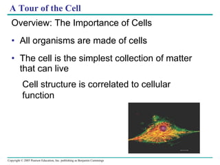
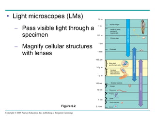
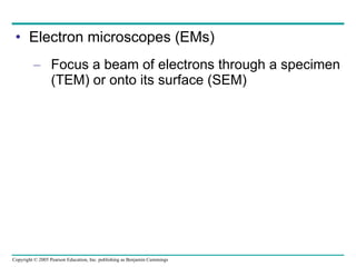
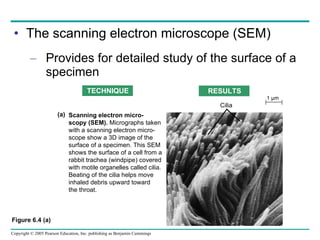
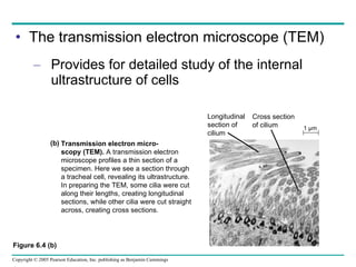
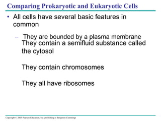
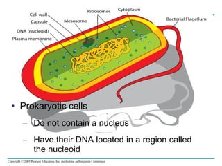
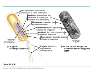
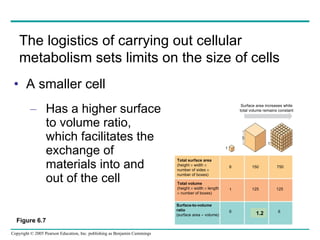
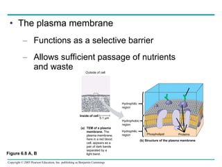
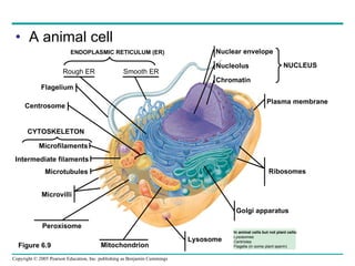
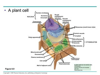
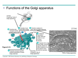
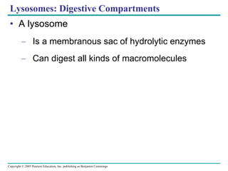
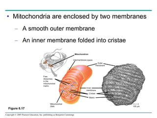
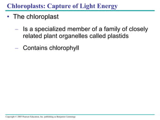
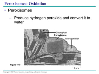
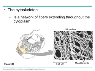
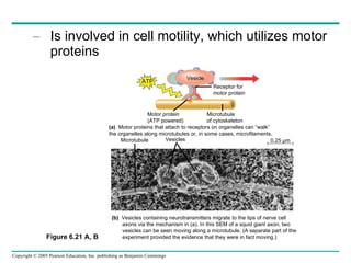
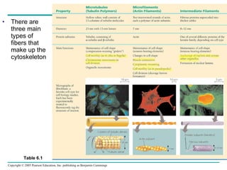
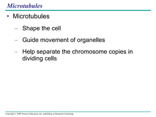
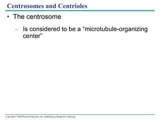
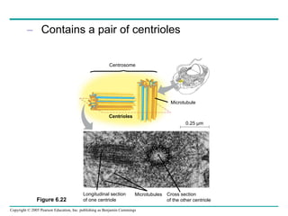
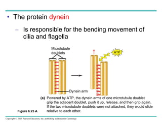
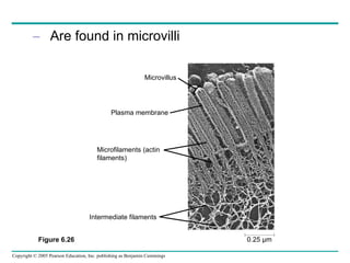
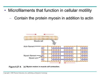
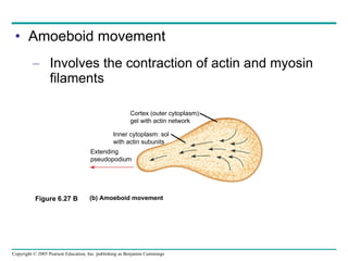
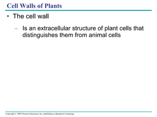
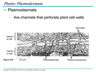
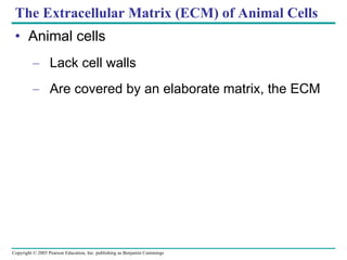
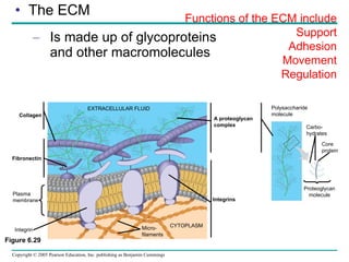
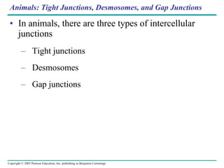
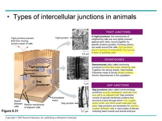
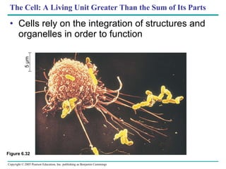
A Tour of the Cell Overview: The Importance of Cells All organisms are made of cells The cell is the simplest collection of matter  that can live Cell structure is correlated to cellular function
Cell Theory The three main parts of the Cell theory are: #1 All organisms are made up of one or more cells.  #2 The cell is the fundamental unit of structure and function in living things.  #3 All cells are essentially the same in chemical composition.
Light microscopes (LMs) Pass visible light through a specimen Magnify cellular structures with lenses 1 m 0.1 nm 10 m 0.1 m 1 cm 1 mm 100 µm 10 µ m 1 µ m 100 nm 10 nm 1 nm Length of some nerve and  muscle cells Chicken egg Frog egg Most plant  and Animal  cells Smallest bacteria Viruses Ribosomes Proteins Lipids Small molecules Atoms Nucleus Most bacteria Mitochondrion Light microscope Electron microscope Electron microscope Figure 6.2 Human height
Use different methods for enhancing visualization of cellular structures TECHNIQUE RESULT Brightfield (unstained specimen).  Passes light directly through specimen.  Unless cell is naturally pigmented or  artificially stained, image has little  contrast. [Parts (a)–(d) show a  human cheek epithelial cell.] (a) Brightfield (stained specimen).   Staining with various dyes enhances  contrast, but most staining procedures  require that cells be fixed (preserved). (b) Phase-contrast.  Enhances contrast  in unstained cells by amplifying  variations in density within specimen;  especially useful for examining living,  unpigmented cells. (c) 50 µm Figure 6.3
Differential-interference-contrast (Nomarski).   Like  phase-contrast microscopy, it uses optical  modifications to exaggerate differences in density, making the image appear almost 3D. Fluorescence.  Shows the locations of specific  molecules in the cell by tagging the molecules  with fluorescent dyes or antibodies. These  fluorescent substances absorb ultraviolet  radiation and emit visible light, as shown  here in a cell from an artery. Confocal.  Uses lasers and special optics for  “ optical sectioning” of fluorescently-stained  specimens. Only a single plane of focus is  illuminated; out-of-focus fluorescence above  and below the plane is subtracted by a computer.  A sharp image results, as seen in stained nervous  tissue (top), where nerve cells are green, support  cells are red, and regions of overlap are yellow. A  standard fluorescence micrograph (bottom) of this  relatively thick tissue is blurry. 50 µm 50 µm (d) (e) (f)
Electron microscopes (EMs) Focus a beam of electrons through a specimen (TEM) or onto its surface (SEM)
The scanning electron microscope (SEM) Provides for detailed study of the surface of a specimen Figure 6.4 (a) TECHNIQUE RESULTS Scanning electron micro- scopy (SEM).  Micrographs taken with a scanning electron micro- scope show a 3D image of the  surface of a specimen. This SEM  shows the surface of a cell from a  rabbit trachea (windpipe) covered  with motile organelles called cilia.  Beating of the cilia helps move inhaled debris upward toward  the throat. (a) Cilia 1 µm
The transmission electron microscope (TEM) Provides for detailed study of the internal ultrastructure of cells Figure 6.4 (b) Transmission electron micro- scopy (TEM).  A transmission electron  microscope profiles a thin section of a  specimen. Here we see a section through  a tracheal cell, revealing its ultrastructure.  In preparing the TEM, some cilia were cut  along their lengths, creating longitudinal  sections, while other cilia were cut straight  across, creating cross sections. (b) Longitudinal section of cilium Cross section of cilium 1 µm
Isolating Organelles by Cell Fractionation Cell fractionation Takes cells apart and separates the major organelles from one another The centrifuge Is used to fractionate cells into their component parts Tissue cells Homogenization Homogenate 1000  g (1000 times the force of gravity) 10 min Differential centrifugation Supernatant poured into next tube 20,000  g 20 min Pellet rich in nuclei and cellular debris Pellet rich in mitochondria (and chloro- plasts if cells are from a  plant) Pellet rich in “ microsomes” (pieces of  plasma mem- branes and cells’ internal membranes) Pellet rich in ribosomes 150,000  g 3 hr 80,000  g 60 min
Comparing Prokaryotic and Eukaryotic Cells All cells have several basic features in common They are bounded by a plasma membrane They contain a semifluid substance called the cytosol They contain chromosomes They all have ribosomes
Prokaryotic cells Do not contain a nucleus Have their DNA located in a region called  the nucleoid
Figure 6.6 A, B (b) A thin section through the bacterium  Bacillus coagulans (TEM) Pili:  attachment structures on the surface of some prokaryotes Nucleoid:  region where the cell’s DNA is located (not enclosed by a membrane) Ribosomes:  organelles that synthesize proteins Plasma membrane:  membrane enclosing the cytoplasm Cell wall:  rigid structure outside the plasma membrane Capsule:  jelly-like outer coating of many prokaryotes Flagella:  locomotion organelles of some bacteria (a) A typical rod-shaped bacterium  0.5 µm Bacterial chromosome
Eukaryotic cells Contain a true nucleus, bounded by a membranous nuclear envelope Are generally quite a bit bigger than prokaryotic cells
A smaller cell Has a higher surface to volume ratio, which facilitates the exchange of materials into and out of the cell Figure 6.7 The logistics of carrying out cellular metabolism sets limits on the size of cells  1.2 Surface area increases while total volume remains constant 5 1 1 Total surface area  (height    width     number of sides     number of boxes) Total volume  (height    width    length     number of boxes) Surface-to-volume  ratio  (surface area    volume) 6 1 6 150 125 12 750 125 6
The plasma membrane Functions as a selective barrier Allows sufficient passage of nutrients  and waste Carbohydrate side chain Figure 6.8 A, B Outside of cell Inside of cell Hydrophilic region Hydrophobic region Hydrophilic region (b) Structure of the plasma membrane  Phospholipid Proteins TEM of a plasma membrane.  The plasma membrane, here in a red blood cell, appears as a pair of dark bands separated by a light band. (a) 0.1 µm
A animal cell Figure 6.9 Rough ER Smooth ER Centrosome CYTOSKELETON Microfilaments Microtubules Microvilli Peroxisome Lysosome Golgi apparatus Ribosomes In animal cells but not plant cells: Lysosomes Centrioles Flagella (in some plant sperm) Nucleolus Chromatin NUCLEUS Flagelium Intermediate filaments ENDOPLASMIC RETICULUM (ER) Mitochondrion Nuclear envelope Plasma membrane
A plant cell In plant cells but not animal cells: Chloroplasts Central vacuole and tonoplast Cell wall Plasmodesmata CYTOSKELETON Figure 6.9 Ribosomes (small brwon dots) Central vacuole Microfilaments Intermediate  filaments Microtubules Rough  endoplasmic  reticulum Smooth  endoplasmic  reticulum Chromatin NUCLEUS Nuclear envelope Nucleolus Chloroplast Plasmodesmata Wall of adjacent cell Cell wall Golgi apparatus Peroxisome Tonoplast Centrosome Plasma membrane Mitochondrion
Concept 6.3: The eukaryotic cell’s genetic instructions are housed in the nucleus and carried out by the ribosomes
The nuclear envelope Encloses the nucleus, separating its contents from the cytoplasm Figure 6.10 Nucleus Nucleus Nucleolus Chromatin Nuclear envelope: Inner membrane Outer membrane Nuclear pore Rough ER Pore complex Surface of nuclear  envelope. Pore complexes (TEM).  Nuclear lamina (TEM).   Close-up of  nuclear envelope Ribosome 1 µm 1 µm 0.25 µm
Ribosomes: Protein Factories in the Cell Ribosomes Are particles made of ribosomal RNA  and protein
Carry out protein synthesis ER Endoplasmic reticulum (ER) Figure 6.11 Ribosomes Cytosol Free ribosomes Bound ribosomes Large subunit Small subunit TEM showing ER and ribosomes Diagram of a ribosome 0.5 µm
The Endoplasmic Reticulum: Biosynthetic Factory The endoplasmic reticulum (ER) Accounts for more than half the total membrane in many eukaryotic cells Is continuous with the nuclear envelope Smooth ER Rough ER ER lumen Cisternae Ribosomes Transport vesicle Smooth ER Transitional ER Rough ER 200 µm Nuclear envelope
There are two distinct regions of ER Smooth ER, which lacks ribosomes Rough ER, which contains ribosomes
Functions of Smooth ER The smooth ER Synthesizes lipids Metabolizes carbohydrates Stores calcium Detoxifies poison
Functions of Rough ER The rough ER Has bound ribosomes Produces proteins and membranes, which are distributed by transport vesicles
The Golgi apparatus Receives many of the transport vesicles produced in the rough ER Consists of flattened membranous sacs called cisternae The Golgi Apparatus: Shipping and  Receiving Center Functions of the Golgi apparatus include: Modification of the products of the rough ER Manufacture of certain macromolecules
Functions of the Golgi apparatus Golgi apparatus TEM of Golgi apparatus Figure 6.13 cis  face (“receiving” side of Golgi apparatus) Vesicles move from ER to Golgi Vesicles also  transport certain proteins back to ER Vesicles coalesce to form new  cis  Golgi cisternae Cisternal maturation: Golgi cisternae move in a  cis - to- trans direction Vesicles form and leave Golgi, carrying specific proteins to other locations or to the plasma mem- brane for secretion Vesicles transport specific proteins backward to newer Golgi cisternae Cisternae trans  face (“shipping” side of Golgi apparatus) 0.1 0 µm 1 6 5 2 3 4
Lysosomes: Digestive Compartments A lysosome Is a membranous sac of hydrolytic enzymes Can digest all kinds of macromolecules
Lysosomes carry out intracellular digestion by Phagocytosis Figure 6.14 A (a) Phagocytosis: lysosome digesting food 1 µm Lysosome contains active hydrolytic enzymes Food vacuole  fuses with  lysosome Hydrolytic enzymes digest food particles Digestion Food vacuole Plasma membrane Lysosome Digestive enzymes Lysosome Nucleus
Food vacuoles Are formed by phagocytosis Contractile vacuoles Pump excess water out of protist cells
Central vacuoles Are found in plant cells Hold reserves of important organic compounds and water Figure 6.15 Central vacuole Cytosol Tonoplast Central vacuole Nucleus Cell wall Chloroplast 5 µm
Relationships among organelles of the endomembrane system Figure 6.16 Plasma membrane expands by fusion of vesicles; proteins are secreted from cell Transport vesicle carries proteins to plasma  membrane for secretion Lysosome available for fusion with another vesicle for digestion 4 5 6 Nuclear envelope is connected  to rough ER,  which is also continuous with smooth ER Nucleus Rough ER Smooth ER cis  Golgi trans Golgi Membranes and proteins produced by the ER flow in the form of transport vesicles to the Golgi Nuclear envelop Golgi pinches off transport  Vesicles and other vesicles  that give rise to lysosomes and  Vacuoles  1 3 2 Plasma membrane
Mitochondria and chloroplasts change energy from one form to another Mitochondria Are the sites of cellular respiration Chloroplasts Found only in plants, are the sites of photosynthesis
Mitochondria: Chemical Energy Conversion Mitochondria Are found in nearly all eukaryotic cells
Mitochondria are enclosed by two membranes A smooth outer membrane An inner membrane folded into cristae Figure 6.17 Mitochondrion Intermembrane space Outer membrane Free ribosomes in the  mitochondrial matrix Mitochondrial DNA Inner membrane Cristae Matrix 100 µm
Chloroplasts: Capture of Light Energy The chloroplast Is a specialized member of a family of closely related plant organelles called plastids Contains chlorophyll
Chloroplasts Are found in leaves and other green organs of plants and in algae Figure 6.18 Chloroplast Chloroplast DNA Ribosomes Stroma Inner and outer membranes Thylakoid 1 µm Granum
Chloroplast structure includes Thylakoids, membranous sacs Stroma, the internal fluid
Peroxisomes: Oxidation Peroxisomes Produce hydrogen peroxide and convert it to water Figure 6.19 Chloroplast Peroxisome Mitochondrion 1 µm
The cytoskeleton Is a network of fibers extending throughout the cytoplasm Figure 6.20 Microtubule 0.25 µm Microfilaments Figure 6.20
Is involved in cell motility, which utilizes motor proteins Vesicle ATP Receptor for motor protein Motor protein (ATP powered) Microtubule of cytoskeleton (a)   Motor proteins that attach to receptors on organelles can “walk” the organelles along microtubules or, in some cases, microfilaments. Microtubule Vesicles 0.25 µm (b)   Vesicles containing neurotransmitters migrate to the tips of nerve cell  axons via the mechanism in (a). In this SEM of a squid giant axon, two     vesicles can be seen moving along a microtubule. (A separate part of the    experiment provided the evidence that they were in fact moving.) Figure 6.21 A, B
There are three main types of fibers that make up the cytoskeleton Table 6.1
Microtubules Microtubules Shape the cell Guide movement of organelles Help separate the chromosome copies in dividing cells
Centrosomes and Centrioles The centrosome Is considered to be a “microtubule-organizing center”
Contains a pair of centrioles Centrosome Microtubule Centrioles 0.25 µm Longitudinal section of one centriole Microtubules Cross section of the other centriole Figure 6.22
Cilia and Flagella Cilia and flagella Contain specialized arrangements of microtubules Are locomotor appendages of some cells (a) Motion of flagella.  A flagellum usually undulates, its snakelike motion driving a cell in the same direction as the axis of the flagellum. Propulsion of a human sperm cell is an example of  flagellatelocomotion (LM).  1 µm Direction of swimming (b) Motion of cilia.  Cilia have a back- and-forth motion that moves the  cell in a direction perpendicular  to the axis of the cilium. A dense  nap of cilia, beating at a rate of  about 40 to 60 strokes a second,  covers this  Colpidium,  a freshwater  protozoan (SEM). Figure 6.23 B
The protein  dynein Is responsible for the bending movement of cilia and flagella Microtubule doublets ATP Dynein arm Powered by ATP, the dynein arms of one microtubule doublet  grip the adjacent doublet, push it up, release, and then grip again.  If the two microtubule doublets were not attached, they would slide  relative to each other. (a) Figure 6.25 A
Microfilaments (Actin Filaments) Microfilaments Are built from molecules of the protein actin
Are found in microvilli 0.25 µm Microvillus Plasma membrane Microfilaments (actin filaments) Intermediate filaments Figure 6.26
Microfilaments that function in cellular motility Contain the protein myosin in addition to actin Actin filament Myosin filament Myosin motors in muscle cell contraction.  (a) Muscle cell Myosin arm Figure 6.27 A
Amoeboid movement Involves the contraction of actin and myosin filaments Figure 6.27 B Cortex (outer cytoplasm): gel with actin network Inner cytoplasm: sol  with actin subunits Extending pseudopodium (b) Amoeboid movement
Intermediate Filaments Intermediate filaments Support cell shape Fix organelles in place
Cell Walls of Plants The cell wall Is an extracellular structure of plant cells that distinguishes them from animal cells
Plant cell walls Are made of  cellulose  fibers embedded in other polysaccharides and protein May have multiple layers Central  vacuole of cell  Plasma membrane Secondary cell wall Primary cell wall Middle lamella 1 µm Central vacuole of cell  Central vacuole  Cytosol Plasma membrane Plant cell walls Plasmodesmata Figure 6.28
Plants: Plasmodesmata Plasmodesmata Are channels that perforate plant cell walls Interior of cell Interior of cell 0.5 µm Plasmodesmata Plasma membranes Cell walls Figure 6.30
The Extracellular Matrix (ECM) of Animal Cells Animal cells Lack cell walls Are covered by an elaborate matrix, the ECM
The ECM Is made up of glycoproteins and other macromolecules Functions of the ECM include Support Adhesion Movement Regulation Collagen Fibronectin Plasma membrane EXTRACELLULAR FLUID Micro- filaments CYTOPLASM Integrins Polysaccharide molecule Carbo- hydrates Proteoglycan molecule Core protein Integrin Figure 6.29 A proteoglycan  complex
Animals: Tight Junctions, Desmosomes, and Gap Junctions In animals, there are three types of intercellular junctions Tight junctions Desmosomes Gap junctions
Types of intercellular junctions in animals Tight junctions prevent  fluid from moving  across a layer of cells Tight junction 0.5 µm 1 µm Space between cells Plasma membranes of adjacent cells Extracellular matrix Gap junction Tight junctions 0.1 µm Intermediate filaments Desmosome Gap junctions At  tight junctions , the membranes of neighboring cells are very tightly pressed against each other, bound together by specific proteins (purple). Forming continu- ous seals around the cells, tight junctions prevent leakage of extracellular fluid across A layer of epithelial cells. Desmosomes  (also called  anchoring junctions ) function like rivets, fastening cells Together into strong sheets. Intermediate Filaments made of sturdy keratin proteins Anchor desmosomes in the cytoplasm. Gap junctions  (also  called communicating junctions ) provide cytoplasmic channels from one cell to an adjacent cell. Gap junctions  consist of special membrane proteins that  surround a pore through which ions, sugars, amino acids, and other small molecules may pass. Gap junctions are necessary for commu- nication between cells in many types of tissues, including heart muscle and animal embryos. TIGHT JUNCTIONS DESMOSOMES GAP JUNCTIONS Figure 6.31
The Cell: A Living Unit Greater Than the Sum of Its Parts Cells rely on the integration of structures and organelles in order to function 5 µm Figure 6.32
Irreducible Complexity  or WPP  of organ systems and body tissues Irreducible complexity  (Popularized by Michael Behe) is the argument that certain biological systems are absolutely too complex to have evolved naturally from simpler, or "less complete" predecessors.   An “irreducibly complex system" is defined as  "one that could not function if it were any simpler",  and therefore, could not possibly have been formed by successive additions to a precursor system with the same functionality. WPP – Whole Package Phenomenon

06 cell text

  • 1.
    A Tour ofthe Cell Overview: The Importance of Cells All organisms are made of cells The cell is the simplest collection of matter that can live Cell structure is correlated to cellular function
  • 2.
    Cell Theory Thethree main parts of the Cell theory are: #1 All organisms are made up of one or more cells. #2 The cell is the fundamental unit of structure and function in living things. #3 All cells are essentially the same in chemical composition.
  • 3.
    Light microscopes (LMs)Pass visible light through a specimen Magnify cellular structures with lenses 1 m 0.1 nm 10 m 0.1 m 1 cm 1 mm 100 µm 10 µ m 1 µ m 100 nm 10 nm 1 nm Length of some nerve and muscle cells Chicken egg Frog egg Most plant and Animal cells Smallest bacteria Viruses Ribosomes Proteins Lipids Small molecules Atoms Nucleus Most bacteria Mitochondrion Light microscope Electron microscope Electron microscope Figure 6.2 Human height
  • 4.
    Use different methodsfor enhancing visualization of cellular structures TECHNIQUE RESULT Brightfield (unstained specimen). Passes light directly through specimen. Unless cell is naturally pigmented or artificially stained, image has little contrast. [Parts (a)–(d) show a human cheek epithelial cell.] (a) Brightfield (stained specimen). Staining with various dyes enhances contrast, but most staining procedures require that cells be fixed (preserved). (b) Phase-contrast. Enhances contrast in unstained cells by amplifying variations in density within specimen; especially useful for examining living, unpigmented cells. (c) 50 µm Figure 6.3
  • 5.
    Differential-interference-contrast (Nomarski). Like phase-contrast microscopy, it uses optical modifications to exaggerate differences in density, making the image appear almost 3D. Fluorescence. Shows the locations of specific molecules in the cell by tagging the molecules with fluorescent dyes or antibodies. These fluorescent substances absorb ultraviolet radiation and emit visible light, as shown here in a cell from an artery. Confocal. Uses lasers and special optics for “ optical sectioning” of fluorescently-stained specimens. Only a single plane of focus is illuminated; out-of-focus fluorescence above and below the plane is subtracted by a computer. A sharp image results, as seen in stained nervous tissue (top), where nerve cells are green, support cells are red, and regions of overlap are yellow. A standard fluorescence micrograph (bottom) of this relatively thick tissue is blurry. 50 µm 50 µm (d) (e) (f)
  • 6.
    Electron microscopes (EMs)Focus a beam of electrons through a specimen (TEM) or onto its surface (SEM)
  • 7.
    The scanning electronmicroscope (SEM) Provides for detailed study of the surface of a specimen Figure 6.4 (a) TECHNIQUE RESULTS Scanning electron micro- scopy (SEM). Micrographs taken with a scanning electron micro- scope show a 3D image of the surface of a specimen. This SEM shows the surface of a cell from a rabbit trachea (windpipe) covered with motile organelles called cilia. Beating of the cilia helps move inhaled debris upward toward the throat. (a) Cilia 1 µm
  • 8.
    The transmission electronmicroscope (TEM) Provides for detailed study of the internal ultrastructure of cells Figure 6.4 (b) Transmission electron micro- scopy (TEM). A transmission electron microscope profiles a thin section of a specimen. Here we see a section through a tracheal cell, revealing its ultrastructure. In preparing the TEM, some cilia were cut along their lengths, creating longitudinal sections, while other cilia were cut straight across, creating cross sections. (b) Longitudinal section of cilium Cross section of cilium 1 µm
  • 9.
    Isolating Organelles byCell Fractionation Cell fractionation Takes cells apart and separates the major organelles from one another The centrifuge Is used to fractionate cells into their component parts Tissue cells Homogenization Homogenate 1000 g (1000 times the force of gravity) 10 min Differential centrifugation Supernatant poured into next tube 20,000 g 20 min Pellet rich in nuclei and cellular debris Pellet rich in mitochondria (and chloro- plasts if cells are from a plant) Pellet rich in “ microsomes” (pieces of plasma mem- branes and cells’ internal membranes) Pellet rich in ribosomes 150,000 g 3 hr 80,000 g 60 min
  • 10.
    Comparing Prokaryotic andEukaryotic Cells All cells have several basic features in common They are bounded by a plasma membrane They contain a semifluid substance called the cytosol They contain chromosomes They all have ribosomes
  • 11.
    Prokaryotic cells Donot contain a nucleus Have their DNA located in a region called the nucleoid
  • 12.
    Figure 6.6 A,B (b) A thin section through the bacterium Bacillus coagulans (TEM) Pili: attachment structures on the surface of some prokaryotes Nucleoid: region where the cell’s DNA is located (not enclosed by a membrane) Ribosomes: organelles that synthesize proteins Plasma membrane: membrane enclosing the cytoplasm Cell wall: rigid structure outside the plasma membrane Capsule: jelly-like outer coating of many prokaryotes Flagella: locomotion organelles of some bacteria (a) A typical rod-shaped bacterium 0.5 µm Bacterial chromosome
  • 13.
    Eukaryotic cells Containa true nucleus, bounded by a membranous nuclear envelope Are generally quite a bit bigger than prokaryotic cells
  • 14.
    A smaller cellHas a higher surface to volume ratio, which facilitates the exchange of materials into and out of the cell Figure 6.7 The logistics of carrying out cellular metabolism sets limits on the size of cells 1.2 Surface area increases while total volume remains constant 5 1 1 Total surface area (height  width  number of sides  number of boxes) Total volume (height  width  length  number of boxes) Surface-to-volume ratio (surface area  volume) 6 1 6 150 125 12 750 125 6
  • 15.
    The plasma membraneFunctions as a selective barrier Allows sufficient passage of nutrients and waste Carbohydrate side chain Figure 6.8 A, B Outside of cell Inside of cell Hydrophilic region Hydrophobic region Hydrophilic region (b) Structure of the plasma membrane Phospholipid Proteins TEM of a plasma membrane. The plasma membrane, here in a red blood cell, appears as a pair of dark bands separated by a light band. (a) 0.1 µm
  • 16.
    A animal cellFigure 6.9 Rough ER Smooth ER Centrosome CYTOSKELETON Microfilaments Microtubules Microvilli Peroxisome Lysosome Golgi apparatus Ribosomes In animal cells but not plant cells: Lysosomes Centrioles Flagella (in some plant sperm) Nucleolus Chromatin NUCLEUS Flagelium Intermediate filaments ENDOPLASMIC RETICULUM (ER) Mitochondrion Nuclear envelope Plasma membrane
  • 17.
    A plant cellIn plant cells but not animal cells: Chloroplasts Central vacuole and tonoplast Cell wall Plasmodesmata CYTOSKELETON Figure 6.9 Ribosomes (small brwon dots) Central vacuole Microfilaments Intermediate filaments Microtubules Rough endoplasmic reticulum Smooth endoplasmic reticulum Chromatin NUCLEUS Nuclear envelope Nucleolus Chloroplast Plasmodesmata Wall of adjacent cell Cell wall Golgi apparatus Peroxisome Tonoplast Centrosome Plasma membrane Mitochondrion
  • 18.
    Concept 6.3: Theeukaryotic cell’s genetic instructions are housed in the nucleus and carried out by the ribosomes
  • 19.
    The nuclear envelopeEncloses the nucleus, separating its contents from the cytoplasm Figure 6.10 Nucleus Nucleus Nucleolus Chromatin Nuclear envelope: Inner membrane Outer membrane Nuclear pore Rough ER Pore complex Surface of nuclear envelope. Pore complexes (TEM). Nuclear lamina (TEM). Close-up of nuclear envelope Ribosome 1 µm 1 µm 0.25 µm
  • 20.
    Ribosomes: Protein Factoriesin the Cell Ribosomes Are particles made of ribosomal RNA and protein
  • 21.
    Carry out proteinsynthesis ER Endoplasmic reticulum (ER) Figure 6.11 Ribosomes Cytosol Free ribosomes Bound ribosomes Large subunit Small subunit TEM showing ER and ribosomes Diagram of a ribosome 0.5 µm
  • 22.
    The Endoplasmic Reticulum:Biosynthetic Factory The endoplasmic reticulum (ER) Accounts for more than half the total membrane in many eukaryotic cells Is continuous with the nuclear envelope Smooth ER Rough ER ER lumen Cisternae Ribosomes Transport vesicle Smooth ER Transitional ER Rough ER 200 µm Nuclear envelope
  • 23.
    There are twodistinct regions of ER Smooth ER, which lacks ribosomes Rough ER, which contains ribosomes
  • 24.
    Functions of SmoothER The smooth ER Synthesizes lipids Metabolizes carbohydrates Stores calcium Detoxifies poison
  • 25.
    Functions of RoughER The rough ER Has bound ribosomes Produces proteins and membranes, which are distributed by transport vesicles
  • 26.
    The Golgi apparatusReceives many of the transport vesicles produced in the rough ER Consists of flattened membranous sacs called cisternae The Golgi Apparatus: Shipping and Receiving Center Functions of the Golgi apparatus include: Modification of the products of the rough ER Manufacture of certain macromolecules
  • 27.
    Functions of theGolgi apparatus Golgi apparatus TEM of Golgi apparatus Figure 6.13 cis face (“receiving” side of Golgi apparatus) Vesicles move from ER to Golgi Vesicles also transport certain proteins back to ER Vesicles coalesce to form new cis Golgi cisternae Cisternal maturation: Golgi cisternae move in a cis - to- trans direction Vesicles form and leave Golgi, carrying specific proteins to other locations or to the plasma mem- brane for secretion Vesicles transport specific proteins backward to newer Golgi cisternae Cisternae trans face (“shipping” side of Golgi apparatus) 0.1 0 µm 1 6 5 2 3 4
  • 28.
    Lysosomes: Digestive CompartmentsA lysosome Is a membranous sac of hydrolytic enzymes Can digest all kinds of macromolecules
  • 29.
    Lysosomes carry outintracellular digestion by Phagocytosis Figure 6.14 A (a) Phagocytosis: lysosome digesting food 1 µm Lysosome contains active hydrolytic enzymes Food vacuole fuses with lysosome Hydrolytic enzymes digest food particles Digestion Food vacuole Plasma membrane Lysosome Digestive enzymes Lysosome Nucleus
  • 30.
    Food vacuoles Areformed by phagocytosis Contractile vacuoles Pump excess water out of protist cells
  • 31.
    Central vacuoles Arefound in plant cells Hold reserves of important organic compounds and water Figure 6.15 Central vacuole Cytosol Tonoplast Central vacuole Nucleus Cell wall Chloroplast 5 µm
  • 32.
    Relationships among organellesof the endomembrane system Figure 6.16 Plasma membrane expands by fusion of vesicles; proteins are secreted from cell Transport vesicle carries proteins to plasma membrane for secretion Lysosome available for fusion with another vesicle for digestion 4 5 6 Nuclear envelope is connected to rough ER, which is also continuous with smooth ER Nucleus Rough ER Smooth ER cis Golgi trans Golgi Membranes and proteins produced by the ER flow in the form of transport vesicles to the Golgi Nuclear envelop Golgi pinches off transport Vesicles and other vesicles that give rise to lysosomes and Vacuoles 1 3 2 Plasma membrane
  • 33.
    Mitochondria and chloroplastschange energy from one form to another Mitochondria Are the sites of cellular respiration Chloroplasts Found only in plants, are the sites of photosynthesis
  • 34.
    Mitochondria: Chemical EnergyConversion Mitochondria Are found in nearly all eukaryotic cells
  • 35.
    Mitochondria are enclosedby two membranes A smooth outer membrane An inner membrane folded into cristae Figure 6.17 Mitochondrion Intermembrane space Outer membrane Free ribosomes in the mitochondrial matrix Mitochondrial DNA Inner membrane Cristae Matrix 100 µm
  • 36.
    Chloroplasts: Capture ofLight Energy The chloroplast Is a specialized member of a family of closely related plant organelles called plastids Contains chlorophyll
  • 37.
    Chloroplasts Are foundin leaves and other green organs of plants and in algae Figure 6.18 Chloroplast Chloroplast DNA Ribosomes Stroma Inner and outer membranes Thylakoid 1 µm Granum
  • 38.
    Chloroplast structure includesThylakoids, membranous sacs Stroma, the internal fluid
  • 39.
    Peroxisomes: Oxidation PeroxisomesProduce hydrogen peroxide and convert it to water Figure 6.19 Chloroplast Peroxisome Mitochondrion 1 µm
  • 40.
    The cytoskeleton Isa network of fibers extending throughout the cytoplasm Figure 6.20 Microtubule 0.25 µm Microfilaments Figure 6.20
  • 41.
    Is involved incell motility, which utilizes motor proteins Vesicle ATP Receptor for motor protein Motor protein (ATP powered) Microtubule of cytoskeleton (a) Motor proteins that attach to receptors on organelles can “walk” the organelles along microtubules or, in some cases, microfilaments. Microtubule Vesicles 0.25 µm (b) Vesicles containing neurotransmitters migrate to the tips of nerve cell axons via the mechanism in (a). In this SEM of a squid giant axon, two  vesicles can be seen moving along a microtubule. (A separate part of the experiment provided the evidence that they were in fact moving.) Figure 6.21 A, B
  • 42.
    There are threemain types of fibers that make up the cytoskeleton Table 6.1
  • 43.
    Microtubules Microtubules Shapethe cell Guide movement of organelles Help separate the chromosome copies in dividing cells
  • 44.
    Centrosomes and CentriolesThe centrosome Is considered to be a “microtubule-organizing center”
  • 45.
    Contains a pairof centrioles Centrosome Microtubule Centrioles 0.25 µm Longitudinal section of one centriole Microtubules Cross section of the other centriole Figure 6.22
  • 46.
    Cilia and FlagellaCilia and flagella Contain specialized arrangements of microtubules Are locomotor appendages of some cells (a) Motion of flagella. A flagellum usually undulates, its snakelike motion driving a cell in the same direction as the axis of the flagellum. Propulsion of a human sperm cell is an example of flagellatelocomotion (LM). 1 µm Direction of swimming (b) Motion of cilia. Cilia have a back- and-forth motion that moves the cell in a direction perpendicular to the axis of the cilium. A dense nap of cilia, beating at a rate of about 40 to 60 strokes a second, covers this Colpidium, a freshwater protozoan (SEM). Figure 6.23 B
  • 47.
    The protein dynein Is responsible for the bending movement of cilia and flagella Microtubule doublets ATP Dynein arm Powered by ATP, the dynein arms of one microtubule doublet grip the adjacent doublet, push it up, release, and then grip again. If the two microtubule doublets were not attached, they would slide relative to each other. (a) Figure 6.25 A
  • 48.
    Microfilaments (Actin Filaments)Microfilaments Are built from molecules of the protein actin
  • 49.
    Are found inmicrovilli 0.25 µm Microvillus Plasma membrane Microfilaments (actin filaments) Intermediate filaments Figure 6.26
  • 50.
    Microfilaments that functionin cellular motility Contain the protein myosin in addition to actin Actin filament Myosin filament Myosin motors in muscle cell contraction. (a) Muscle cell Myosin arm Figure 6.27 A
  • 51.
    Amoeboid movement Involvesthe contraction of actin and myosin filaments Figure 6.27 B Cortex (outer cytoplasm): gel with actin network Inner cytoplasm: sol with actin subunits Extending pseudopodium (b) Amoeboid movement
  • 52.
    Intermediate Filaments Intermediatefilaments Support cell shape Fix organelles in place
  • 53.
    Cell Walls ofPlants The cell wall Is an extracellular structure of plant cells that distinguishes them from animal cells
  • 54.
    Plant cell wallsAre made of cellulose fibers embedded in other polysaccharides and protein May have multiple layers Central vacuole of cell Plasma membrane Secondary cell wall Primary cell wall Middle lamella 1 µm Central vacuole of cell Central vacuole Cytosol Plasma membrane Plant cell walls Plasmodesmata Figure 6.28
  • 55.
    Plants: Plasmodesmata PlasmodesmataAre channels that perforate plant cell walls Interior of cell Interior of cell 0.5 µm Plasmodesmata Plasma membranes Cell walls Figure 6.30
  • 56.
    The Extracellular Matrix(ECM) of Animal Cells Animal cells Lack cell walls Are covered by an elaborate matrix, the ECM
  • 57.
    The ECM Ismade up of glycoproteins and other macromolecules Functions of the ECM include Support Adhesion Movement Regulation Collagen Fibronectin Plasma membrane EXTRACELLULAR FLUID Micro- filaments CYTOPLASM Integrins Polysaccharide molecule Carbo- hydrates Proteoglycan molecule Core protein Integrin Figure 6.29 A proteoglycan complex
  • 58.
    Animals: Tight Junctions,Desmosomes, and Gap Junctions In animals, there are three types of intercellular junctions Tight junctions Desmosomes Gap junctions
  • 59.
    Types of intercellularjunctions in animals Tight junctions prevent fluid from moving across a layer of cells Tight junction 0.5 µm 1 µm Space between cells Plasma membranes of adjacent cells Extracellular matrix Gap junction Tight junctions 0.1 µm Intermediate filaments Desmosome Gap junctions At tight junctions , the membranes of neighboring cells are very tightly pressed against each other, bound together by specific proteins (purple). Forming continu- ous seals around the cells, tight junctions prevent leakage of extracellular fluid across A layer of epithelial cells. Desmosomes (also called anchoring junctions ) function like rivets, fastening cells Together into strong sheets. Intermediate Filaments made of sturdy keratin proteins Anchor desmosomes in the cytoplasm. Gap junctions (also called communicating junctions ) provide cytoplasmic channels from one cell to an adjacent cell. Gap junctions consist of special membrane proteins that surround a pore through which ions, sugars, amino acids, and other small molecules may pass. Gap junctions are necessary for commu- nication between cells in many types of tissues, including heart muscle and animal embryos. TIGHT JUNCTIONS DESMOSOMES GAP JUNCTIONS Figure 6.31
  • 60.
    The Cell: ALiving Unit Greater Than the Sum of Its Parts Cells rely on the integration of structures and organelles in order to function 5 µm Figure 6.32
  • 61.
    Irreducible Complexity or WPP of organ systems and body tissues Irreducible complexity (Popularized by Michael Behe) is the argument that certain biological systems are absolutely too complex to have evolved naturally from simpler, or "less complete" predecessors. An “irreducibly complex system" is defined as "one that could not function if it were any simpler", and therefore, could not possibly have been formed by successive additions to a precursor system with the same functionality. WPP – Whole Package Phenomenon