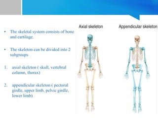
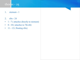
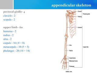
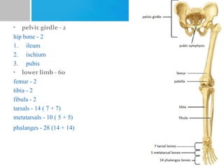
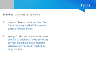
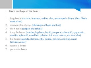
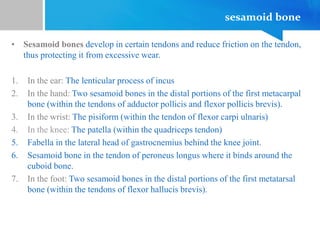
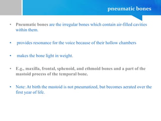
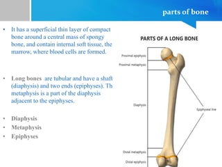
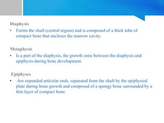
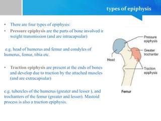
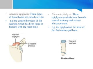
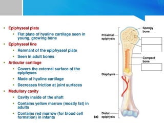
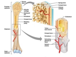
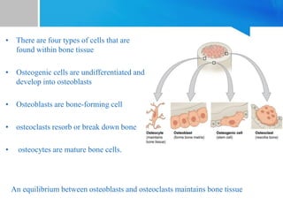
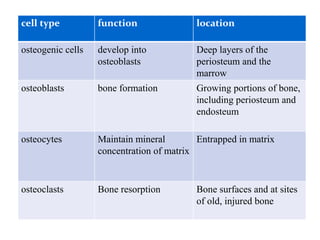
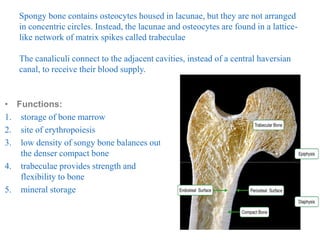
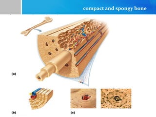
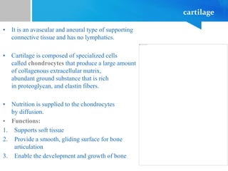
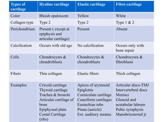
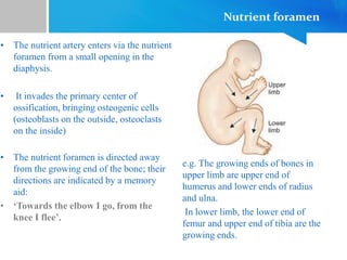
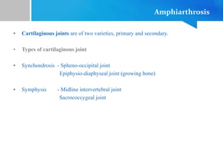
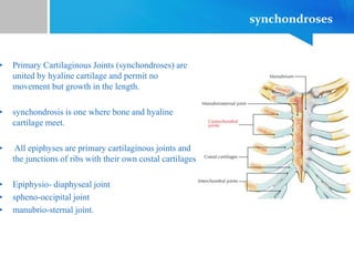
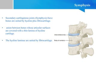
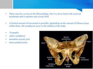
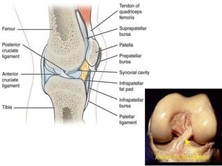
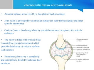
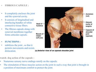
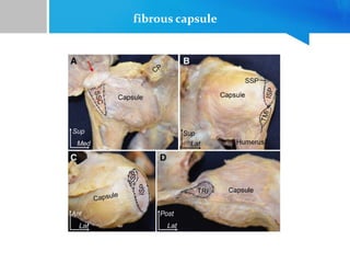
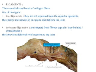
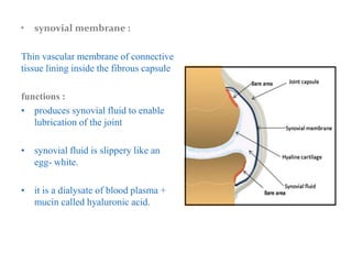
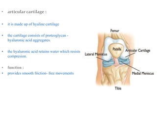
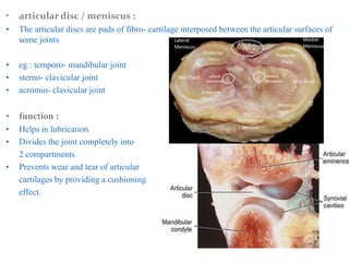
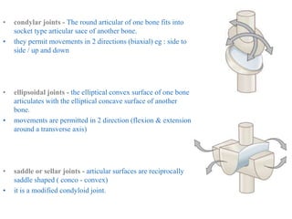
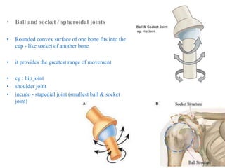
SKELETAL SYSTEM
• The skeletal system consists of bone
and cartilage.
• The skeleton can be divided into 2
subgroups
1. axial skeleton ( skull, vertebral
column, thorax)
2. appendicular skeleton ( pectoral
girdle, upper limb, pelvic girdle,
lower limb)
axial skeleton
skull - 29
1. Cranium - 8 (frontal, occipital, sphenoid, ethamoid, parietal-2, temporal-2)
2. Facial bones - 14 (zygomatic bone -2, maxilla-2, palatine-2, lacrimal
bone- 2, nasal bone-2, inferior nasal oncha-2, vomer-1, mandile-1)
3. Hyoid - 1
4. Ear ossicles - 6 (malleus-2, stapes-2, incus-2)
VERTEBRAL COLUMN - 26
• cervical vertebrae - 7
• thoracic vertebrae - 12
• lumbar vertebrae - 5
• sacrum - 1
• coccyx -1
thorax - 25
1. sternum - 1
2. ribs - 24
• 1 - 7 ( attaches directly to sternum)
• 8 - 10 ( attaches to 7th rib)
• 11 - 12 ( floating ribs)
appendicular skeleton
pectoral girdle- 4
clavicle - 2
scapula - 2
upper limb - 60
humerus - 2
radius - 2
ulna - 2
carpals - 16 ( 8 + 8)
metacarpals - 10 (5 + 5)
phalanges - 28 (14 + 14)
• pelvic girdle - 2
hip bone - 2
1. ileum
2. ischium
3. pubis
• lower limb - 60
femur - 2
tibia - 2
fibula - 2
tarsals - 14 ( 7 + 7)
metatarsals - 10 ( 5 + 5)
phalanges - 28 (14 + 14)
bone
• Bone is a calcified and rigid connective tissue consisting of cells
(osteocytes) embedded in a matrix of ground substance and collagen
fibers.
• highly vascular tissue with high cell density
• low metabolic rate
• supportive sturctures of the body
• protectors of vital organs
• resoervoirs of calcium and phosphorus
• biochemical levers on which muscles act to produce movement
• containers for blood producing cells
types
Based on structure of the bone :
1. compact bone - is a dense bone that
forms the outer shell of all bones or
cortex of mature bones.
2. spongy/trabecular/cancellous bone-
consists of spicules of bone enclosing
cavities containing blood- forming
cells (marrow) or honeycombed by
large cavities
• Based on shape of the bone :
1. long bones (clavicle, humerus, radius, ulna, metacarpals, femur, tibia, fibula,
metatarsals)
2. miniature long bones (phalanges of hand and foot)
3. short bones (carpals and tarsals)
4. irregular bones (vetebra, hip bone, hyoid, temporal, ethamoid, zygomatic,
maxilla, sphenoid, mandible, palatine, inf. nasal concha, ear osscicles)
5. flat bones (scapula, sternum, ribs, frontal, parietal, occipital, nasal,
lacrimal,vomer)
6. sesamoid bones
7. pneumatic bones
sesamoid bone
• Sesamoid bones develop in certain tendons and reduce friction on the tendon,
thus protecting it from excessive wear.
1. In the ear: The lenticular process of incus
2. In the hand: Two sesamoid bones in the distal portions of the first metacarpal
bone (within the tendons of adductor pollicis and flexor pollicis brevis).
3. In the wrist: The pisiform (within the tendon of flexor carpi ulnaris)
4. In the knee: The patella (within the quadriceps tendon)
5. Fabella in the lateral head of gastrocnemius behind the knee joint.
6. Sesamoid bone in the tendon of peroneus longus where it binds around the
cuboid bone.
7. In the foot: Two sesamoid bones in the distal portions of the first metatarsal
bone (within the tendons of flexor hallucis brevis).
pneumatic bones
• Pneumatic bones are the irregular bones which contain air-filled cavities
within them.
• provides resonance for the voice because of their hollow chambers
• makes the bone light in weight.
• E.g., maxilla, frontal, sphenoid, and ethmoid bones and a part of the
mastoid process of the temporal bone.
• Note: At birth the mastoid is not pneumatized, but becomes aerated over the
first year of life.
parts of bone
• It has a superficial thin layer of compact
bone around a central mass of spongy
bone, and contain internal soft tissue, the
marrow, where blood cells are formed.
• Long bones are tubular and have a shaft
(diaphysis) and two ends (epiphyses). The
metaphysis is a part of the diaphysis
adjacent to the epiphyses.
• Diaphysis
• Metaphysis
• Epiphyses
Diaphysis
• Forms the shaft (central region) and is composed of a thick tube of
compact bone that encloses the marrow cavity.
Metaphysis
• Is a part of the diaphysis, the growth zone between the diaphysis and
epiphysis during bone development.
Epiphyses
• Are expanded articular ends, separated from the shaft by the epiphyseal
plate during bone growth and composed of a spongy bone surrounded by a
thin layer of compact bone.
types of epiphysis
• There are four types of epiphysis:
• Pressure epiphysis are the parts of bone involved in
weight transmission (and are intracapsular)
e.g. head of humerus and femur and condyles of
humerus, femur, tibia etc.
• Traction epiphysis are present at the ends of bones
and develop due to traction by the attached muscles
(and are extracapsular)
e.g. tubercles of the humerus (greater and lesser ), and
trochanters of the femur (greater and lesser). Mastoid
process is also a traction epiphysis.
• Atavistic epiphysis: These types
of fused bones are called atavistic
• e.g. the coracoid process of the
scapula, which has been fused in
humans with the main bone.
• Aberrant epiphysis: These
epiphyses are deviations from the
normal anatomy and are not
always present.
• e.g. the epiphysis at the head of
the first metacarpal bone.
• The periosteum is a membrane that covers the outer surface of all bones,
except at the articular surfaces (i.e. the parts within a joint space) of long
bones.
• Endosteum lines the inner surface of the medullary cavity of all long bones.
• There are four types of cells that are
found within bone tissue
• Osteogenic cells are undifferentiated and
develop into osteoblasts
• Osteoblasts are bone-forming cell
• osteoclasts resorb or break down bone
• osteocytes are mature bone cells.
An equilibrium between osteoblasts and osteoclasts maintains bone tissue
cell type function location
osteogenic cells develop into
osteoblasts
Deep layers of the
periosteum and the
marrow
osteoblasts bone formation Growing portions of bone,
including periosteum and
endosteum
osteocytes Maintain mineral
concentration of matrix
Entrapped in matrix
osteoclasts Bone resorption Bone surfaces and at sites
of old, injured bone
structure of compact bone :
The compact bone is made up of a system of Haversian (longitudinal)
canals connected by Volkmann's (transverse) canals.
haversian system
• Compact bone consists of closely packed
osteons or haversian systems.
• The osteon consists of a central canal called the
osteonic (haversian) canal, which is surrounded by
concentric rings (lamellae) of matrix.
• Between the rings of matrix, the bone
cells (osteocytes) are located in spaces called lacunae.
• Small channels (canaliculi) radiate from the
lacunae to the osteonic (haversian) canal to provide
passageways through the hard matrix.
• The osteonic canals contain blood vessels that are
parallel to the long axis of the bone.
volkmann’s canal
• Volkmann's canals are inside osteons.
• They interconnect the haversian canals
with each other and the periosteum.
• Volkmann's canals are the small
channels in the bone that transmit
blood vessels from the periosteum into
the bone and that communicate with
the haversian canals.
structure of spongy bone :
Spongy bone contains osteocytes housed in lacunae, but they are not arranged
in concentric circles. Instead, the lacunae and osteocytes are found in a lattice-
like network of matrix spikes called trabeculae
The canaliculi connect to the adjacent cavities, instead of a central haversian
canal, to receive their blood supply.
• Functions:
1. storage of bone marrow
2. site of erythropoiesis
3. low density of songy bone balances out
the denser compact bone
4. trabeculae provides strength and
flexibility to bone
5. mineral storage
compact and spongy bone
cartilage
• It is an avascular and aneural type of supporting
connective tissue and has no lymphatics.
• Cartilage is composed of specialized cells
called chondrocytes that produce a large amount
of collagenous extracellular matrix,
abundant ground substance that is rich
in proteoglycan, and elastin fibers.
• Nutrition is supplied to the chondrocytes
by diffusion.
• Functions:
1. Supports soft tissue
2. Provide a smooth, gliding surface for bone
articulation
3. Enable the development and growth of bone
• cells of cartilage are of 3 types
1. chondrogenic cells : found in perichondrium and differentiate into
chondroblasts
2. chondroblasts : young cartilage cells
3. chrondrocytes : mature cartilage cells
• cartilage has 2 types of fibrous cartlage :
1. type 1 collagen fibers (fibro- cartilage)
2. type 2 collagen fibes ( hyaline and elastic cartilage )
ground substance consist of :
1. proteoglycans : chondroitin sulphate + keratan sulphate
2. glycoproteins ( chondronectin, chondrocalcin - a calcium binding protein)
3. water ( tissue fluid)
Types of cartilage
• Hyaline cartilage : matrix contains moderate amount of collagen fibers
• Example : Connection between ribs and sternum, nasal cartilage and
articular cartilage (which covers opposing bone surfaces in many joints).
• Elastic cartilage : matrix contains collagen fibers along with large number
of elastic fibers
• Example : the auricle of the outer ear.
• Fibro- cartilage : matrix contains a limited number of ground substance
amidst a substantial amount of collagen fibers
• Example : Intervertebral discs
Types of
cartilage
Hyaline cartilage Elastic cartilage Fibro cartilage
Color Bluish opalescent Yellow White
Collagen type Type 2 Type 2 Type 1 & 2
Perichondrium Present ( except at
epiphysis and
articular cartilage)
Present Absent
Calcification Occurs with old age No calcification Occurs only with
bone repair
Cells Chondrocytes &
chondroblasts
Chondrocytes &
chondroblasts
Chondrocytes &
fibroblasts
Fibers Thin collagen Elastic fibers Thick collagen
Examples Cricoid cartilage
Thyroid cartilage
Trachea & bronchi
Articular cartilage of
bone
Epiphyseal plate
Costal Cartilage
(ribs)
Apices of arytenoid
Epiglottis
Corniculate cartilage
Cuneiform cartilages
Eustachian tube
Pinna (auricle)
Ext. auditory meatus
Articular discs-TMJ
Intervertebral discs
Menisci
Glenoid and
acetabular labrum
Pubic symphysis
Manubriosternal jt
ossification
• Ossification is the process of laying down new bone material by cells
called osteoblasts.
• It is of two types:
• Membranous ossification is the direct laying down of bone into the mesenchyme
(embryonic connective tissue).
Example: some flat bones of the skull and face
• Endochondral ossification involves osteogenesis in a precursor model of cartilage.
Example: The bones of the extremities, vertebral column and thoracic cage.
• Membrano-cartilaginous ossification, in which bones are initially formed in
membrane but later partly in cartilage
Example: clavicle.
Types of Cartilaginous ossification
• The point from where ossification begins is called a center of ossification.
• Cartilaginous ossification involves two centres of ossification.
• Primary center of ossification
• Secondary center of ossification
Primary center of ossification
• In long bones, bone tissue first appears in the
diaphysis (middle of shaft).
• Primary centers starts appearing at 6th week of
intrauterine life.
• Chondrocytes multiply and form trabeculae.
• cartilage is progressively eroded and replaced by
bone, extending towards the epiphysis.
• A perichondrium layer surrounding the cartilage forms
the periosteum, which generates osteogenic cells that
make a collar to encircles the exterior of the bone and
remodels the medullary cavity on the inside.
Nutrient foramen
• The nutrient artery enters via the nutrient
foramen from a small opening in the
diaphysis.
• It invades the primary center of
ossification, bringing osteogenic cells
(osteoblasts on the outside, osteoclasts
on the inside)
e.g. The growing ends of bones in
upper limb are upper end of
humerus and lower ends of radius
and ulna.
In lower limb, the lower end of
femur and upper end of tibia are the
growing ends.
• The nutrient foramen is directed away
from the growing end of the bone; their
directions are indicated by a memory
aid:
• ‘Towards the elbow I go, from the
knee I flee’.
Secondary center of ossification
• The secondary centres generally appear at the
ends (epiphysis) of long bones.
• Secondary ossification mostly occurs after birth
• The epiphyseal arteries and osteogenic cells
invade the epiphysis, depositing osteoblasts and
osteoclasts which erode the cartilage and build
bone
radiograph of ossification of bones
LAWS OF OSSIFICATION
• Primary ossification center is single and appears before birth.
• Exceptions : carpal bones ossify after birth
Clavicle has 2 primary ossification centers
• Secondary ossification centers can be single or multiple and appear
after birth.
• Exceptions : secondary center for lower end of femur appears before birth
(9th month of fetal life).
Proximal epiphysis of tibia may appear before birth in some individuals
• Secondary centers of ossification which appear first are last to unite.
• Exceptions : secondary center at the lower end of fibula appears during
1st yr (before the upper end which appears in 3rd or 4th yr) but fuses between
15th and 17th yr (before the fusion of upper end between 17th and 19th year)
LAWS OF OSSIFICATION
• The secondary ossification centers fuse together to form a single
epiphysis, which then fuses with diaphysis.
• Exceptions : the 3 secondary centers at the upper end of femur
( head - 1st yr, greater trochanter - 4th yr, lesser trochanter - 12th yr) fuse
independently with the shaft.
• The direction of the nutrient artery is opposite to the direction of the
growing end of the bone.
Time period Bones affected
Second month of fetal
development
Ossification of long bones begins
Fourth month Most primary ossification centers have appeared in the
diaphyses of bone.
Birth to 5 years Secondary ossification centers appear in the epiphyses
5 years to 12 years in females,
5 to 14 years in males
Ossification is spreading rapidly from the ossification
centers and various bones
are becoming ossified
17 to 20 years Bone of upper limbs and scapulae becoming
completely ossified
18 to 23 years Bone of the lower limbs and os coxae become
completely ossified
23 to 25 years Bone of the sternum, clavicles, and vertebrae become
completely ossified
By 25 years Nearly all bones are completely ossified
blood supply
• The long bone is supplied by
four set of arteries
1. nutrient artery
2. periosteal artery
3. metaphyseal artery
4. epiphyseal artery
• Nutrient artery enters the middle of
shaft through nutrient foramen.
• divides into ascending and descending
branches in medullary cavity
• each branches subdivides into smaller
paralle branches and enters into
metaphysis
• this forms the hair- pin loops / bends
• these loops anastomose with other
arteries
joints
• The site where two skeletal structures come together are termed as joints
• Union between bones can be in one of three types: by fibrous tissue; by
cartilage; or by synovial joints
• structural classificaion :
1. Fibrous joints
2. Cartilaginous joint
3. Synovial joints
• regional classifiation :
1. skull type : immovable
2. vertebral type : slightly movable
3. limb type : freely movable
Functional classification :
1. Synarthrosis (immovable)
2. Amphiarthrosis (slight mobile)
3. Diarthrosis (freely mobile)
According to the number of articulating bones :
1. simple joint - Two bones articulate (eg : interphalangeal joints )
2. compound joint - More than two bones articulate within one capsule. (eg
: wrist / elbow joint )
3. complex joint - the joint cavity is divided by an intra- articular disc
( eg : temporo- mandibular joint )
classification of joints
Synarthrosis
• Fibrous joints occur where bones are separated only by connective tissue and
movement between them is negligible.
• Types of fibrous joint
1. Suture
2. Gomphosis - Tooth and socket joints
3. Syndesmosis - The bones are connected by the interosseous
ligament (eg. Middle radioulnar joint and Inferior radioulnar joint)
sutures
• Sutures: These are peculiar to skull, and are
immovable.
• According to the shape of bony margins, the
sutures can be:
1. Plane, e.g. internasal suture
2. Serrate, e.g. interparietal suture
3. Squamous, e.g. temporo-parietal suture
4. Denticulate, e.g. lambdoid suture
5. Schindylesis type, e.g. between rostrum of
sphenoid and upper border of vomer
types of sutures
Amphiarthrosis
• Cartilaginous joints are of two varieties, primary and secondary.
• Types of cartilaginous joint
• Synchondrosis - Spheno-occipital joint
Epiphysio-diaphyseal joint (growing bone)
• Symphysis - Midline intervertebral joint
Sacrococcygeal joint
synchondroses
• Primary Cartilaginous Joints (synchondroses) are
united by hyaline cartilage and permit no
movement but growth in the length.
• synchondrosis is one where bone and hyaline
cartilage meet.
• All epiphyses are primary cartilaginous joints and
the junctions of ribs with their own costal cartilages
• Epiphysio- diaphyseal joint
• spheno-occipital joint
• manubrio-sternal joint.
Symphysis
• Secondary cartilaginous joints (Symphysis) have
bones are united by hyaline plus fibrocartilage.
• union between bones whose articular surfaces
are covered with a thin lamina of hyaline
cartilage.
• The hyaline laminae are united by fibrocartilage.
• There may be a cavity in the fibrocartilage, but it is never lined with synovial
membrane and it contains only tissue fluid.
• A limited amount of movement is possible, depending on the amount of fibrous tissue
within them. All symphyses occur in the midline of the body.
• Examples
• pubic symphysis
• manubrio-sternal joint
• intervertebral joints.
Diarthrosis
• Synovial joints are freely mobile joints.
• connections between skeletal components where the bones involved are
seperated by a narrow aticular cavity
• These joints possess a cavity and articular ends of bones forming joints are
enclosed in a fibrous capsule.
• Narrow cavity is called as articular cavity
• Filled by synovial fluid
• It consist of
1. fibrous capsule
2. synovial membrane
3. articular cartilage
4. ligaments
5. articular disc
6. bursae
7. fat pads
characteristic feature of synovial joints
• Articular surfaces are covered by a thin plate of hyaline catilage.
• Joint cavity is enveloped by an articular capsule (an outer fibrous capsule and inner
synovial membrane)
• Cavity of joint is lined everywhere by synovial membrane except over the articular
cartilages.
• The cavity is filled with synovial fluid
( secreted by synovial membrane) which
provides lubrication of articular surfaces
and nutrition
• Sometimes joint cavity is completely
and incompletely divided by articular disc /
meniscus.
• FIBROUS CAPSULE :
• It completely encloses the joint
and the synovial cavity.
• It consists of longitudinal and
interlacing bundles of white
connective tissue fibers.
• The fibrous capsule along with
synovial membrane together
forms articular capsule.
• FUNCTIONS :
• stabilizes the joint , so that it
permits movements and resists
dislocation.
watch- dog action of the capsule :
• Numerous sensory nerve endings ramify on the capsule.
• The stimulation of these muscles action on the joint in such a way that joint is brought in
a position of maximum comfort to protect the joint.
fibrous capsule
• LIGAMENTS :
These are thickened bands of collagen fibers
it is of two types:
• true ligaments : they are not seperated from the capsular ligaments.
they permit movements in one plane and stabilize the joint.
• accessory ligaments : are seperate from fibrous capsule ( may be intra /
extracapsular )
they provide additional reinforcement to the joint
• synovial membrane :
Thin vascular membrane of connective
tissue lining inside the fibrous capsule
functions :
• produces synovial fluid to enable
lubrication of the joint
• synovial fluid is slippery like an
egg- white.
• it is a dialysate of blood plasma +
mucin called hyaluronic acid.
• articular cartilage :
• it is made up of hyaline cartilage
• the cartilage consists of proteoglycan -
hyaluronic acid aggregates.
• the hyaluronic acid retains water which resists
compresion.
• function :
• provides smooth friction- free movements
• articular disc / meniscus :
• The articular discs are pads of fibro- cartilage interposed between the articular surfaces of
some joints
• eg : temporo- mandibular joint
• sterno- clavicular joint
• acromio- clavicular joint
• function :
• Helps in lubrication
• Divides the joint completely into
2 compartments
• Prevents wear and tear of articular
cartilages by providing a cushioning
effect.
• bursae :
• These are pouch like sacs of connective
tissue filled with synovial fluid ( in
synovial joint)
• Commonly located
between tendon and bone /
between muscle and bone /
between skin and bone /
between tendon and skin.
Function :
• cushions certain muscles and to
facilitate the movement of tendon/
muscle over bony surfaces.
• The synovial tendon sheath is a modified bursa that surrounds and lubricates the
tendon of certain muscles ( eg : that crosses the wrist and ankle joint)
• fat pads :
• also kown as haversian glands
• pads of fat placed between synovial membrane and bone
• eg : acetabular fat of hip joint
• infra patellar fat of knee joint
classification of synovial joints
• According to the shape of articular
surfaces :
1. plane joints
2. hinge / ginglymus joints
3. pivot / tricoid joints
4. condylar joints
5. ellipsoidal joints
6. saddle or sellar joints
7. ball and socket / spheroidal joints
• According to the plane of
movement :
1. uniaxial joints
2. biaxial joints
3. multi / polyaxial joints
• According to the number of
articulating bones :
1. simple joints
2. complex joints
• plane joints - The articular surfaces are nearly
flat (plane)
• they permit gliding movements (eg: side to side ,
back to forth )
• hinge / ginglymus joints - the articular surfaces
are pulley shaped
• movements are permitted only in one plane
around transverse axis.
• pivot / tricoid joints - the articular surface of one
bone is rounded and fits into concavity of another
bone
• movement is limited totation around a central axis
• condylar joints - The round articular of one bone fits into
socket type articular sace of another bone.
• they permit movements in 2 directions (biaxial) eg : side to
side / up and down
• ellipsoidal joints - the elliptical convex surface of one bone
articulates with the elliptical concave surface of another
bone.
• movements are permitted in 2 direction (flexion & extension
around a transverse axis)
• saddle or sellar joints - articular surfaces are reciprocally
saddle shaped ( conco - convex)
• it is a modified condyloid joint.
• Ball and socket / spheroidal joints
• Rounded convex surface of one bone fits into the
cup - like socket of another bone
• it provides the greatest range of movement
• eg : hip joint
• shoulder joint
• incudo - stapedial joint (smallest ball & socket
joint)
Types of synovial joint Examples
Plane Acromioclavicular
Intercarpal & Intertarsal
Hinge Elbow & Interphalangeal
Pivot (Trochoid) Atlanto-axial / Superior radio-ulnar & Inferior radio-
ulnar
Condylar Temporo-mandibular & Knee joint
Ellipsoid Atlanto-occipital
Wrist (radio-carpal)
Metacarpo-phalangeal (knuckle)
Saddle Malleus-incus joint
Sternoclavicular
First carpo-metacarpal
Calcaneocuboid
Ball and socket Incus-stapes joint
Shoulder & Hip
Talo-calcaneo-navicular
• According to the plane of movement :
• uniaxial joints - movement occur only in one plane or axis
eg : hinge joints, pivot joints
• biaxial joints - movement occur in two planes or axes
eg : condylar joints, ellipsoidal joints, saddle joints
• multi / polyaxial joints - movements occur in three planes or axes
eg : ball and socket joints
According to the number of articulating bones :
• simple joints - only two bones take part in the formation of a joint
• complex joints - more than two bones take part in the formation of a joint.
• eg : ankle joint, elbow joint, radio- carpal joint
skeletal system and its mobility and function

skeletal system and its mobility and function

  • 1.
  • 2.
    • The skeletalsystem consists of bone and cartilage. • The skeleton can be divided into 2 subgroups 1. axial skeleton ( skull, vertebral column, thorax) 2. appendicular skeleton ( pectoral girdle, upper limb, pelvic girdle, lower limb)
  • 3.
    axial skeleton skull -29 1. Cranium - 8 (frontal, occipital, sphenoid, ethamoid, parietal-2, temporal-2) 2. Facial bones - 14 (zygomatic bone -2, maxilla-2, palatine-2, lacrimal bone- 2, nasal bone-2, inferior nasal oncha-2, vomer-1, mandile-1) 3. Hyoid - 1 4. Ear ossicles - 6 (malleus-2, stapes-2, incus-2)
  • 4.
    VERTEBRAL COLUMN -26 • cervical vertebrae - 7 • thoracic vertebrae - 12 • lumbar vertebrae - 5 • sacrum - 1 • coccyx -1
  • 5.
    thorax - 25 1.sternum - 1 2. ribs - 24 • 1 - 7 ( attaches directly to sternum) • 8 - 10 ( attaches to 7th rib) • 11 - 12 ( floating ribs)
  • 6.
    appendicular skeleton pectoral girdle-4 clavicle - 2 scapula - 2 upper limb - 60 humerus - 2 radius - 2 ulna - 2 carpals - 16 ( 8 + 8) metacarpals - 10 (5 + 5) phalanges - 28 (14 + 14)
  • 7.
    • pelvic girdle- 2 hip bone - 2 1. ileum 2. ischium 3. pubis • lower limb - 60 femur - 2 tibia - 2 fibula - 2 tarsals - 14 ( 7 + 7) metatarsals - 10 ( 5 + 5) phalanges - 28 (14 + 14)
  • 8.
    bone • Bone isa calcified and rigid connective tissue consisting of cells (osteocytes) embedded in a matrix of ground substance and collagen fibers. • highly vascular tissue with high cell density • low metabolic rate • supportive sturctures of the body • protectors of vital organs • resoervoirs of calcium and phosphorus • biochemical levers on which muscles act to produce movement • containers for blood producing cells
  • 9.
    types Based on structureof the bone : 1. compact bone - is a dense bone that forms the outer shell of all bones or cortex of mature bones. 2. spongy/trabecular/cancellous bone- consists of spicules of bone enclosing cavities containing blood- forming cells (marrow) or honeycombed by large cavities
  • 10.
    • Based onshape of the bone : 1. long bones (clavicle, humerus, radius, ulna, metacarpals, femur, tibia, fibula, metatarsals) 2. miniature long bones (phalanges of hand and foot) 3. short bones (carpals and tarsals) 4. irregular bones (vetebra, hip bone, hyoid, temporal, ethamoid, zygomatic, maxilla, sphenoid, mandible, palatine, inf. nasal concha, ear osscicles) 5. flat bones (scapula, sternum, ribs, frontal, parietal, occipital, nasal, lacrimal,vomer) 6. sesamoid bones 7. pneumatic bones
  • 11.
    sesamoid bone • Sesamoidbones develop in certain tendons and reduce friction on the tendon, thus protecting it from excessive wear. 1. In the ear: The lenticular process of incus 2. In the hand: Two sesamoid bones in the distal portions of the first metacarpal bone (within the tendons of adductor pollicis and flexor pollicis brevis). 3. In the wrist: The pisiform (within the tendon of flexor carpi ulnaris) 4. In the knee: The patella (within the quadriceps tendon) 5. Fabella in the lateral head of gastrocnemius behind the knee joint. 6. Sesamoid bone in the tendon of peroneus longus where it binds around the cuboid bone. 7. In the foot: Two sesamoid bones in the distal portions of the first metatarsal bone (within the tendons of flexor hallucis brevis).
  • 12.
    pneumatic bones • Pneumaticbones are the irregular bones which contain air-filled cavities within them. • provides resonance for the voice because of their hollow chambers • makes the bone light in weight. • E.g., maxilla, frontal, sphenoid, and ethmoid bones and a part of the mastoid process of the temporal bone. • Note: At birth the mastoid is not pneumatized, but becomes aerated over the first year of life.
  • 13.
    parts of bone •It has a superficial thin layer of compact bone around a central mass of spongy bone, and contain internal soft tissue, the marrow, where blood cells are formed. • Long bones are tubular and have a shaft (diaphysis) and two ends (epiphyses). The metaphysis is a part of the diaphysis adjacent to the epiphyses. • Diaphysis • Metaphysis • Epiphyses
  • 14.
    Diaphysis • Forms theshaft (central region) and is composed of a thick tube of compact bone that encloses the marrow cavity. Metaphysis • Is a part of the diaphysis, the growth zone between the diaphysis and epiphysis during bone development. Epiphyses • Are expanded articular ends, separated from the shaft by the epiphyseal plate during bone growth and composed of a spongy bone surrounded by a thin layer of compact bone.
  • 15.
    types of epiphysis •There are four types of epiphysis: • Pressure epiphysis are the parts of bone involved in weight transmission (and are intracapsular) e.g. head of humerus and femur and condyles of humerus, femur, tibia etc. • Traction epiphysis are present at the ends of bones and develop due to traction by the attached muscles (and are extracapsular) e.g. tubercles of the humerus (greater and lesser ), and trochanters of the femur (greater and lesser). Mastoid process is also a traction epiphysis.
  • 16.
    • Atavistic epiphysis:These types of fused bones are called atavistic • e.g. the coracoid process of the scapula, which has been fused in humans with the main bone. • Aberrant epiphysis: These epiphyses are deviations from the normal anatomy and are not always present. • e.g. the epiphysis at the head of the first metacarpal bone.
  • 18.
    • The periosteumis a membrane that covers the outer surface of all bones, except at the articular surfaces (i.e. the parts within a joint space) of long bones. • Endosteum lines the inner surface of the medullary cavity of all long bones.
  • 20.
    • There arefour types of cells that are found within bone tissue • Osteogenic cells are undifferentiated and develop into osteoblasts • Osteoblasts are bone-forming cell • osteoclasts resorb or break down bone • osteocytes are mature bone cells. An equilibrium between osteoblasts and osteoclasts maintains bone tissue
  • 21.
    cell type functionlocation osteogenic cells develop into osteoblasts Deep layers of the periosteum and the marrow osteoblasts bone formation Growing portions of bone, including periosteum and endosteum osteocytes Maintain mineral concentration of matrix Entrapped in matrix osteoclasts Bone resorption Bone surfaces and at sites of old, injured bone
  • 22.
    structure of compactbone : The compact bone is made up of a system of Haversian (longitudinal) canals connected by Volkmann's (transverse) canals.
  • 23.
    haversian system • Compactbone consists of closely packed osteons or haversian systems. • The osteon consists of a central canal called the osteonic (haversian) canal, which is surrounded by concentric rings (lamellae) of matrix. • Between the rings of matrix, the bone cells (osteocytes) are located in spaces called lacunae. • Small channels (canaliculi) radiate from the lacunae to the osteonic (haversian) canal to provide passageways through the hard matrix. • The osteonic canals contain blood vessels that are parallel to the long axis of the bone.
  • 24.
    volkmann’s canal • Volkmann'scanals are inside osteons. • They interconnect the haversian canals with each other and the periosteum. • Volkmann's canals are the small channels in the bone that transmit blood vessels from the periosteum into the bone and that communicate with the haversian canals.
  • 25.
  • 26.
    Spongy bone containsosteocytes housed in lacunae, but they are not arranged in concentric circles. Instead, the lacunae and osteocytes are found in a lattice- like network of matrix spikes called trabeculae The canaliculi connect to the adjacent cavities, instead of a central haversian canal, to receive their blood supply. • Functions: 1. storage of bone marrow 2. site of erythropoiesis 3. low density of songy bone balances out the denser compact bone 4. trabeculae provides strength and flexibility to bone 5. mineral storage
  • 27.
  • 28.
    cartilage • It isan avascular and aneural type of supporting connective tissue and has no lymphatics. • Cartilage is composed of specialized cells called chondrocytes that produce a large amount of collagenous extracellular matrix, abundant ground substance that is rich in proteoglycan, and elastin fibers. • Nutrition is supplied to the chondrocytes by diffusion. • Functions: 1. Supports soft tissue 2. Provide a smooth, gliding surface for bone articulation 3. Enable the development and growth of bone
  • 29.
    • cells ofcartilage are of 3 types 1. chondrogenic cells : found in perichondrium and differentiate into chondroblasts 2. chondroblasts : young cartilage cells 3. chrondrocytes : mature cartilage cells • cartilage has 2 types of fibrous cartlage : 1. type 1 collagen fibers (fibro- cartilage) 2. type 2 collagen fibes ( hyaline and elastic cartilage )
  • 30.
    ground substance consistof : 1. proteoglycans : chondroitin sulphate + keratan sulphate 2. glycoproteins ( chondronectin, chondrocalcin - a calcium binding protein) 3. water ( tissue fluid)
  • 31.
    Types of cartilage •Hyaline cartilage : matrix contains moderate amount of collagen fibers • Example : Connection between ribs and sternum, nasal cartilage and articular cartilage (which covers opposing bone surfaces in many joints). • Elastic cartilage : matrix contains collagen fibers along with large number of elastic fibers • Example : the auricle of the outer ear. • Fibro- cartilage : matrix contains a limited number of ground substance amidst a substantial amount of collagen fibers • Example : Intervertebral discs
  • 32.
    Types of cartilage Hyaline cartilageElastic cartilage Fibro cartilage Color Bluish opalescent Yellow White Collagen type Type 2 Type 2 Type 1 & 2 Perichondrium Present ( except at epiphysis and articular cartilage) Present Absent Calcification Occurs with old age No calcification Occurs only with bone repair Cells Chondrocytes & chondroblasts Chondrocytes & chondroblasts Chondrocytes & fibroblasts Fibers Thin collagen Elastic fibers Thick collagen Examples Cricoid cartilage Thyroid cartilage Trachea & bronchi Articular cartilage of bone Epiphyseal plate Costal Cartilage (ribs) Apices of arytenoid Epiglottis Corniculate cartilage Cuneiform cartilages Eustachian tube Pinna (auricle) Ext. auditory meatus Articular discs-TMJ Intervertebral discs Menisci Glenoid and acetabular labrum Pubic symphysis Manubriosternal jt
  • 33.
    ossification • Ossification isthe process of laying down new bone material by cells called osteoblasts. • It is of two types: • Membranous ossification is the direct laying down of bone into the mesenchyme (embryonic connective tissue). Example: some flat bones of the skull and face • Endochondral ossification involves osteogenesis in a precursor model of cartilage. Example: The bones of the extremities, vertebral column and thoracic cage. • Membrano-cartilaginous ossification, in which bones are initially formed in membrane but later partly in cartilage Example: clavicle.
  • 36.
    Types of Cartilaginousossification • The point from where ossification begins is called a center of ossification. • Cartilaginous ossification involves two centres of ossification. • Primary center of ossification • Secondary center of ossification
  • 37.
    Primary center ofossification • In long bones, bone tissue first appears in the diaphysis (middle of shaft). • Primary centers starts appearing at 6th week of intrauterine life. • Chondrocytes multiply and form trabeculae. • cartilage is progressively eroded and replaced by bone, extending towards the epiphysis. • A perichondrium layer surrounding the cartilage forms the periosteum, which generates osteogenic cells that make a collar to encircles the exterior of the bone and remodels the medullary cavity on the inside.
  • 38.
    Nutrient foramen • Thenutrient artery enters via the nutrient foramen from a small opening in the diaphysis. • It invades the primary center of ossification, bringing osteogenic cells (osteoblasts on the outside, osteoclasts on the inside) e.g. The growing ends of bones in upper limb are upper end of humerus and lower ends of radius and ulna. In lower limb, the lower end of femur and upper end of tibia are the growing ends. • The nutrient foramen is directed away from the growing end of the bone; their directions are indicated by a memory aid: • ‘Towards the elbow I go, from the knee I flee’.
  • 39.
    Secondary center ofossification • The secondary centres generally appear at the ends (epiphysis) of long bones. • Secondary ossification mostly occurs after birth • The epiphyseal arteries and osteogenic cells invade the epiphysis, depositing osteoblasts and osteoclasts which erode the cartilage and build bone
  • 41.
  • 42.
    LAWS OF OSSIFICATION •Primary ossification center is single and appears before birth. • Exceptions : carpal bones ossify after birth Clavicle has 2 primary ossification centers • Secondary ossification centers can be single or multiple and appear after birth. • Exceptions : secondary center for lower end of femur appears before birth (9th month of fetal life). Proximal epiphysis of tibia may appear before birth in some individuals • Secondary centers of ossification which appear first are last to unite. • Exceptions : secondary center at the lower end of fibula appears during 1st yr (before the upper end which appears in 3rd or 4th yr) but fuses between 15th and 17th yr (before the fusion of upper end between 17th and 19th year)
  • 43.
    LAWS OF OSSIFICATION •The secondary ossification centers fuse together to form a single epiphysis, which then fuses with diaphysis. • Exceptions : the 3 secondary centers at the upper end of femur ( head - 1st yr, greater trochanter - 4th yr, lesser trochanter - 12th yr) fuse independently with the shaft. • The direction of the nutrient artery is opposite to the direction of the growing end of the bone.
  • 44.
    Time period Bonesaffected Second month of fetal development Ossification of long bones begins Fourth month Most primary ossification centers have appeared in the diaphyses of bone. Birth to 5 years Secondary ossification centers appear in the epiphyses 5 years to 12 years in females, 5 to 14 years in males Ossification is spreading rapidly from the ossification centers and various bones are becoming ossified 17 to 20 years Bone of upper limbs and scapulae becoming completely ossified 18 to 23 years Bone of the lower limbs and os coxae become completely ossified 23 to 25 years Bone of the sternum, clavicles, and vertebrae become completely ossified By 25 years Nearly all bones are completely ossified
  • 45.
    blood supply • Thelong bone is supplied by four set of arteries 1. nutrient artery 2. periosteal artery 3. metaphyseal artery 4. epiphyseal artery
  • 46.
    • Nutrient arteryenters the middle of shaft through nutrient foramen. • divides into ascending and descending branches in medullary cavity • each branches subdivides into smaller paralle branches and enters into metaphysis • this forms the hair- pin loops / bends • these loops anastomose with other arteries
  • 47.
    joints • The sitewhere two skeletal structures come together are termed as joints • Union between bones can be in one of three types: by fibrous tissue; by cartilage; or by synovial joints • structural classificaion : 1. Fibrous joints 2. Cartilaginous joint 3. Synovial joints • regional classifiation : 1. skull type : immovable 2. vertebral type : slightly movable 3. limb type : freely movable
  • 48.
    Functional classification : 1.Synarthrosis (immovable) 2. Amphiarthrosis (slight mobile) 3. Diarthrosis (freely mobile) According to the number of articulating bones : 1. simple joint - Two bones articulate (eg : interphalangeal joints ) 2. compound joint - More than two bones articulate within one capsule. (eg : wrist / elbow joint ) 3. complex joint - the joint cavity is divided by an intra- articular disc ( eg : temporo- mandibular joint )
  • 49.
  • 50.
    Synarthrosis • Fibrous jointsoccur where bones are separated only by connective tissue and movement between them is negligible. • Types of fibrous joint 1. Suture 2. Gomphosis - Tooth and socket joints 3. Syndesmosis - The bones are connected by the interosseous ligament (eg. Middle radioulnar joint and Inferior radioulnar joint)
  • 51.
    sutures • Sutures: Theseare peculiar to skull, and are immovable. • According to the shape of bony margins, the sutures can be: 1. Plane, e.g. internasal suture 2. Serrate, e.g. interparietal suture 3. Squamous, e.g. temporo-parietal suture 4. Denticulate, e.g. lambdoid suture 5. Schindylesis type, e.g. between rostrum of sphenoid and upper border of vomer
  • 52.
  • 54.
    Amphiarthrosis • Cartilaginous jointsare of two varieties, primary and secondary. • Types of cartilaginous joint • Synchondrosis - Spheno-occipital joint Epiphysio-diaphyseal joint (growing bone) • Symphysis - Midline intervertebral joint Sacrococcygeal joint
  • 55.
    synchondroses • Primary CartilaginousJoints (synchondroses) are united by hyaline cartilage and permit no movement but growth in the length. • synchondrosis is one where bone and hyaline cartilage meet. • All epiphyses are primary cartilaginous joints and the junctions of ribs with their own costal cartilages • Epiphysio- diaphyseal joint • spheno-occipital joint • manubrio-sternal joint.
  • 56.
    Symphysis • Secondary cartilaginousjoints (Symphysis) have bones are united by hyaline plus fibrocartilage. • union between bones whose articular surfaces are covered with a thin lamina of hyaline cartilage. • The hyaline laminae are united by fibrocartilage.
  • 57.
    • There maybe a cavity in the fibrocartilage, but it is never lined with synovial membrane and it contains only tissue fluid. • A limited amount of movement is possible, depending on the amount of fibrous tissue within them. All symphyses occur in the midline of the body. • Examples • pubic symphysis • manubrio-sternal joint • intervertebral joints.
  • 59.
    Diarthrosis • Synovial jointsare freely mobile joints. • connections between skeletal components where the bones involved are seperated by a narrow aticular cavity
  • 60.
    • These jointspossess a cavity and articular ends of bones forming joints are enclosed in a fibrous capsule. • Narrow cavity is called as articular cavity • Filled by synovial fluid • It consist of 1. fibrous capsule 2. synovial membrane 3. articular cartilage 4. ligaments 5. articular disc 6. bursae 7. fat pads
  • 63.
    characteristic feature ofsynovial joints • Articular surfaces are covered by a thin plate of hyaline catilage. • Joint cavity is enveloped by an articular capsule (an outer fibrous capsule and inner synovial membrane) • Cavity of joint is lined everywhere by synovial membrane except over the articular cartilages. • The cavity is filled with synovial fluid ( secreted by synovial membrane) which provides lubrication of articular surfaces and nutrition • Sometimes joint cavity is completely and incompletely divided by articular disc / meniscus.
  • 64.
    • FIBROUS CAPSULE: • It completely encloses the joint and the synovial cavity. • It consists of longitudinal and interlacing bundles of white connective tissue fibers. • The fibrous capsule along with synovial membrane together forms articular capsule. • FUNCTIONS : • stabilizes the joint , so that it permits movements and resists dislocation. watch- dog action of the capsule : • Numerous sensory nerve endings ramify on the capsule. • The stimulation of these muscles action on the joint in such a way that joint is brought in a position of maximum comfort to protect the joint.
  • 65.
  • 66.
    • LIGAMENTS : Theseare thickened bands of collagen fibers it is of two types: • true ligaments : they are not seperated from the capsular ligaments. they permit movements in one plane and stabilize the joint. • accessory ligaments : are seperate from fibrous capsule ( may be intra / extracapsular ) they provide additional reinforcement to the joint
  • 67.
    • synovial membrane: Thin vascular membrane of connective tissue lining inside the fibrous capsule functions : • produces synovial fluid to enable lubrication of the joint • synovial fluid is slippery like an egg- white. • it is a dialysate of blood plasma + mucin called hyaluronic acid.
  • 68.
    • articular cartilage: • it is made up of hyaline cartilage • the cartilage consists of proteoglycan - hyaluronic acid aggregates. • the hyaluronic acid retains water which resists compresion. • function : • provides smooth friction- free movements
  • 69.
    • articular disc/ meniscus : • The articular discs are pads of fibro- cartilage interposed between the articular surfaces of some joints • eg : temporo- mandibular joint • sterno- clavicular joint • acromio- clavicular joint • function : • Helps in lubrication • Divides the joint completely into 2 compartments • Prevents wear and tear of articular cartilages by providing a cushioning effect.
  • 71.
    • bursae : •These are pouch like sacs of connective tissue filled with synovial fluid ( in synovial joint) • Commonly located between tendon and bone / between muscle and bone / between skin and bone / between tendon and skin. Function : • cushions certain muscles and to facilitate the movement of tendon/ muscle over bony surfaces. • The synovial tendon sheath is a modified bursa that surrounds and lubricates the tendon of certain muscles ( eg : that crosses the wrist and ankle joint)
  • 72.
    • fat pads: • also kown as haversian glands • pads of fat placed between synovial membrane and bone • eg : acetabular fat of hip joint • infra patellar fat of knee joint
  • 73.
    classification of synovialjoints • According to the shape of articular surfaces : 1. plane joints 2. hinge / ginglymus joints 3. pivot / tricoid joints 4. condylar joints 5. ellipsoidal joints 6. saddle or sellar joints 7. ball and socket / spheroidal joints • According to the plane of movement : 1. uniaxial joints 2. biaxial joints 3. multi / polyaxial joints • According to the number of articulating bones : 1. simple joints 2. complex joints
  • 74.
    • plane joints- The articular surfaces are nearly flat (plane) • they permit gliding movements (eg: side to side , back to forth ) • hinge / ginglymus joints - the articular surfaces are pulley shaped • movements are permitted only in one plane around transverse axis. • pivot / tricoid joints - the articular surface of one bone is rounded and fits into concavity of another bone • movement is limited totation around a central axis
  • 75.
    • condylar joints- The round articular of one bone fits into socket type articular sace of another bone. • they permit movements in 2 directions (biaxial) eg : side to side / up and down • ellipsoidal joints - the elliptical convex surface of one bone articulates with the elliptical concave surface of another bone. • movements are permitted in 2 direction (flexion & extension around a transverse axis) • saddle or sellar joints - articular surfaces are reciprocally saddle shaped ( conco - convex) • it is a modified condyloid joint.
  • 76.
    • Ball andsocket / spheroidal joints • Rounded convex surface of one bone fits into the cup - like socket of another bone • it provides the greatest range of movement • eg : hip joint • shoulder joint • incudo - stapedial joint (smallest ball & socket joint)
  • 78.
    Types of synovialjoint Examples Plane Acromioclavicular Intercarpal & Intertarsal Hinge Elbow & Interphalangeal Pivot (Trochoid) Atlanto-axial / Superior radio-ulnar & Inferior radio- ulnar Condylar Temporo-mandibular & Knee joint Ellipsoid Atlanto-occipital Wrist (radio-carpal) Metacarpo-phalangeal (knuckle) Saddle Malleus-incus joint Sternoclavicular First carpo-metacarpal Calcaneocuboid Ball and socket Incus-stapes joint Shoulder & Hip Talo-calcaneo-navicular
  • 80.
    • According tothe plane of movement : • uniaxial joints - movement occur only in one plane or axis eg : hinge joints, pivot joints • biaxial joints - movement occur in two planes or axes eg : condylar joints, ellipsoidal joints, saddle joints • multi / polyaxial joints - movements occur in three planes or axes eg : ball and socket joints According to the number of articulating bones : • simple joints - only two bones take part in the formation of a joint • complex joints - more than two bones take part in the formation of a joint. • eg : ankle joint, elbow joint, radio- carpal joint