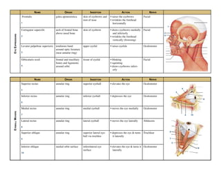
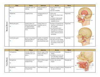
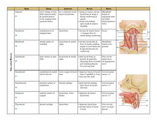
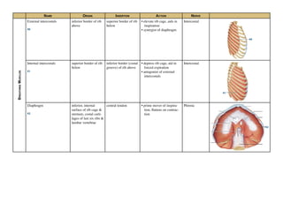
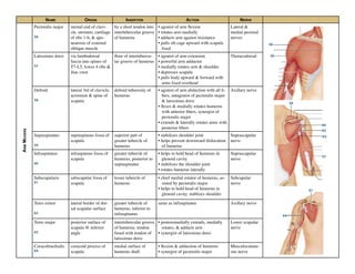
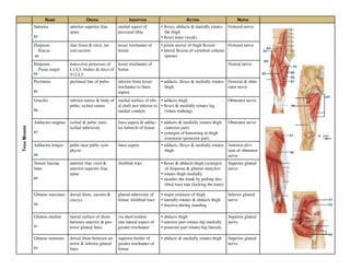
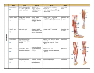
This document describes the origin, insertion, action, and nerve innervation of 55 muscles of the head, neck, back, abdomen, and upper limbs. The muscles control functions like facial expression, eye movement, swallowing, breathing, posture, and shoulder and arm movement. They originate on bones like the skull, vertebrae, ribs, and clavicle, and insert on structures like the eyebrows, eyelids, mouth, hyoid bone, scapula, and humerus. Each muscle has a specific action, such as raising the eyebrows, depressing the eyeballs, or extending the vertebral column. They are innervated by cranial nerves like the facial nerve or spinal nerves.
Lower Limb Human Anatomy ( Muscles )
by DR RAI M. AMMAR
www.facebook.com/drraiammar
www.twitter.com/drraiammar
www.instagram.com/drraiammar
www.linkedin.com/in/drraiammar
www.themedicall.com/blog/auther/drraiammar/
For Any Book or Notes Visit Our Website:
www.allmedicaldata.wordpress.com
www.drraiammar.blogspot.com
YOUTUBE CHANNEL :
https://www.youtube.com/channel/UCu-oR9V3OdFNTJW5yqXWXxA
ANY QUESTION ??
Get in touch with us at Any of the Above Social Media or Email at
drraiammar@gmail.com
allmedicaldata@gmail.com
Radial Nerve is very important topic for first year MBBS Students and as well as for day today clinical practice. This slide gives you full course & relations with clear diagrams as well as applied anatomy with clinical Co-relation.
A summary for learning the muscles of the upper limb including their attachments, innervation, etc., without having to have too many books open. Resources: "Gray’s Anatomy", "Taschenatlas der Anatomie" and Wikipedia. Awaiting further proof-reading!
Head and Neck Human Anatomy (Muscles)
by DR RAI M. AMMAR
www.facebook.com/drraiammar
www.twitter.com/drraiammar
www.instagram.com/drraiammar
www.linkedin.com/in/drraiammar
www.themedicall.com/blog/auther/drraiammar/
For Any Book or Notes Visit Our Website:
www.allmedicaldata.wordpress.com
www.drraiammar.blogspot.com
YOUTUBE CHANNEL :
https://www.youtube.com/channel/UCu-oR9V3OdFNTJW5yqXWXxA
ANY QUESTION ??
Get in touch with us at Any of the Above Social Media or Email at
drraiammar@gmail.com
allmedicaldata@gmail.com
As we know that the muscles play an important role in stability and support of a prosthesis,hence we should be well learned about their peripheries and actions.
Read| The latest issue of The Challenger is here! We are thrilled to announce that our school paper has qualified for the NATIONAL SCHOOLS PRESS CONFERENCE (NSPC) 2024. Thank you for your unwavering support and trust. Dive into the stories that made us stand out!
Model Attribute Check Company Auto PropertyCeline George
In Odoo, the multi-company feature allows you to manage multiple companies within a single Odoo database instance. Each company can have its own configurations while still sharing common resources such as products, customers, and suppliers.
We all have good and bad thoughts from time to time and situation to situation. We are bombarded daily with spiraling thoughts(both negative and positive) creating all-consuming feel , making us difficult to manage with associated suffering. Good thoughts are like our Mob Signal (Positive thought) amidst noise(negative thought) in the atmosphere. Negative thoughts like noise outweigh positive thoughts. These thoughts often create unwanted confusion, trouble, stress and frustration in our mind as well as chaos in our physical world. Negative thoughts are also known as “distorted thinking”.
The Roman Empire A Historical Colossus.pdfkaushalkr1407
The Roman Empire, a vast and enduring power, stands as one of history's most remarkable civilizations, leaving an indelible imprint on the world. It emerged from the Roman Republic, transitioning into an imperial powerhouse under the leadership of Augustus Caesar in 27 BCE. This transformation marked the beginning of an era defined by unprecedented territorial expansion, architectural marvels, and profound cultural influence.
The empire's roots lie in the city of Rome, founded, according to legend, by Romulus in 753 BCE. Over centuries, Rome evolved from a small settlement to a formidable republic, characterized by a complex political system with elected officials and checks on power. However, internal strife, class conflicts, and military ambitions paved the way for the end of the Republic. Julius Caesar’s dictatorship and subsequent assassination in 44 BCE created a power vacuum, leading to a civil war. Octavian, later Augustus, emerged victorious, heralding the Roman Empire’s birth.
Under Augustus, the empire experienced the Pax Romana, a 200-year period of relative peace and stability. Augustus reformed the military, established efficient administrative systems, and initiated grand construction projects. The empire's borders expanded, encompassing territories from Britain to Egypt and from Spain to the Euphrates. Roman legions, renowned for their discipline and engineering prowess, secured and maintained these vast territories, building roads, fortifications, and cities that facilitated control and integration.
The Roman Empire’s society was hierarchical, with a rigid class system. At the top were the patricians, wealthy elites who held significant political power. Below them were the plebeians, free citizens with limited political influence, and the vast numbers of slaves who formed the backbone of the economy. The family unit was central, governed by the paterfamilias, the male head who held absolute authority.
Culturally, the Romans were eclectic, absorbing and adapting elements from the civilizations they encountered, particularly the Greeks. Roman art, literature, and philosophy reflected this synthesis, creating a rich cultural tapestry. Latin, the Roman language, became the lingua franca of the Western world, influencing numerous modern languages.
Roman architecture and engineering achievements were monumental. They perfected the arch, vault, and dome, constructing enduring structures like the Colosseum, Pantheon, and aqueducts. These engineering marvels not only showcased Roman ingenuity but also served practical purposes, from public entertainment to water supply.
Synthetic Fiber Construction in lab .pptxPavel ( NSTU)
Synthetic fiber production is a fascinating and complex field that blends chemistry, engineering, and environmental science. By understanding these aspects, students can gain a comprehensive view of synthetic fiber production, its impact on society and the environment, and the potential for future innovations. Synthetic fibers play a crucial role in modern society, impacting various aspects of daily life, industry, and the environment. ynthetic fibers are integral to modern life, offering a range of benefits from cost-effectiveness and versatility to innovative applications and performance characteristics. While they pose environmental challenges, ongoing research and development aim to create more sustainable and eco-friendly alternatives. Understanding the importance of synthetic fibers helps in appreciating their role in the economy, industry, and daily life, while also emphasizing the need for sustainable practices and innovation.
The Indian economy is classified into different sectors to simplify the analysis and understanding of economic activities. For Class 10, it's essential to grasp the sectors of the Indian economy, understand their characteristics, and recognize their importance. This guide will provide detailed notes on the Sectors of the Indian Economy Class 10, using specific long-tail keywords to enhance comprehension.
For more information, visit-www.vavaclasses.com
Palestine last event orientationfvgnh .pptxRaedMohamed3
An EFL lesson about the current events in Palestine. It is intended to be for intermediate students who wish to increase their listening skills through a short lesson in power point.
1. NAME ORIGIN INSERTION ACTION NERVE
EYEEXPRESSIONS
Frontalis
1
galea aponeurotica skin of eyebrows and
root of nose
• raises the eyebrows
• wrinkles the forehead
horizontally
Facial
Corrugator supercilii
2
arch of frontal bone
above nasal bone
skin of eyebrow • draws eyebrows medially
and inferiorly
• wrinkles the forehead
vertically (frowning)
Facial
Levator palpebrae superioris
3
tendinous band
around optic foramen
(near annular ring)
upper eyelid • raises eyelids Oculomotor
Orbicularis oculi
4
frontal and mazillary
bones and ligaments
around orbit
tissue of eyelid • blinking
• squinting
• draws eyebrows inferi-
orly
Facial
4
2
NAME ORIGIN INSERTION ACTION NERVE
EYEBALLMOVERS
Superior rectus
5
annular ring superior eyeball • elevates the eye Oculomotor
Inferior rectus
6
annular ring inferior eyeball • depresses the eye Oculomotor
Medial rectus
7
annular ring medial eyeball • moves the eye medially Oculomotor
Lateral rectus
8
annular ring lateral eyeball • moves the eye laterally Abducens
Superior oblique
9
annular ring superior lateral eye-
ball via trochlea
• depresses the eye & turns
it laterally
Trochlear
Inferior oblique
10
medial orbit surface inferolateral eye
surface
• elevates the eye & turns it
laterally
Ocolomotor
5
5
6
8
7
8
9
9
10
1
2. NAME ORIGIN INSERTION ACTION NERVE
MOUTHMOVERS
Levator labii superioris
11
zygomatic bone &
infraorbital margin of
maxilla
skin & muscle of up-
per lip
• raises & furrows the up-
per lip
Facial
Zygomaticus minor
12
zygomatic bone skin & muscle @
corner of mouth
• raises lateral corners of
mouth (smiling)
Facial
Zygomaticus major
13
zygomatic bone skin & muscle @
corner of mouth
• raises lateral corners of
mouth (smiling)
Facial
Risorius
14
lateral facia assoc.
with masseter muscle
skin @ angle of
mouth
• draws corner of lip later-
ally
• tense of lips
• synergist of zygomaticus
Facial
Buccinator
15
molar region of max-
illa and mandible
orbicularis oris • draws corner of mouth
laterally
• compresses cheek (suck-
ing)
• holds food between teeth
during chewing
Facial
Depressor anguli oris
16
body of mandible
below incisors
skin & muscle @
angle of mouth
(below insertion of
zygomaticus)
• draws corner of mouth
laterally & downward
• antagonist of zygomati-
cus
Facial
Depressor labii inferioris
17
body and mandible
lateral to its midline
skin & muscle of
lower lip
• draws lower lip inferiorly
(pout)
Facial
Orbicularis oris
18
arises directly from
maxilla & mandible
encircles mouth;
inserts into muscle
& skin @ angles of
mouth
• closes lips
• purses and protrues lips
• kissing & whistling
Facial
Platysma
19
fascia of chest (over
pectoral muscle &
deltoid)
lower margin of
mandible, and skin &
muscle @ corner of
mouth
• depresses mandible
• pulls lower lip back &
down
Facial
12
11
14
15
16
17
18
19
13
3. NAME ORIGIN INSERTION ACTION NERVE
MANDIBLEMOVERS
Masseter
20
zygomatic arch and
maxilla
angle & ramus of
mandible
• prime mover of jaw
closure
• elevates mandible
Trigeminal
Temporalis
21
temporal fossa coronoid process of
mandible
• closes jaw
• elevates & retracts man-
dible
• synergist of pterygoids
• maintains position of
mandible at rest
Trigeminal
Medial pterygoid
22
medial surface of
lateral pterygoid plate
of sphenoid bone,
maxilla & palatine
bone
medial surface of
mandible near its
angle
•synergist of temporalis &
masseter in elevation of
the mandible
• act with lateral ptery-
goid muscle to protrude
mandible of to promote
side-to-side movements
(grinding)
Trigeminal
Lateral pterygoid
23
greater wing & lateral
pterygoid plate of
sphenoid bone
condyle of mandible
and capsule of tem-
poromandibular joint
• protrudes mandible
• provides forward sliding
and side-to-side grinding
movements of the lower
teeth
Trigeminal
NAME ORIGIN INSERTION ACTION NERVE
TONGUEMOVERS
Genioglossus
24
internal surface of
mandible near sym-
physis
inferior aspect of the
tongue and body of
hyoid bone
• primarily protrudes
tongue
• can depress or act in con-
cert with other extrinsic
muscles to retract tongue
Hypoglossal
Styloglossus
25
styloid process of
temporal bone
lateral inferior aspect
of tongue
• retracts (& elevates)
tongue
Hypoglossal
Hyoglossus
26
body & greater horn
of hyoid bone
inferolateral tongue • depresses tongue & draws
its sides downward
Hypoglossal
20
21
22
23
24
25
26
4. NAME ORIGIN INSERTION ACTION NERVE
SWALLOWINGMUSCLES
Digastric
27
lower margin of man-
dible (anterior belly)
& mastoid process
of the temporal bone
(posterior belly)
by a connective tissue
loop to hyoid bone
• acting in concert, elevate
hyoid bone & steady it
during swallowing &
speech
• acting from behind,
open mouth & depress
mandible
Mandibular
branch of
trigeminal (ante-
rior belly)
Facial (posterior
belly)
Stylohyoid
28
styloid process of
temporal bone
hyoid bone • elevates & retracts hyoid
-> elongate floor of
mouth during swallowing
Facial
Mylohyoid
29
medial surface of
mandible
hyoid bone & medial
raphe
• elevates hyoid bone &
floor of mouth, enabling
tongue to exert backward
& upward pressure for
swallowing
Mandibular
branch of tri-
geminal
Geniohyoid
30
inner surface of man-
dible
hyoid bone & medial
raphe
• pulls hyoid bone su-
periorly & anteriorly,
shortening floor of mouth
and widening pharynx
for receiving food
First cervial
spinal nerve
via hypoglossal
nerve
Sternohyoid
31
manubrium & medial
end of clavicle
lower margin of hyoid
bone
• depresses larynx & hyoid
bone if mandible is fixed
• may also flex skull
Cervical spinal
nerves 1-3
Sternothyroid
32
posterior surface of
manubrium
thyroid cartilage • pulls thyroid cartilage
(plus larynx & hyoid)
inferiorly
Cervical spinal
nerves 1-3
Omohyoid
33
superior surface of
scapula
hyoid bone, lower
border
• depresses & retracts
hyoid bone
Cervical spinal
nerves 1-3
Thyrohyoid
34
thyroid cartilage hyoid bone • depresses hyoid bone
• elevates larynx if hyoid
is fixed
First cervical
nerve via hypo-
glossal
27
28
29
30
31
3233
34
34
5. NAME ORIGIN INSERTION ACTION NERVE
WHOLEHEADMOVERS
Sternocleidomastoid
35
manubrium of ster-
num & medial portion
of clavicle
mastoid process of
temporal bone &
superior nuchal line
of occipital bone
• prime mover of active
head flexion (when act
together)
• rotates head toward op-
posite side (when act
singularly
Accesory nerve
Splenius (capitis)
36
ligamentum nuchae,
spinous process of
vertebrae C7-T6
mastoid process of
temporal bone & oc-
cipital bone (capitis)
• extend or hyperextend
head when act together
• rotate and bend head lat-
erally toward same side
when act on one side
Cervical spinal
nerves (dorsal
rami)
Semispinalis capitis
37
transverse process of
C7-T12
occipital bone (ca-
pitis)
• extends head and move it
to opposite side
• synergist with sterno-
cleidomastoid of oppo-
site side
Spinal nerves
(dorsal rami)
35
36
37
6. NAME ORIGIN INSERTION ACTION NERVE
TORSOEXTENDERS
Erector spinae
Iliocostalis
38-A
iliac crests (luborum)
inferior 6 ribs (tho-
racis)
ribs 3-6 (cervicis)
angle of ribs (luborum
& thoracis)
cervical vertebrae C6-
C4 (cervicis)
• extend vertebral column,
maintain posture
• bend vertebral column to
same side when act on
one side
Spinal nerves
(dorsal rami)
Erector spinae
Longissimus
38-B
transverse process of
lumbar through cervi-
cal vertebrae
transverse process of
thoracic or cervical
vertebrae and to ribs
superior to origin;
mastoid process for
capitis
• thoracis & cervicis act to-
gether to extend vertebral
column
• act on one side, bend it
laterally
• capitis extends heads and
turns the face toward
same side
Spinal nerves
(dorsal rami)
Erector spinae
Spinalis
38-C
spines of upper lum-
bat & lower thoracic
vertebrae
spines of upper
thoracic & cervical
vertebrae
• extends vertebral column Spinal nerves
(dorsal rami)
Quadratus lumborum
39
iliac crest & lumbar
fascia
transverse process of
upper lumbar verte-
brae & lower margin
of 12th rib
• flexes vertebral column
laterally when alone
• when together extends
lumbar spine & fixes
12th rib
• maintains upright posture
• assists in forced breathing
T12 & upper
lumber spinal
nerves (ventral
rami)
38-A
38-B
38-C
39
7. NAME ORIGIN INSERTION ACTION NERVE
BREATHINGMUSCLES
External intercostals
40
inferior border of rib
above
superior border of rib
below
• elevate rib cage, aids in
inspiration
• synergist of diaphragm
Intercostal
Internal intercostals
41
superior border of rib
below
inferior border (costal
groove) of rib above
• depress rib cage, aid in
forced expiration
• antagonist of external
intercostals
Intercostal
Diaphragm
42
inferior, internal
surface of rib cage &
sternum, costal carti-
lages of last six ribs &
lumbar vertebrae
central tendon • prime mover of inspira-
tion, flattens on contrac-
tion
Phrenic
40
41
42
8. NAME ORIGIN INSERTION ACTION NERVE
ABDOMINALMUSCLES
Rectus Abdominis
43
pubic crest & sym-
physis
xyphoid process &
costal cartilages of
ribs 5-7
• flex & rotate lumbar re-
gion of vertebral column
• fix & depress ribs
• stabilize pelvis during
walking
• increase intra-abdominal
pressure
Intercostals
External oblique
44
outer surface of lower
8 ribs
linea alba via aponeu-
rosis
• when together, synergist
to rectus abdominis, flex
vertebral column & com-
press abdominal wall
• when alone, synergist to
muscles of back, roate &
lateral flexion of trunk
Intercostals
Internal oblique
45
lumbar fascia, iliac
crest, & inguinal liga-
ment
linea alba, pubic
crest, last 3 or 4 ribs,
& costal margin
• same as external oblique Intercostals
Transverse abdominis
46
inguinal ligament,
lumbar fascia, carti-
lages of last 6 ribs,
iliac crest
linea alba, pubic crest • compresses abdominal
contents
Intercostals
43
NAME ORIGIN INSERTION ACTION NERVE
PELVICFLOORMUSCLES
Levator ani
47
inside pevis from pu-
bis to ischial spine
inner surface of coc-
cyx
• supports & maintains
pelvic viscera
• resists downward thrusts
• forms sphincters at ano-
rectal junction & vagina
• lifts anal canal during
defecation
S4 & inferior
rectal
Coccygeus
48
spine of ischium sacrum & coccyx • supports pelvic viscera
• supports coccyx & pulls
it forward
S4 & S5
47
47
47
48
44
45
46
43 4645
44
9. NAME ORIGIN INSERTION ACTION NERVE
SHOULDERMOVERS
Pectoralis minor
49
anterior surface of
ribs 3-5 (or 2-4)
coracoid process of
scapula
• draws scapula forward &
downward (ribs fixed)
• draws rib cage superiorly
(scaupla fixed)
Both pectoral
nerves
Subclavius
50
costal cartilage of
rib 1
groove on inferior
surface of clavicle
• stablizes & depresses
pectoral girdle
Nerve to subcla-
vius
Serratus anterior
(boxer’s muscle)
51
by series of muscle
slips from ribs 1-9
anterior surface of
vertebral border of
scapula
• agonist to protract & hold
scapula against rib cage
• rotates scapula (infe-
rior angle laterally &
upward)
• abduct & raise arm &
horizontal arm move-
ments
Long thoracic
nerve
Trapezius
52
occipital bone,
ligamentum nuchae,
spines of C7 - T12
spine & acromion of
scapula, lateral 3rd of
clavicle
• stablizes, raises, retracts
& rotates scapula
• adducts & retracts
scapula (middle)
• elevates scapula or syner-
gist to head extension
(suprior)
• depresses scapula &
shoulder (inferior)
Accesory nerve
Levator scapulae
53
transverse processes
of C1-C4
medial border of
scapula, superior to
spine
• elevates & adducts
scapula (synergist to
trapezius)
• tilts glenoid cavity down,
flexes neck to same side
(fixed scapula)
Cervical spinal
nerves & dorsal
scapular nerve
Rhomboid minor
54
spinous processes of
C7 & T1
medial border of
scapula
• retract scapula (squar-
ing shoulders), synergist
with middle fibers of
Trapezius
• rotate glenoid cavity
downward (lowering arm
against resistence)
• stablize scapula
Dorsal scapular
nerve
Rhomboid major
55
spinous processes of
T2-T5
medial border of
scapula
49
50
51
52
53
54
55
10. NAME ORIGIN INSERTION ACTION NERVE
ARMMOVERS
Pectoralis major
56
sternal end of clavi-
cle, sternum, cartilage
of ribs 1-6, & apo-
neurosis of external
oblique muscle
by a short tendon into
intertubercular groove
of humerus
• agonist of arm flexion
• rotates arm medially
• adducts arm against resistance
• pulls rib cage upward with scapula
fixed
Lateral &
medial pectoral
nerves
Latissimus dorsi
57
via lumbodorsal
fascia into spines of
T7-L5, lower 4 ribs &
iliac crest
floor of intertubercu-
lar groove of humerus
• agonist of arm extension
• powerful arm adductor
• medially rotates arm & shoulder
• depresses scapula
• pulls body upward & forward with
arms fixed overhead
Thoracodorsal
Deltoid
58
lateral 3rd of clavicle,
acromion & spine of
scapula
deltoid tuberosity of
humerus
• agonist of arm abduction with all fi-
bers, antagonist of pectoralis major
& latissimus dorsi
• flexes & medially rotates humerus
with anterior fibers, synergist of
pectoralis major
• extends & laterally rotates arms with
posterior fibers
Axillary nerve
Supraspinatus
59
supraspinous fossa of
scapula
superior part of
greater tubercle of
humerus
• stabilizes shoulder joint
• helps prevent downward dislocation
of humerus
Suprascapular
nerve
Infraspinatus
60
infraspinous fossa of
scapula
greater tubercle of
humerus, posterior to
supraspinatus
• helps to hold head of humerus in
glenoid cavity
• stabilizes the shoulder joint
• rotates humerus laterally
Suprascapular
nerve
Subscapularis
61
subscapular fossa of
scapula
lesser tubercle of
humerus
• chief medial rotator of humerus, as-
sisted by pectoralis major
• helps to hold head of humerus in
glenoid cavity, stablizes shoulder
Subcapular
nerve
Teres minor
62
lateral border of dor-
sal scapular surface
greater tubercle of
humerus, inferior to
infraspinatus
same as infraspinatus Axillary nerve
Teres major
63
posterior surface of
scapula @ inferior
angle
intertubercular groove
of humerus, tendon
fused with tendon of
latissimus dorsi
• posteromedially extends, medially
rotates, & adducts arm
• synergist of latissimus dorsi
Lower scapular
nerve
Coracobrachialis
64
coracoid process of
scapula
medial surface of
humerus shaft
• flexion & adduction of humerus
• synergist of pectoralis major
Musculocutane-
ous nerve
56
57
58
59
60
61
62
63
64
11. NAME ORIGIN INSERTION ACTION NERVE
FOREARMMOVERS
Biceps brachii
65
long head (65-1): tuber-
cle above glenoid cav-
ity and lip of glenoid
cavity of scapula
short head: (65-2): cora-
coid process of scapula
by common tendon to
radial tuberosity
• flexes elbor joint & supinates
forearm (usually at the same
time)
• weak flexor of arm @ shoulder
Musculocutane-
ous nerve
Brachialis
66
front of distal humerus coronoid process of
ulna
• major forearm flexor, synergist
with biceps brachii
Musculocutane-
ous nerve
Brachioradialis
67
lateral supracondylar
ridge @ distal end of
humerus
base of styloid pro-
cess of radius
• synergist in forearm flexion, best
when forearm is partially flexed
• stablizes the elbow during rapid
flexion & extension
Radial nerve
Triceps brachii
68
lateral head (68-1): pos-
terior shaft of humerus
long head (68-2) :
infraglenoid tubercle
of scapula
medial head (68-3):
posterior humeral shaft
distal to radial groove
by common tendon
into olacrenon pro-
cess of ulna
• agonist of forearm extension
(medial head)
• antagonist of forearm flexors
• stablizes shoulder joint & assist in
arm adduction (long head tendon)
Radial nerve
Anconeus
69
lateral epicondyle of
humerus
lateral aspect of ola-
cranon process
• abducts ulna during forearm
pronation
• synergist of triceps brachii in
elbow extension
Radial nerve
FOREARMROTATORS
Pronator teres
70
medial epicondyle of hu-
merus, coronoid process
of ulna
by common tendon
into lateral radius,
midshaft
• pronates forearm
• weak flexor of elbow
Median nerve
Supinator
71
lateral epicondyle of hu-
merus, radial collateral
& annular ligaments,
supinator fossa & crest
of ulna
lateral, anterior &
posterior surfaces of
proximal 1/3 of radius
• forcibly supinates forearm with
biceps brachii
• weakly supinates forearm work-
ing along
• antagonist of Pronator teres
Posterior inter-
osseous nerve
65
68-1
65-1
65-2
66
66
67
68-2
68-3
68-1
68-2
69
70
71
12. NAME ORIGIN INSERTION ACTION NERVE
WRISTFLEXORS
Flexor carpi
radialis
72
medial epicondyle of
humerus
base of 2nd & 3rd
metacarpals (anterior)
• powerful flexor of wrist
• abducts the hand
• weak syngergist of elbow flexion
Median nerve
Palmaris longus
73
medial epicondyle of
humerus
palmar aponeurosis,
skin & fascia of palm
• weak wrist flexor
• weak synergist of elbow flexion
• tenses skin of palm during hand
movements
Median nerve
Flexor carpi
ulnaris
74
medial epicondyle of
humerus, olecranon pro-
cess & posterior surface
of ulna
pisiform & hamate
bones & base of 5th
metacarpal (anterior)
• powerful flexor of writs
• adducts hand with extensor carpi
ulnaris
• stablized wrist during finger
extension
Ulnar nerve
WRISTEXTENSORS
Extensor carpi
radialis longus
75
lateral supracondylar
ridge of humerus
base of 2nd metacar-
pal (posterior)
• extends wrist with extensor carpi
ulnaris
• abducts write with flexor carpi
radialis
Radial nerve
Extensor carpis
ulnaris
76
lateral epicondyle of
humerus & posterior
border of ulna
base of 5th metacar-
pal (posterior)
• extends & adducts wrist Deep branch of
radial nerve
FINGERMOVERS
Flexor digitorum
superficialis
77
medial epicondyle of hu-
merus, coronoid process
of ulna, shaft of radius
by four tendons into
middle phalanges of
fingers 2-5
• flexes wrist & middle phalanges
of fingers 2-5
Median nerve
Extensor
digitorum
78
lateral epicondyle of
humerus
by four tendons into
extensor expansions
& distal phalanges of
fingers 2-5
• prime mover of finger extension
• extends wrist
• can abduct (flare) fingers
Posterior inter-
osseous nerve, a
branch of radial
nerve
Extensor digiti
minimi
79
lateral epicondyle of
humerus
extensor expansion of
5th digit
• extends 5th digit Posterior inter-
osseous nerve,
deep branch of
radial nerve
Extensor pollicis
longus
80
posterior surface of
middle 3rd of ulna
base of distal phallanx
of thumb
• extends thumb Posterior inter-
osseous nerve
Abductor pollicis
longus
81
posterior surface of
radius & ulna
base of 1st metacarpal
& trapezium
• abducts & extends thumb Posterior inter-
osseous nerve
75
77
72
73
74
78
80
81
76
79
13. NAME ORIGIN INSERTION ACTION NERVE
THIGHMOVERS
Satorius
82
anterior superior iliac
spine
medial aspect of
proximal tibia
• flexes, abducts & laterally rotates
the thigh
• flexes knee (weak)
Femoral nerve
Iliopsoas
Iliacus
83
iliac fossa & crest, lat-
eral sacrum
lesser trochanter of
femur
• prime mover of thigh flexion
• lateral flexion of vertebral column
(psoas)
Femoral nerve
Iliopsoas
Psoas major
84
transverse processes of
L1-L5, bodies & discs of
T12-L5
lesser trochanter of
femur
Ventral nerve
Pectineus
85
pectineal line of pubis inferior from lesser
trochanter to linea
aspera
• adducts, flexes & medially rotates
thigh
Femoral & obtu-
rator nerve
Gracilis
86
inferior ramus & body of
pubis, ischial ramus
medial surface of tibi-
al shaft just inferior to
medial condyle
• adducts thigh
• flexes & medially rotates leg
(when walking)
Obturator nerve
Adductor magnus
87
ischial & pubic rami,
ischial tuberosity
linea aspera & adduc-
tor tubercle of femur
• adducts & medially rotates thigh
(anterior part)
• synergist of hamstring in thigh
extension (posterior part)
Obturator nerve
Adductor longus
88
pubic near pubic sym-
physis
linea aspera • adducts, flexes & medially rotates
thigh
Anterior divi-
sion of obturator
nerve
Tensor fasciae
latae
89
anterior iliac crest &
anterior superior iliac
spine
iliotibial tract • flexes & abducts thigh (synergist
of iliopsoas & gluteus muscles)
• rotates thigh medially
• steadies the trunk by pulling ilio-
tibial tract taut (locking the knee)
Superior gluteal
nerve
Gluteus maximus
90
dorsal ilium, sacrum &
coccyx
gluteal tuberosity of
femur, iliotibial tract
• major extensor of thigh
• laterally rotates & abducts thigh
• inactive during standing
Inferior gluteal
nerve
Gluteus medius
91
lateral surface of ilium
between anterior & pos-
terior gluteal lines
via short tendon
into lateral aspect of
greater trochanter
• abducts thigh
• anterior part rotates hip medially
• posterior part rotates hip lateraly
Superior gluteal
nerve
Gluteus minimus
92
dorsal ilium between an-
terior & inferior gluteal
lines
superior border of
greater trochanter of
femur
• abducts & medially rotates thigh Superior gluteal
nerve
82
83
84
85
86
87
88
87
88
89
90
91
91
92
90
14. NAME ORIGIN INSERTION ACTION NERVE
HAMSTRINGS
Biceps femoris
93
long head (a): ischial
tuberosity
short head (b): linea
aspera & distal femur
by common tendon
into head of fibula
& lateral condyle of
tibia
• extends thigh & flexes knee
• laterally rotates leg when knee
is flexed
Sciatic nerve
Semitendinosus
94
iscial tuberosity medial aspect of up-
per tibial shaft
• extends thigh & flexes knee
• medially rotates leg with semi-
membranosus
Sciatic nerve
Semimembranosus
95
ischial tuberosity medial condyle of
tibia
• extends thigh & flexes knee
• medially rotates leg
Sciatic nerve
Popliteus
96
lateral condyle of femur proximal tibia (poste-
rior surface)
• unlocks knee by flexes & ro-
tates leg medially
• rotates thigh laterally with tibia
fixed
Tibial nerve
QUADRICEPS
Rectus femoris
97
anterior inferior iliac
spine & superior margin
of acetabulum
patella & tibial
tuberosity via patella
ligament
• extends knee
• flexes thigh @ hip
Femoral nerve
Vastus lateralis
98
greater trochanter, inter-
trochanteric line, linea
aspera
• extends & stablizes knee Femoral nerve
Vastus medialis
99
linea aspera, intertro-
chanteric line
• extends knee
• stablizes patella (inferior fibers)
Femoral nerve
Vastus intermedius
100
anterior & lateral sur-
faces of proximal femur
• extends knee Femoral nerve
93
94
97
96
98
99
99
98
100
95
15. NAME ORIGIN INSERTION ACTION NERVE
FOOTMOVERS
Tibialis anterior
101
lateral condyle & upper
2/3 of tibial shaft, inter-
osseous membrane
by tendon into
inferior surface of
medial cuneiform &
1st metatarsal
• prime mover of dorsiflexion
• inverts foot
• assist in supporting medial longi-
tudinal arch
Deep fibular
nerve
Fibularis longus
102
head & upper portion of
lateral fibula
by long tendon
underfoot into 1st
metatarsal & medial
cuneiform
• plantar flexes & everts foot
• may help keep foot flat on ground
Superficial fibu-
lar nerve
Fibularis brevis
103
distal fibula shaft by tendon behind
lateral malleolus into
base of 5th metatarsal
• plantar flexes & everts foot Superficial fibu-
lar nerve
Gastrocnemius
104
by two heads from me-
dial & lateral condyles
of femur
posterior calcaneus
via calcaneal tendon
(Achilles)
• plantar flexes foot when knee is
extended
• flexes knee when foot is dorsi-
flexed
Tibial nerve
Soleus
105
superior tibia, fibula &
inerosseous membrane
• plantar flexes foot
• important locomoter & postural
muscle
Tibial nerve
Plantaris
106
posterior femur above
lateral condyle
via a long, thin ten-
don into calcaneus or
calcaneal tendon
• assists in knee flexion
• plantar flexion of foot
Tibial nerve
102
103
101
104
104
105
105
106
102
101
104
105
105
106
96
16. NAME ORIGIN INSERTION ACTION NERVE
TOEMOVERS
Extensor hallucis
longus
107
anteromedial fibula shaft
& interosseous mem-
brane
distal phalanx of big
toe
• extends big toe
• dorsiflexes foot
Deep fibular
nerve
Extensor
digitorum longus
108
lateral condyle of tibia,
proximal 3/4 of fibula,
interosseous membrane
middle & distal pha-
langes of toes 2-5 via
extensor expansion
• prime mover of toe extension
(mainly @ metatarsal joints)
• dorsiflexes foot (with tibialis an-
terior & extensor hallucis longus)
Deep fibular
nerve
Flexor hallucis
longus
109
mid-shaft of fibula, inter-
osseous membrane
tendon runs underfoot
to distal phalanx of
big toe
• plantar flexes & inverts foot
• flexes big toe (push-off muscle
when walking)
Tibial nerve
Flexor digitorum
longus
110
posterior tibia tendon runs behind
medial malleolus &
insert into distal pha-
lanx of toes 2-5
• plantar flexes & inverts foot
• flexes toes
• helps foot “grip” ground
Tibial nerve
107
108
110
109
110
109