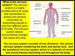
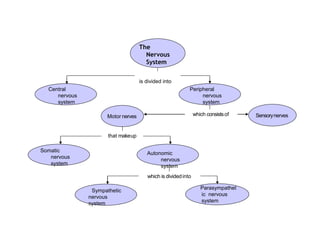
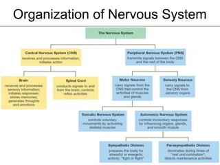
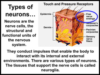
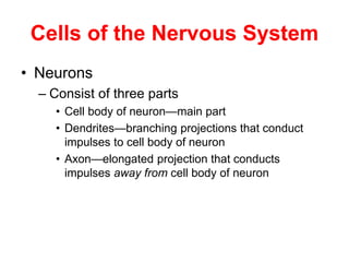
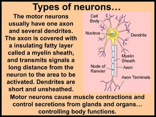
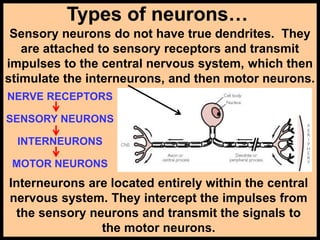
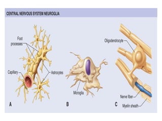
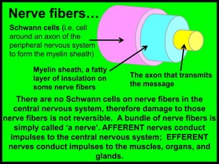
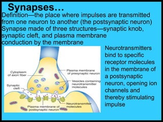
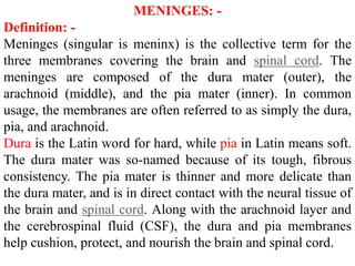
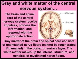
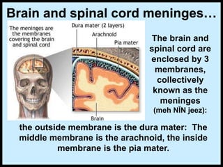
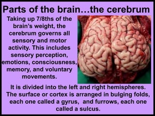
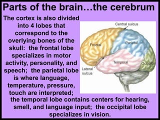
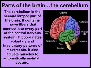
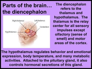
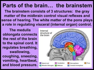
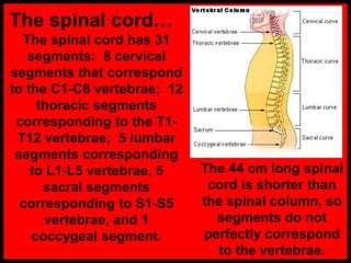
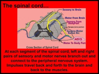
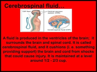
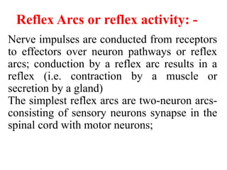
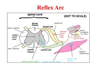
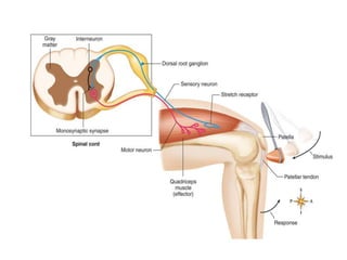
The nervous system is divided into the central nervous system and peripheral nervous system. The central nervous system consists of the brain and spinal cord and contains gray matter and white matter. The peripheral nervous system is made up of nerves that branch throughout the body. The nervous system detects environmental changes and coordinates the body's actions and sensory information through transmitting signals via neurons. It is composed of neurons, which are the basic functional units, and neuroglia, which provide structure and support. The nervous system functions through nerve impulses that travel along neurons via action potentials and neurotransmitters to transmit signals between neurons.
Escort Service Call Girls In Sarita Vihar,, 99530Ā°56974 Delhi NCR
Ā
Nervous System Anatomy
1. Unit-I: - Nervous System
Represented By,
Mr. Audumbar Mali,
(Asst. Prof.)
Sahyadri College of Pharmacy
Methwade
BP201T. Human Anatomy And
Physiology-II
2. What is the nervous
system? The nervous
system is a highly
complex part of an body
that coordinates its
actions and sensory
information by
transmitting signals to
and from different parts
of its body. The nervous
system detects
environmental changes
that impact the body.
The nervous system consists of two divisions: the central
nervous system containing the brain and spinal cord, and
the peripheral nervous system which is a network of nerves
and neural tissues branching out throughout the body.
3. which consistsof
that makeup
which is dividedinto
The
Nervous
System
is divided into
Sensorynerves
Motor nerves
Autonomic
nervous
system
Somatic
nervous
system
Central
nervous
system
Peripheral
nervous
system
Sympathetic
nervous
system
Parasympathet
ic nervous
system
5. Types of
neuronsā¦
Neurons are the
nerve cells, the
structural and
functional units of
the nervous
system.
They conduct impulses that enable the body to
interact with its internal and external
environments. There are various types of neurons.
The tissues that support the nerve cells is called
neuroglia.
6. Cells of the Nervous System
ā¢ Neurons
ā Consist of three parts
ā¢ Cell body of neuronāmain part
ā¢ Dendritesābranching projections that conduct
impulses to cell body of neuron
ā¢ Axonāelongated projection that conducts
impulses away from cell body of neuron
7. Types of neuronsā¦
The motor neurons
usually have one axon
and several dendrites.
The axon is covered with
a insulating fatty layer
called a myelin sheath,
and transmits signals a
long distance from the
neuron to the area to be
activated. Dendrites are
short and unsheathed.
Motor neurons cause muscle contractions and
control secretions from glands and organsā¦
controlling body functions.
8. Types of neuronsā¦
Sensory neurons do not have true dendrites. They
are attached to sensory receptors and transmit
impulses to the central nervous system, which then
stimulate the interneurons, and then motor neurons.
Interneurons are located entirely within the central
nervous system. They intercept the impulses from
the sensory neurons and transmit the signals to
the motor neurons.
NERVE RECEPTORS
SENSORY NEURONS
INTERNEURONS
MOTOR NEURONS
9. Neuroglia (Glia ): -
Support cells, bringing the cells of nervous
tissue together structurally and functionally.
Three main types of connective tissue cells of
the CNS :-
Astrocytesāstar-shaped cells that anchor
small blood vessels to neurons
Microgliaāsmall cells that move in
inflamed brain tissue carrying on
phagocytosis.
Oligodendrocytesāform myelin sheaths on
axons in the CNS (Schwann cells form
myelin sheaths in PNS only)
10.
11. Nerve fibersā¦
There are no Schwann cells on nerve fibers in the
central nervous system, therefore damage to those
nerve fibers is not reversible. A bundle of nerve fibers is
simply called āa nerveā. AFFERENT nerves conduct
impulses to the central nervous system; EFFERENT
nerves conduct impulses to the muscles, organs, and
glands.
Schwann cells (i.e. cell
around an axon of the
peripheral nervous system
to form the myelin sheath)
Myelin sheath, a fatty
layer of insulation on
some nerve fibers
The axon that transmits
the message
12. Definitionāthe place where impulses are transmitted
from one neuron to another (the postsynaptic neuron)
Synapse made of three structuresāsynaptic knob,
synaptic cleft, and plasma membrane
conduction by the membrane
Synapsesā¦
Neurotransmitters
bind to specific
receptor molecules
in the membrane of
a postsynaptic
neuron, opening ion
channels and
thereby stimulating
impulse
13. Some Neurotransmitters
Neurotransmitter Location Some Functions
Acetylcholine Neuron-to-muscle synapse Activates muscles
Dopamine Mid-brain Control of movement
Epinephrine Sympathetic system Stress response
Serotonin Midbrain, pons, medulla Mood, sleep
Endorphins Brain, spine Mood, pain reduction
Nitric Oxide Brain Memory storage
14. Action Potential or Nerve Impulses: -
Definitionāself-propagating wave of
electrical disturbance that travels
along the surface of a neuron
membrane is called action potentials.
15. Mechanism: -
At rest, the neuronās membrane is
slightly positive on the outsideā
polarizedāfrom a slight excess of Na+
on the outside
A stimulus triggers the opening of Na+
channels in the plasma membrane of the
neuron
16. Mechanism: -
Inward movement of Na+ depolarizes the
membrane by making the inside more
positive than the outside at the stimulated
point; this depolarization is a nerve
impulse (action potential)
The stimulated section of membrane
immediately repolarizes, but by that time
the depolarization has already triggered
the next section of membrane to
depolarize, thus propagating a wave of
electrical disturbances (depolarizations)
all the way down the membrane
17. Central Nervous System
The central nervous system (CNS) controls most
functions of the body and mind. It consists of two parts:
the brain and the spinal cord. The brain is the center of
our thoughts, the interpreter of our external environment,
and the origin of control over body movement.
Divisions of the brain :-
Brain stem:-
Consists of three parts, named in ascending order:
medulla oblongata, pons (lies between the medulla
oblongata and the midbrain), and midbrain
Structureāwhite matter with bits of gray matter
scattered through it
18. MENINGES: -
Definition: -
Meninges (singular is meninx) is the collective term for the
three membranes covering the brain and spinal cord. The
meninges are composed of the dura mater (outer), the
arachnoid (middle), and the pia mater (inner). In common
usage, the membranes are often referred to as simply the dura,
pia, and arachnoid.
Dura is the Latin word for hard, while pia in Latin means soft.
The dura mater was so-named because of its tough, fibrous
consistency. The pia mater is thinner and more delicate than
the dura mater, and is in direct contact with the neural tissue of
the brain and spinal cord. Along with the arachnoid layer and
the cerebrospinal fluid (CSF), the dura and pia membranes
help cushion, protect, and nourish the brain and spinal cord.
19. Gray and white matter of the central
nervous systemā¦
The brain and spinal
cord of the central
nervous system receive
impulses, process the
information, and
respond with the
appropriate action.
Gray matter of the brain and spinal cord consists
of unsheathed nerve fibers (cannot be regenerated
if damaged) in the cortex or surface layer. The
white matter makes up the internal structure, and
consists of myelinated nerve fibers.
20. Brain and spinal cord meningesā¦
The brain and
spinal cord are
enclosed by 3
membranes,
collectively
known as the
meninges
(meh NĬN jeez):
the outside membrane is the dura mater: The
middle membrane is the arachnoid, the inside
membrane is the pia mater.
21.
22. Parts of the brainā¦the cerebrum
Taking up 7/8ths of the
brainās weight, the
cerebrum governs all
sensory and motor
activity. This includes
sensory perception,
emotions, consciousness,
memory, and voluntary
movements.
It is divided into the left and right hemispheres.
The surface or cortex is arranged in bulging folds,
each one called a gyrus, and furrows, each one
called a sulcus.
23. Parts of the brainā¦the cerebrum
The cortex is also divided
into 4 lobes that
correspond to the
overlying bones of the
skull: the frontal lobe
specializes in motor
activity, personality, and
speech; the parietal lobe
is where language,
temperature, pressure,
touch are interpreted;
the temporal lobe contains centers for hearing,
smell, and language input; the occipital lobe
specializes in vision.
24. Parts of the brainā¦the cerebellum
The cerebellum is the
second largest part of
the brain. It contains
nerve fibers that
connect it to every part
of the central nervous
system. It coordinates
voluntary and
involuntary patterns of
movements. It also
adjusts muscles to
automatically maintain
posture.
25. Parts of the brainā¦
the diencephalon
The diencephalon
refers to the
thalamus and
hypothalamus. The
thalamus is the relay
center for all sensory
impulses except
olfactory (sense of
smell) and motor
areas of the cortex.
The hypothalamus regulates behavior and emotional
expression, body temperature, and many metabolic
activities. Attached to the pituitary gland, it also
controls hormonal secretions of this gland.
26. Parts of the brainā¦ the brainstem
The brainstem consists of 3 structures: the gray
matter of the midbrain control visual reflexes and
sense of hearing. The white matter of the pons plays
a role in regulating visceral (internal organ) control.
The medulla
oblongata connects
the rest of the brain
to the spinal cord. It
regulates breathing,
swallowing,
coughing, sneezing,
vomiting, heartbeat,
and blood pressure.
27. The spinal cordā¦
The spinal cord has 31
segments: 8 cervical
segments that correspond
to the C1-C8 vertebrae; 12
thoracic segments
corresponding to the T1-
T12 vertebrae; 5 lumbar
segments corresponding
to L1-L5 vertebrae, 5
sacral segments
corresponding to S1-S5
vertebrae, and 1
coccygeal segment.
The 44 cm long spinal
cord is shorter than
the spinal column, so
segments do not
perfectly correspond
to the vertebrae.
28. structure
ā¢ The spinal cord is
protected by the
vertebrae.
ā¢ Gray matter
contains cell
bodies; white
matter contains
myelinated fibers.
ā¢ PNS nerves
extend outside of
the vertebrae.
29. The spinal cordā¦
At each segment of the spinal cord, left and right
pairs of sensory and motor nerves branch out and
connect to the peripheral nervous system.
Impulses travel back and forth to the brain and
back to the muscles.
30. Cerebrospinal fluidā¦
A fluid is produced in the ventricles of the brain; it
surrounds the brain and spinal cord. It is called
cerebrospinal fluid, and it cushions (i. e. something
providing support) the brain and cord from shocks
that could cause injury. It is maintained at a level
around 1/2 - 2/3 cup.
31. CSF acts as a cushion or buffer, providing
basic mechanical and immunological
protection to the brain inside the
skull. CSF also serves a vital function in
the cerebral auto regulation of cerebral
blood flow.
Cerebrospinal fluid has three main
functions: CSF protects brain and spinal
cord from trauma. CSF
supplies nutrients to nervous system tissue.
CSF removes waste products from cerebral
metabolism.
32. Reflex Arcs or reflex activity: -
Nerve impulses are conducted from receptors
to effectors over neuron pathways or reflex
arcs; conduction by a reflex arc results in a
reflex (i.e. contraction by a muscle or
secretion by a gland)
The simplest reflex arcs are two-neuron arcs-
consisting of sensory neurons synapse in the
spinal cord with motor neurons;
33. these-neuron arcs consist of sensory
neurons synapsing in the spinal cord with
interneurons that synapse with motor
neurons.
The simplest neural pathway is the reflex
arc.
This involves one or more sensory
neurons, association neurons in the spine,
and motor neurons, which carry out the
reflex entirely before the brain is aware of
the response.
36. References:
ļ¶Human Anatomy and Physiology, By
Dr. S. B. Bhise, Dr. A. V. Yadav. Nirali
Prakashan, As per PCI Syllabus,
Page No. 1.1-1.24.
ļ¶ Human Anatomy and Physiology-II, By
Dr. Mahesh Prasad, Dr. Antesh Kumar
Jha, Mr. Ritesh Kumar Srivastav, Nirali
Prakashan, As per PCI Syllabus.