This document provides an overview of muscle physiology, including:
1. The objectives are to learn about muscle functions, types, structures, contraction mechanisms, and differences between muscle fiber types.
2. Muscles have general functions like body movement, stabilization, substance transport, heat generation, and respiration.
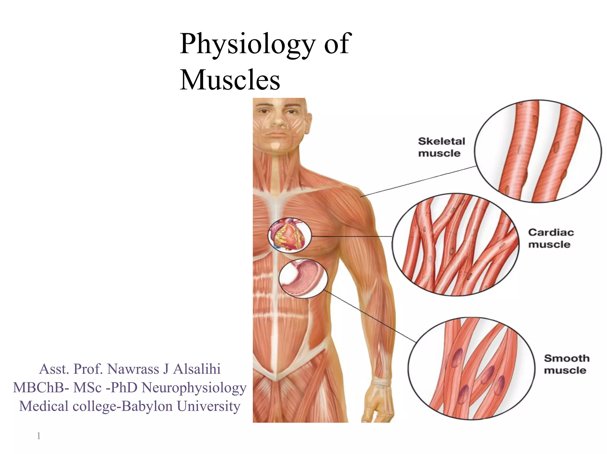
3. Muscles are classified by control (voluntary or involuntary), location, and striation. The three main types are skeletal, cardiac, and smooth muscle.
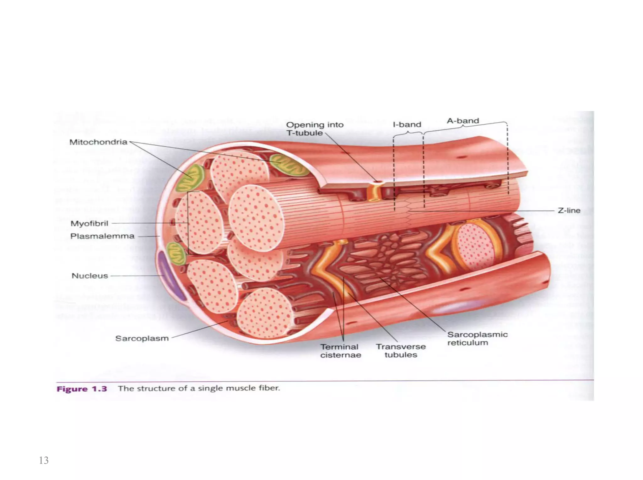
4. Skeletal muscle contraction occurs via the sliding filament theory where the cross-bridge cycling of actin and myosin fibers causes sarcomere shortening and muscle contraction.
2. Objectives
1-Learn major functions of the muscles.
2-Teach types of muscles.
3-Understande the detailed structures of muscles.
4-Learn mechanism of muscle contraction.
5-understanding the differences between red and
white muscle fibers.
6-Knowing what is the meaning of muscle tone
and muscle fatigue.
2
3. General functions of
Muscles
1-Produces body
movements.
2- Stabilizes body
positions.
3- Stores and moves
substances within the body.
4- Generates heat.
5-Respiration
6-Communication
3
4. General properties of Muscle Tissue
1-Electrical excitability:
a property of both muscle and nerve cells to respond to certain
stimuli by producing electrical signals called action potentials
2-Contractility:
ability of muscular tissue to contract forcefully when stimulated by
an action potential.
3- Extensibility:
ability of muscular tissue to stretch without being damaged.
Extensibility allows a muscle to contract forcefully even if it is
already stretched.
4-Elasticity :
ability of muscular tissue to return to its original length and shape
4
5. after contraction .
Classification of Muscles
• Muscles are classified based on three different factors:
I. Depending upon the control :
1- Voluntary muscle :
is controlled by our own will.
Skeletal muscles are the voluntary muscles.
These muscles are innervated by somatic nerves.
2. Involuntary Muscle
Muscle that cannot be controlled by the will .
Cardiac muscle and smooth muscle are involuntary
muscles
5
6. II. Depending upon the situation
• Skeletal muscles
• Cardiac muscle and
• Smooth muscle
III. Depending upon the presence or absence of
striations
1. Striated muscle (Skeletal and Cardiac
muscle)
2. Non-striated muscle (Smooth muscle)
6
7. • Situated in association with bones forming the skeletal system.
• Form 40% to 50% of body mass
• Voluntary and striated.
• Supplied by somatic nerves.
• Fibers of the skeletal muscles are arranged in parallel.
• Muscle fibers are attached to tendons on either end.
• Skeletal muscles are anchored to the bones by the tendons.
• Function : Motion, posture, heat production, and protection.
Skeletal
muscle
7
8. Structure of skeletal muscle
• The muscle consists of numerous fibres called muscle fiber and
each muscle fiber is individual muscle cell(myocyte), these cells
extend the entire length of the muscle, each muscle fiber is
covered by a connective tissue layer called the endomysium.
• Hundreds to thousands of muscle fiber are arranged forming
bundle called fascicles that covered by a connective tissue
layer called the perimysium.
• The fascicles are arranged to form the whole muscle mass, that is
surrounded by a layer of connective tissue continuous with that
surrounds the tendon, called the epimysium.
8
Peri=around
Endo=inside
10. *What is Epimysium?
1.A fibrous connective tissue.
2. The most outer layer that covers the muscle
fibers.
3. It's continuous with tendons where it becomes
thicker.
4. It divides the muscle into columns or bundles
called fascicles and these fascicles are surrounded
by Perimysium.
10
11. Muscle Fiber
• Each muscle cell or muscle fiber is cylindrical in shape.
• Average length of the fiber is 3 cm (1-4 cm, depending upon the
length of the muscle).
• The diameter of the muscle fiber varies from 10 µ to 100 µ and it
varies in a single muscle.
• Each muscle fiber is enclosed by a cell membrane called plasma
membrane, that lies beneath the endomysium. It is also called
sarcolemma.
• Cytoplasm of the muscle fiber is known as sarcoplasm.
11
12. Structures embedded within the sarcoplasm are:
1. Multiple nuclei
2. Myofibril- rod-like elements
3. Golgi apparatus
4. Mitochondria
5-Ribosomes
6. Glycogen droplets.
7. Sarcoplasmic reticulum
8. Transverse tubules (T tubules), which are continuous
with the sarcolemma and penetrate into the cell’s interior.
12
14. 14
Sarcotubular system:
• is a system composed of T-tubules and sarcoplasmic
reticulum.
• it surrounds the myofibrils embedded in the
sarcoplasm.
• It play important roles in the activation of muscle
contractions:
T-tubules help transmit signals from the sarcolemma to the
myofibrils, enabling a muscle cell to respond to neural input. The
function of the sarcoplasmic reticulum is to store calcium ions (Ca),
Calcium ions are released in response to electrical signals that
travel from the sarcolemma to the T tubules, and they serve as
chemical messengers that carry these signals to the myofibrils to
initiate contraction.
16. Myofibril
• Each muscle fiber contains several hundred to several thousand
myofibrils.
• Myofibrils (myofibrillae) are the fine parallel filaments present
in sarcoplasm of the muscle cell.
• The myofibrils contain 2 kinds of protein filaments (contractile
proteins )
Thick filaments - composed of myosin .
Thin filaments - composed of Actin, troponin and
tropomyosin
• Striations are produced by alternating light and dark filaments.
• Each myofibril is composed of about 1500 adjacent myosin
filaments and 3000 actin filaments.
16
17. The ends of actin filaments are attached to Z discs and from this
disc actin extend in both directions to interdigitate with myosin.
The area of muscle between two Z discs are called sarcomere
which constitute contractile unit of the muscle.
A band : is dark, area contains thick filaments (mostly myosin)
H band : Light area at center of A band , it is area where actin and
myosin don't overlap.
I band : is light area , contains thin filaments (only actin)
Z line/disc : At center of I band is where actins attach.
17
18. • Molecular Characteristics of the Contractile Filaments
• Myosin filament
• Each thick filament (myosin filament) is made of 200 myosin
molecules, each of which looks a bit like two golf clubs
wrapped around each other.
• Each myosin molecule is a dimer consisting of two
intertwined subunits, each having a long tail and a fat
protruding head.
• Myosin head functions as adenosin triphosphate enzyme
(ATPase) that cleaves the ATP molecule and provide energy
necessary for muscle contraction.
18
19. Actin filament
• It is the thin filament.
• It composed of three proteins, Actin, Troponin and
Tropomyosin
• Actin protein is composed of double stranded
protein that wrap around each other as a helix.
• Troponin and tropomyosin are two proteins present
on the actin molecule and play important role in
regulating muscle contraction and cross-bridge
cycle.
• Tropomyosin ,wrapped spirally around the sides of
the actin helix.
- In the resting state, the tropomyosin molecules lie on
top of the active sites of the actin strands, so that
attraction cannot occur between the actin and myosin
19
22. The Sliding-Filament theory of contraction
Muscles contract because the thin filaments of the
myofibrils slide past the thick filament bringing either
end of a sarcomere move closer together, thereby
shortening the sarcomere.
As sarcomeres shorten, myofibrils also shorten, as do
muscle fibers and ultimately whole muscles.
The mechanism that drives the sliding of thick and thin
filaments past one another is called the cross-bridge
cycle.
22
23. The Cross-bridge Cycle
Each cross-bridge cycle involves the following five steps:
1) Binding of myosin to actin.
In this state where ADP and Pi (inorganic phosphate) are bound to
the ATPase site of the myosin head, myosin has a high affinity for
actin, and the myosin head binds to an actin monomer in the
adjacent thin filament.
2) Power stroke.
The binding of myosin to actin triggers the release of the Pi from
the ATPase site. During this process, the myosin head reel toward
the middle of the sarcomere, pulling the thin filament along with it.
23
24. 3) Rigor.
As the power stroke ends, ADP is released from the myosin head and
the myosin molecule goes into its low-energy state. In this state,
myosin and actin are tightly bound together, a condition called rigor.
4) Unbinding of myosin and actin.
A new ATP enters the ATPase site on the myosin head, triggering a
conformational change in the head, which decreases the affinity of
myosin for actin, so the myosin detaches from the actin.
24
25. 5) Cocking of the myosin head.
-Soon after it binds to myosin’s ATPase site, ATP is split by
hydrolysis into ADP and Pi, which releases energy. Some of the
energy is captured by the myosin molecule as it goes into its high-
energy conformation.
-The ADP and Pi remain bound to the ATPase site. If calcium is
present, the cycle will continue by return to step 1.
25
27. • When the cross-bridge cycle stops and the contraction ends, the
thin filaments passively slide back to their original position.
• The cross-bridge cycle could continue indefinitely, so long as
there is sufficient ATP. To prevent this never-ending cycle from
happening, the regulatory proteins troponin and tropomyosin
interact with calcium, controlling the availability of myosin-
binding sites on actin and thereby regulating the cross-bridge
cycle.
27
29. 29
Motor Unit
A single motor neuron with muscle fibers (few, hundreds
or thousands of muscle fibers) that innervates.
Motor Unit Parts
1-Motor neuron
A specialized nerve that transmits an impulse to a muscle,
stimulates a muscle cell causing contraction
2- Neuromuscular junction
- A specialized point of contact between a nerve ending and the
muscle fiber it innervates.
- Chemicals are released in response to a nervous impulse that
result in a contraction.
3- Muscle fibers.
32. 32
Fast Versus Slow Muscle Fibers.
• Every muscle of the body is composed of a mixture of so-
called
fast and slow muscle fibers, with still other fibers gradated
between these two extremes.
• Muscles that react rapidly, including the anterior tibialis, are
composed mainly of “fast” fibers with only small numbers of
the slow variety.
Conversely, muscles such as soleus that respond slowly but
with prolonged contraction are composed mainly of “slow”
fibers.
33. 33
Slow Fibers (Type 1, Red Muscle).
1.Slow fibers are smaller than fast fibers.
2-Slow fibers are also innervated by smaller nerve fibers.
3-Compared with fast fibers, slow fibers have a more extensive
blood vessel system and more capillaries to supply extra amounts of
oxygen.
4. Slow fibers have greatly increased numbers of mitochondria to
support high levels of oxidative metabolism.
5. Slow fibers contain large amounts of myoglobin, an iron-
containing protein similar to hemoglobin in red blood cells.
Myoglobin combines with oxygen and stores it until needed, which
also greatly speeds oxygen transport to the mitochondria. The
myoglobin gives the slow muscle a reddish appearance and hence
the name red muscle.
34. 34
Fast Fibers (Type II, White Muscle).
1. Fast fibers are large for great strength of contraction.
2. An extensive sarcoplasmic reticulum is present for rapid
release of calcium ions to initiate contraction.
3. Large amounts of glycolytic enzymes are present for rapid
release of energy by the glycolytic process.
4. Fast fibers have a less extensive blood supply than do slow
fibers because oxidative metabolism is of secondary
importance.
5. Fast fibers have fewer mitochondria than do slow fibers, also
because oxidative metabolism is secondary.
6. A deficit of red myoglobin in fast muscle gives it the name
white muscle.
35. 35
Skeletal Muscle Tone.
• Is the resting tension in a skeletal system.
• It occurs because there are always a few motor units
contracting in a resting muscle. These contractions
do not cause enough tension to produce movement.
• Resting muscle tone is important for maintaining
normal posture, and provides support for the joints to
stabilize their position and help prevent sudden
changes in the position
36. 36
Muscle Fatigue.
• Prolonged and strong contraction of a muscle leads to the well-
known state of muscle fatigue.
• Studies shown that muscle fatigue increases in direct proportion
to the rate of depletion of muscle glycogen. Therefore, fatigue
results mainly from inability of the contractile and metabolic
processes of the muscle fibers to continue supplying the same
work output.
• However, experiments have also shown that transmission of the
nerve signal through the neuromuscular junction, diminish at least
a small amount after intense prolonged muscle activity, thus
further diminishing muscle contraction. Interruption of blood flow
through a contracting
muscle leads to almost complete muscle fatigue within 1
37. 37
Rigor Mortis
-Several hours after death, all the muscles of the body go into a
state of contracture called "rigor mortis"; that is, the muscles
contract and become rigid, even without action potentials.
-This rigidity results from loss of all the ATP, which is required to
cause separation of the cross bridges from the actin filaments
during the relaxation process.
-The muscles remain in rigor until the muscle proteins deteriorate
about 15 to 25 hours later, which presumably results from
autolysis caused by enzymes released from lysosomes..... All
these events occur more rapidly at higher temperatures.