Downloaded 274 times
![Sexual Dimorphism in Sacrum
Features Male sacrum Female sacrum
1. Length More Less
2. Ratio between the transverse
width of body of 1st sacral
vertebra and the entire width of
sacral base.
More than 1/3rd. Less thn 1/3rd.
3. Auricular surface Relatively longer, upper three
segments.
Smaller, occupies only upper two
segments of sacrum.
4. Anterior surface of sacrum Shallower Deeper
5. Sacral Index
[Breadth of the base X 100]
Length
Lesser Greater
6. Width Relatively narrower Wider
7. Curvature Uniformly curved Flattened in the upper part but
sharply curved in the lower part.](https://image.slidesharecdn.com/sacrum-190714080537/85/Sacrum-15-320.jpg)


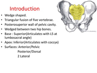
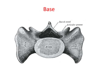
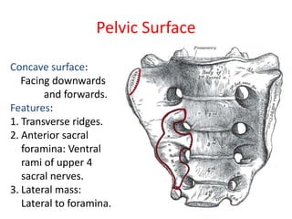
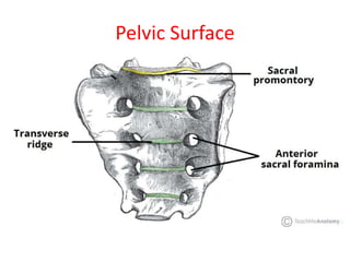
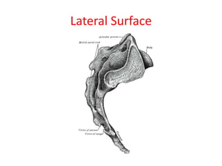
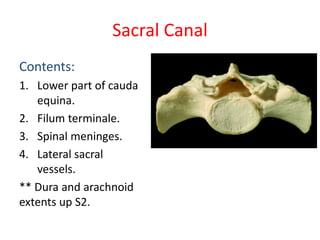
1. The sacrum is a triangular bone formed by the fusion of 5 vertebrae located between the hip bones at the base of the spine. 2. It has anterior and posterior surfaces, with the anterior surface facing downward and forward into the pelvis. 3. The sacrum articulates superiorly with L5 and inferiorly with the coccyx and contains the sacral canal which houses the cauda equina and other structures.


