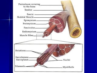
1. Skeletal muscle is composed of bundles of muscle fibers surrounded by connective tissue layers.
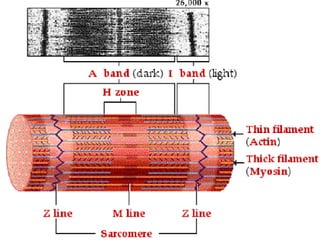
2. Each muscle fiber contains protein filaments that slide past each other when stimulated, causing contraction.
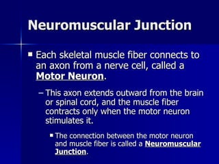
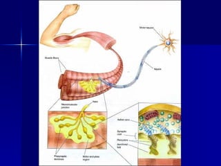
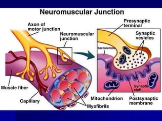
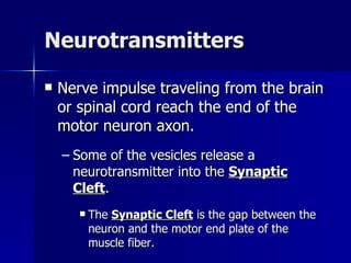
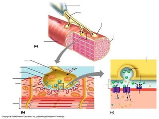
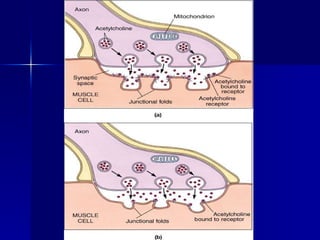
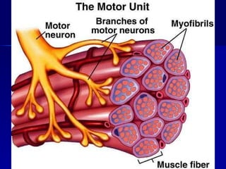
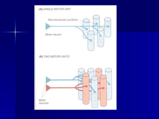
3. Motor neurons connect to muscle fibers at neuromuscular junctions and release neurotransmitters that stimulate contraction when an impulse is received from the brain or spinal cord.

![Structure of a Skeletal Muscle The skeletal muscle structure is composed of four different types of tissues: 1. Skeletal Muscle Tissue 2. Nervous Tissue 3. Blood Tissue [connective] 4. Connective Tissue](https://image.slidesharecdn.com/structureofskeletalmuscle-100914194435-phpapp01/85/Structure-of-skeletal_muscle-4-320.jpg)