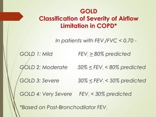
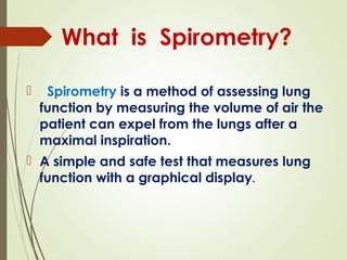
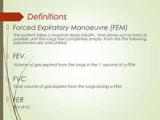
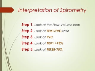
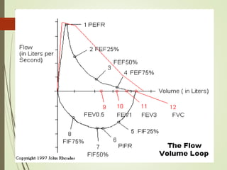
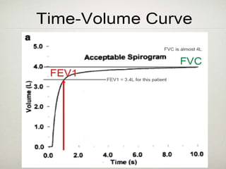
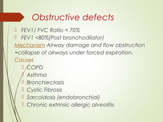
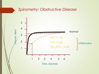
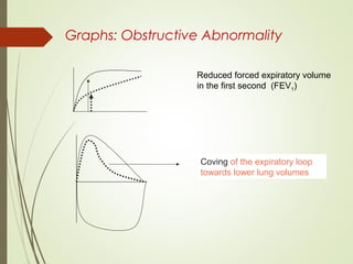
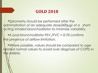
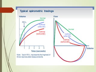
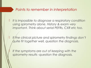
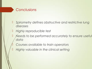
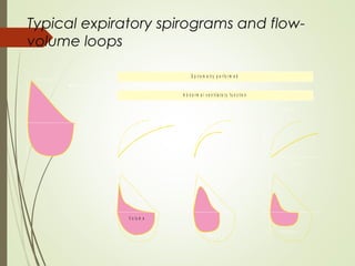
Simplifying
Spirometry Dr KUMAR UTSAV MD
CLINICIAN
WHY DO WE NEED SPIROMETRY…
 Spirometry is a method of assessing lung
function by measuring the volume of air the
patient can expel from the lungs after a
maximal inspiration.
 A simple and safe test that measures lung
function with a graphical display.
What is Spirometry?
spirometry is like the
ECG of lungs
Eur Respir J 2005; 25:587-588
Level of FEV1 as a predictor of all-cause
and cardiovascular mortality: an effect
beyond smoking and physical fitness?
J. Sunyer1 and C. S. Ulrik2
LUNG HEALTH STUDY
Chest. 2005;127:1952-1959
The Relationship Between Reduced Lung
Function and Cardiovascular Mortality
A Population-Based Study
Don D. Sin, MD, MPH, FCCP; LieLing Wu, MSc and S. F. Paul Man, MD
Every 10% decrease in FEV1 leads to –
14% Increase in all cause mortality
28% increase in Cardio-Vascular mortality
COPD is powerful independent risk
factor for Cardio-Vascular mortality
LUNG HEALTH STUDY
In fact, approximately 50% patients of COPD
have ischemic heart disease as the primary
cause of hospitalization.
COPD & CVD:
Chest. 2005;127:1952-1959
LUNG HEALTH STUDY
Why should every doctor know
about spirometry?
More than half of the population of India
suffer from respiratory disease or are the
potential patients of these diseases
Therefore any doctor involved in care of
asthma, COPD or pulmonary fibrosis should
understand the basics of pulmonary function
testing
Why do physicians not do
spirometry ?
 Don’t ever think about it
 Too busy
 Feel the test is complex
 Don’t know what to ask for in the spirogram
 Time consuming
 Not cost effective
 Results don’t affect treatment
Indications
 Diagnosis of obstructive & restrictive lung diseases
 To screen people at risk of pulmonary diseases
 To assess preoperative risk
 To assess prognosis
 To assess therapeutic interventions
 To assess patients as part of a rehabilitation program
 Epidemiologic surveys for pulmonary diseases
Contraindications:
 Within 6 weeks since the last
exacerbation.
 Recent MI less than 3-6 months
ago.
 Unstable angina in last 24 hours.
 Haemoptysis of unknown origin.
 Recent eye surgery less than 3-6
months.
 Abdominal surgery within last 3-6
months.
 Recent CVA less than 3-6 months.
 Diagnosis of Tuberculosis unless special
precautions used.
 Current chest infection or within in last 6
weeks.
 Current chest pain with no diagnosis.
 Pulmonary embolism (PE) within last 3-6
months.
 Ear infection.
 Spontaneous pneumothorax.
 Aortic aneurysm.
DONT’S
 Smoking (1 hour prior)
 Consumption of alcohol (4 hours prior)
 Vigorous exercise (30 minutes prior)
 Wearing tight clothes.
 Eating a substantial meal (at least 2 hours prior)
 Test should be performed when the patient is
clinically stable and free from infection.
 Drinking fluids containing caffeine (12 hours prior)
Withholding bronchodilators
(ARTP)
Inhaled bronchodilators Duration of
action (hours)
Short-acting 4 to 8
Long-acting 24
Anticholinergics 6
Oral short-acting bronchodilators 8
Sustained release β2
agonists 24
Theophylline
Twice-daily preparations 24
Once-daily preparations 48
Definitions
 Forced Expiratory Manoeuvre (FEM)
The patient takes a maximal deep breath , then blows out as hard as
possible until the lungs feel completely empty. From this the following
parameters are calculated:
 FEV1
Volume of gas expired from the lungs in the 1st
second of a FEM
 FVC
Total volume of gas expired from the lungs during a FEM
 FER
FEV1/FVC
Spirometric
Interpretation
Interpretation of Spirometry
Step 1. Look at the Flow-Volume loop
Step 2. Look at FEV1/FVC ratio
Step 3. Look at FVC
Step 4. Look at FEV1 >75%
Step 5. Look at FEF25-75%
Criteria for Normal
Post-bronchodilator Spirometry
 FEV1: % predicted > 80%
 FVC: % predicted > 80%
 FEV1/FVC: > 70
Two basic types of pulmonary function
abnormalities are described using the basic
spirometric parameters:
Obstructive and Restrictive
Obstructive defects
 FEV1/ FVC Ratio < 70%
 FEV1 <80%(Post bronchodilator)
Mechanism Airway damage and flow obstruction
+collapse of airways under forced expiration.
Causes
 COPD
 Asthma
 Bronchiectasis
 Cystic Fibrosis
 Sarcoidosis (endobronchial)
 Chronic extrinsic allergic alveolitis
Spirometry: Obstructive Disease
Time, seconds
Volume,liters
Time, seconds
5
4
3
2
1
1 2 3 4 5 6
FEV1 = 1.8L
FVC = 3.2L
FEV1/FVC = 0.56
Normal
Obstructive
Graphs: Obstructive Abnormality
Coving of the expiratory loop
towards lower lung volumes
Reduced forced expiratory volume
in the first second (FEV1)
Severity
Post bronchodilator
FEV1
(% of predicted value)
Mild airflow obstruction > 80%
Moderate airflow
obstruction
79-50%
Severe airflow obstruction <30 – 49%
V. Severe airflow
obstruction
<30%
Measuring Reversibility of Airflow
Obstruction
 To measure the degree of reversibility of airflow
obstruction, perform spirometry before and 10
to 15 minutes after administering a
bronchodilator by metered dose inhaler or
nebuliser
 Beta2
agonists (e.g. salbutamol) are generally
considered the benchmark bronchodilator
Obstructive with reversibility
 If you get a COPD picture (FEV1/FVC < 70% and
FEV1 <80%) but there is a good degree of
reversibility-
- If > 200ml, then more likely to be asthma than
COPD.
- If < 200ml, it could be a combination of
asthma and COPD or another diagnosis
too. (Go back to history, exam and other inv).
Spirometry For COPD
SYMPTOMS
chronic cough
shortness of breath
EXPOSURE TO RISK
FACTORS
tobacco
occupation
indoor/outdoor pollution
SPIROMETRY: Required to establish
diagnosis
GOLD 2018
sputum
GOLD 2018
Spirometry should be performed after the
administration of an adequate dose(400µg) of a short-
acting inhaled bronchodilator to minimize variability.
A post-bronchodilator FEV1/FVC < 0.70 confirms
the presence of airflow limitation.
Where possible, values should be compared to age-
related normal values to avoid over diagnosis of COPD in
the elderly.
GOLD
Classification of Severity of Airflow
Limitation in COPD*
In patients with FEV1/FVC < 0.70 -
GOLD 1: Mild FEV1 > 80% predicted
GOLD 2: Moderate 50% < FEV1 < 80% predicted
GOLD 3: Severe 30% < FEV1 < 50% predicted
GOLD 4: Very Severe FEV1 < 30% predicted
*Based on Post-Bronchodilator FEV1
Division of lung volumes in normal subjects
and patients with COPD
T L C
IC
F R C
R V
N o rm a l C O P D
R V
VOLUME
T L C
IC
F R C
Spirometry for Asthma
Restrictive defects
 FEV1/FVC Ratio normal i.e. >70%
 Both FEV1 & FVC reduced(But no text says below
what level)
Mechanism-Reduction in all lung volumes
and reduced lung compliance.
 Causes
 Intrinsic - Interstitial lung diseases
 Extrinsic - Kyphoscoliosis, Ankylosing spondylitis,
Muscular dystrophies, Diffuse pleural thickening
Spirometry: Restrictive Disease
Volume,liters
Time, seconds
FEV1 = 1.9L
FVC = 2.0L
FEV1/FVC = 0.95
1 2 3 4 5 6
5
4
3
2
1
Normal
Restrictive
Graphs: Restrictive Abnormality
Tall narrow graph
Reduced flow,
miniature graph
Categorisation of restriction.
VC or TLC- % predicted
 Normal ≥ 81
 Mild restriction 66 - 80
 Moderate restriction 51 - 65
 Severe restriction ≤ 50
Mixed Obstructive/Restrictive
 FEV1: % predicted < 80%
 FVC: % predicted < 80%
 FEV1 /FVC: < 70
Mixed Obstructive and Restrictive
Restrictive and mixed obstructive-restrictive are difficult to diagnose by
spirometry alone; full respiratory function tests are usually required (e.g.,
body plethysmography, etc)
Volume,liters
Time, seconds
FEV1 = 0.5L
FVC = 1.5L
FEV1/FVC = 0.30
Normal
Obstructive - Restrictive
FEV1/FVC ratio
(Post bronchodilator)
Normal
FEV1
Normal
spirometry
Normal FEV1 reduced
Look at FVC
Restrictive defect
FVC reduced
Reduced (<70%)
Obstructive
defect
Points to remember in interpretation
 It is impossible to diagnose a respiratory condition
using spirometry alone. History & exam very
important. Think about serial PEFRs, CXR etc too.
 If the clinical picture and spirometry findings don’t
quite fit together well, question the diagnosis.
 If the symptoms are out of keeping with the
spirometry results -question the diagnosis.
Conclusions
 Spirometry defines obstructive and restrictive lung
diseases
 Highly reproducible test
 Needs to be performed accurately to ensure useful
data
 Courses available to train operators
 Highly valuable in the clinical setting
spirometry is like the
ECG of lungs
Thank you
Complications
 Syncope, dizziness, light headedness
 Pneumothorax
 Chest pain
 Coughing
 Bronchospasm
 Contraction of infection
Severe COPD
A
a
A
a
A
a
A
a
Case 1
Case 2
Pulmonary Fibrosis (Asbestosis) A
A
A
A
a
Case 3
?Severe COPD (with reversibility)
But history very important in this one
A
a
A
a
A
a
A
a
Case 4
Bronchiectasis(but could easily be Asthma or COPD with
reversibility)
A
A
A
A
A
A
Case 5
Idiopathic Pulmonary Fibrosis
A
A
Case 6
Idiopathic Pulmonary Fibrosis
Case 7
A
a
A
a
Muscular dystrophy
Typical expiratory spirograms and flow-
volume loops
Volume
T im e
O b s tr u c t io n
Volume
T im e
R e s tr ic tio n
Volume
T im e
M ix e dFlow
V o lu m e
Flow
V o lu m e
Flow
V o lu m e
N o r m a l ( ? )
A b n o r m a l v e n t ila t o r y f u n c t io n
S p ir o m e tr y p e r f o r m e d

simplyfying spirometry

  • 2.
  • 3.
  • 4.
    WHY DO WENEED SPIROMETRY…
  • 5.
     Spirometry isa method of assessing lung function by measuring the volume of air the patient can expel from the lungs after a maximal inspiration.  A simple and safe test that measures lung function with a graphical display. What is Spirometry?
  • 6.
    spirometry is likethe ECG of lungs
  • 7.
    Eur Respir J2005; 25:587-588 Level of FEV1 as a predictor of all-cause and cardiovascular mortality: an effect beyond smoking and physical fitness? J. Sunyer1 and C. S. Ulrik2 LUNG HEALTH STUDY
  • 8.
    Chest. 2005;127:1952-1959 The RelationshipBetween Reduced Lung Function and Cardiovascular Mortality A Population-Based Study Don D. Sin, MD, MPH, FCCP; LieLing Wu, MSc and S. F. Paul Man, MD Every 10% decrease in FEV1 leads to – 14% Increase in all cause mortality 28% increase in Cardio-Vascular mortality COPD is powerful independent risk factor for Cardio-Vascular mortality LUNG HEALTH STUDY
  • 9.
    In fact, approximately50% patients of COPD have ischemic heart disease as the primary cause of hospitalization. COPD & CVD: Chest. 2005;127:1952-1959 LUNG HEALTH STUDY
  • 10.
    Why should everydoctor know about spirometry? More than half of the population of India suffer from respiratory disease or are the potential patients of these diseases Therefore any doctor involved in care of asthma, COPD or pulmonary fibrosis should understand the basics of pulmonary function testing
  • 11.
    Why do physiciansnot do spirometry ?  Don’t ever think about it  Too busy  Feel the test is complex  Don’t know what to ask for in the spirogram  Time consuming  Not cost effective  Results don’t affect treatment
  • 12.
    Indications  Diagnosis ofobstructive & restrictive lung diseases  To screen people at risk of pulmonary diseases  To assess preoperative risk  To assess prognosis  To assess therapeutic interventions  To assess patients as part of a rehabilitation program  Epidemiologic surveys for pulmonary diseases
  • 13.
    Contraindications:  Within 6weeks since the last exacerbation.  Recent MI less than 3-6 months ago.  Unstable angina in last 24 hours.  Haemoptysis of unknown origin.  Recent eye surgery less than 3-6 months.  Abdominal surgery within last 3-6 months.  Recent CVA less than 3-6 months.  Diagnosis of Tuberculosis unless special precautions used.  Current chest infection or within in last 6 weeks.  Current chest pain with no diagnosis.  Pulmonary embolism (PE) within last 3-6 months.  Ear infection.  Spontaneous pneumothorax.  Aortic aneurysm.
  • 14.
    DONT’S  Smoking (1hour prior)  Consumption of alcohol (4 hours prior)  Vigorous exercise (30 minutes prior)  Wearing tight clothes.  Eating a substantial meal (at least 2 hours prior)  Test should be performed when the patient is clinically stable and free from infection.  Drinking fluids containing caffeine (12 hours prior)
  • 15.
    Withholding bronchodilators (ARTP) Inhaled bronchodilatorsDuration of action (hours) Short-acting 4 to 8 Long-acting 24 Anticholinergics 6 Oral short-acting bronchodilators 8 Sustained release β2 agonists 24 Theophylline Twice-daily preparations 24 Once-daily preparations 48
  • 16.
    Definitions  Forced ExpiratoryManoeuvre (FEM) The patient takes a maximal deep breath , then blows out as hard as possible until the lungs feel completely empty. From this the following parameters are calculated:  FEV1 Volume of gas expired from the lungs in the 1st second of a FEM  FVC Total volume of gas expired from the lungs during a FEM  FER FEV1/FVC
  • 17.
  • 18.
    Interpretation of Spirometry Step1. Look at the Flow-Volume loop Step 2. Look at FEV1/FVC ratio Step 3. Look at FVC Step 4. Look at FEV1 >75% Step 5. Look at FEF25-75%
  • 23.
    Criteria for Normal Post-bronchodilatorSpirometry  FEV1: % predicted > 80%  FVC: % predicted > 80%  FEV1/FVC: > 70
  • 24.
    Two basic typesof pulmonary function abnormalities are described using the basic spirometric parameters: Obstructive and Restrictive
  • 25.
    Obstructive defects  FEV1/FVC Ratio < 70%  FEV1 <80%(Post bronchodilator) Mechanism Airway damage and flow obstruction +collapse of airways under forced expiration. Causes  COPD  Asthma  Bronchiectasis  Cystic Fibrosis  Sarcoidosis (endobronchial)  Chronic extrinsic allergic alveolitis
  • 26.
    Spirometry: Obstructive Disease Time,seconds Volume,liters Time, seconds 5 4 3 2 1 1 2 3 4 5 6 FEV1 = 1.8L FVC = 3.2L FEV1/FVC = 0.56 Normal Obstructive
  • 27.
    Graphs: Obstructive Abnormality Covingof the expiratory loop towards lower lung volumes Reduced forced expiratory volume in the first second (FEV1)
  • 28.
    Severity Post bronchodilator FEV1 (% ofpredicted value) Mild airflow obstruction > 80% Moderate airflow obstruction 79-50% Severe airflow obstruction <30 – 49% V. Severe airflow obstruction <30%
  • 29.
    Measuring Reversibility ofAirflow Obstruction  To measure the degree of reversibility of airflow obstruction, perform spirometry before and 10 to 15 minutes after administering a bronchodilator by metered dose inhaler or nebuliser  Beta2 agonists (e.g. salbutamol) are generally considered the benchmark bronchodilator
  • 30.
    Obstructive with reversibility If you get a COPD picture (FEV1/FVC < 70% and FEV1 <80%) but there is a good degree of reversibility- - If > 200ml, then more likely to be asthma than COPD. - If < 200ml, it could be a combination of asthma and COPD or another diagnosis too. (Go back to history, exam and other inv).
  • 31.
  • 32.
    SYMPTOMS chronic cough shortness ofbreath EXPOSURE TO RISK FACTORS tobacco occupation indoor/outdoor pollution SPIROMETRY: Required to establish diagnosis GOLD 2018 sputum
  • 33.
    GOLD 2018 Spirometry shouldbe performed after the administration of an adequate dose(400µg) of a short- acting inhaled bronchodilator to minimize variability. A post-bronchodilator FEV1/FVC < 0.70 confirms the presence of airflow limitation. Where possible, values should be compared to age- related normal values to avoid over diagnosis of COPD in the elderly.
  • 34.
    GOLD Classification of Severityof Airflow Limitation in COPD* In patients with FEV1/FVC < 0.70 - GOLD 1: Mild FEV1 > 80% predicted GOLD 2: Moderate 50% < FEV1 < 80% predicted GOLD 3: Severe 30% < FEV1 < 50% predicted GOLD 4: Very Severe FEV1 < 30% predicted *Based on Post-Bronchodilator FEV1
  • 35.
    Division of lungvolumes in normal subjects and patients with COPD T L C IC F R C R V N o rm a l C O P D R V VOLUME T L C IC F R C
  • 36.
  • 38.
    Restrictive defects  FEV1/FVCRatio normal i.e. >70%  Both FEV1 & FVC reduced(But no text says below what level) Mechanism-Reduction in all lung volumes and reduced lung compliance.  Causes  Intrinsic - Interstitial lung diseases  Extrinsic - Kyphoscoliosis, Ankylosing spondylitis, Muscular dystrophies, Diffuse pleural thickening
  • 39.
    Spirometry: Restrictive Disease Volume,liters Time,seconds FEV1 = 1.9L FVC = 2.0L FEV1/FVC = 0.95 1 2 3 4 5 6 5 4 3 2 1 Normal Restrictive
  • 40.
    Graphs: Restrictive Abnormality Tallnarrow graph Reduced flow, miniature graph
  • 41.
    Categorisation of restriction. VCor TLC- % predicted  Normal ≥ 81  Mild restriction 66 - 80  Moderate restriction 51 - 65  Severe restriction ≤ 50
  • 42.
    Mixed Obstructive/Restrictive  FEV1:% predicted < 80%  FVC: % predicted < 80%  FEV1 /FVC: < 70
  • 43.
    Mixed Obstructive andRestrictive Restrictive and mixed obstructive-restrictive are difficult to diagnose by spirometry alone; full respiratory function tests are usually required (e.g., body plethysmography, etc) Volume,liters Time, seconds FEV1 = 0.5L FVC = 1.5L FEV1/FVC = 0.30 Normal Obstructive - Restrictive
  • 44.
    FEV1/FVC ratio (Post bronchodilator) Normal FEV1 Normal spirometry NormalFEV1 reduced Look at FVC Restrictive defect FVC reduced Reduced (<70%) Obstructive defect
  • 45.
    Points to rememberin interpretation  It is impossible to diagnose a respiratory condition using spirometry alone. History & exam very important. Think about serial PEFRs, CXR etc too.  If the clinical picture and spirometry findings don’t quite fit together well, question the diagnosis.  If the symptoms are out of keeping with the spirometry results -question the diagnosis.
  • 46.
    Conclusions  Spirometry definesobstructive and restrictive lung diseases  Highly reproducible test  Needs to be performed accurately to ensure useful data  Courses available to train operators  Highly valuable in the clinical setting
  • 48.
    spirometry is likethe ECG of lungs
  • 49.
  • 50.
    Complications  Syncope, dizziness,light headedness  Pneumothorax  Chest pain  Coughing  Bronchospasm  Contraction of infection
  • 51.
  • 52.
    Case 2 Pulmonary Fibrosis(Asbestosis) A A A A a
  • 53.
    Case 3 ?Severe COPD(with reversibility) But history very important in this one A a A a A a A a
  • 54.
    Case 4 Bronchiectasis(but couldeasily be Asthma or COPD with reversibility) A A A A A A
  • 55.
  • 56.
  • 57.
  • 59.
    Typical expiratory spirogramsand flow- volume loops Volume T im e O b s tr u c t io n Volume T im e R e s tr ic tio n Volume T im e M ix e dFlow V o lu m e Flow V o lu m e Flow V o lu m e N o r m a l ( ? ) A b n o r m a l v e n t ila t o r y f u n c t io n S p ir o m e tr y p e r f o r m e d

Editor's Notes

  • #6 Assesses dynamic flow within the airways Airway calibre Lung compliance
  • #14 (Increases Intraocular,Intrathoracic, Intra-abd and Intracranial pressure)
  • #26 Basic spirometry involves only the measurement of forced vital capacity (FVC) and the forced expired volume in the first second (FEV1). The ratio between the two is a self-controlled statistic which tells if obstruction is present. FVC and FEV1 can be measured against predicted values. PEFR is not reproducible enough measurement for accurate diagnosis, but may be used more for following progress with asthma. FEF 25-75% is a measurement of smaller airway function but this measurement is usually not clinically useful.
  • #28 Some texts suggest its the narrowing of the airways and some suggest it could be the collapse of the airways when air is expelled forcefully that impedes the expiration. In reality , it could be both to some extent.
  • #38 Note the significant increases in total lung capacity (TLC), functional residual capacity (FRC), and residual volume (RV), as well as the increase in the ratio of FRC to TLC and of RV to TLC. Note also the fall in inspiratory capcity (IC