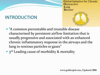
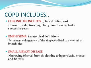
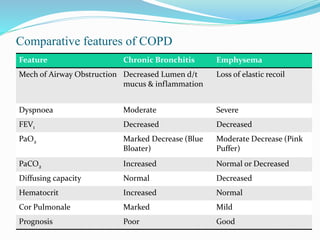
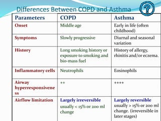
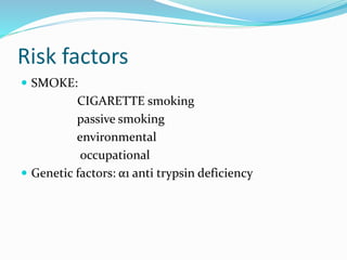
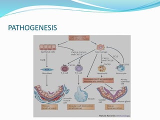
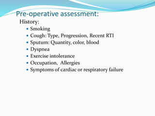
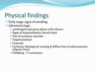
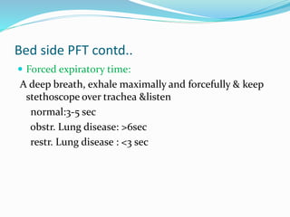
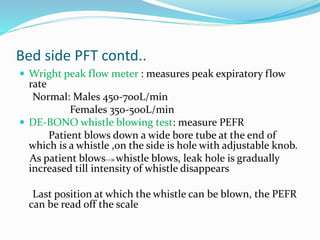
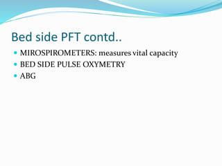
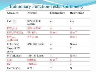
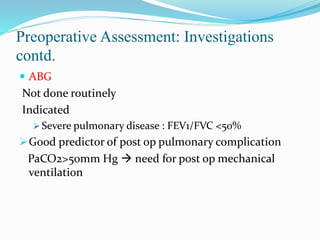
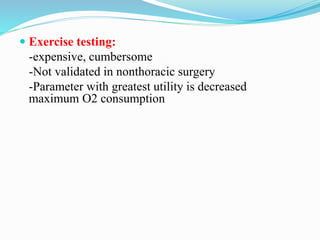
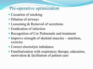
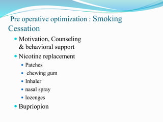
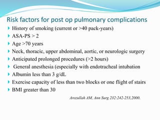
Dr PRASHANTH
Dr GANESH
Dr KARAN
Dr KIRAN
 DEFINITION
 PATHOPHYSIOLOGY
 DIAGNOSIS
 PRE OPERATIVE OPTIMISATION
www.goldcopd.com, Updated 2006
INTRODUCTION
 “A common preventable and treatable disease
characterised by persistent airflow limitation that is
usually progressive and associated with an enhanced
chronic inflammatory response in the airways and the
lung to noxious particles or gases’’
 3rd Leading cause of morbidity & mortality
COPD INCLUDES..
 CHRONIC BRONCHITIS: (clinical definition)
Chronic productive cough for 3 months in each of 2
successive years
 EMPHYSEMA: (anatomical definition)
Permanent enlargement of the airspaces distal to the terminal
bronchioles
 SMALL AIRWAY DISEASE:
Narrowing of small bronchioles due to hyperplasia, mucus
and fibrosis
Comparative features of COPD
Feature Chronic Bronchitis Emphysema
Mech of Airway Obstruction Decreased Lumen d/t
mucus & inflammation
Loss of elastic recoil
Dyspnoea Moderate Severe
FEV1 Decreased Decreased
PaO2 Marked Decrease (Blue
Bloater)
Moderate Decrease (Pink
Puffer)
PaCO2 Increased Normal or Decreased
Diffusing capacity Normal Decreased
Hematocrit Increased Normal
Cor Pulmonale Marked Mild
Prognosis Poor Good
Differences Between COPD and Asthma
Parameters COPD Asthma
Onset Middle age Early in life (often
childhood)
Symptoms Slowly progressive Diurnal and seasonal
variation
History Long smoking history or
exposure to smoking and
bio-mass fuel
History of allergy,
rhinitis and/or eczema.
Inflammatory cells Neutrophils Eosinophils
Airway
hyperresponsivene
ss
++ ++++
Airflow limitation Largely irreversible
usually < 15% or 200 ml
change
Largely reversible
usually > 15% or 200 ml
change. (irreversible in
later stages)
Risk factors
 SMOKE:
CIGARETTE smoking
passive smoking
environmental
occupational
 Genetic factors: α1 anti trypsin deficiency
PATHOGENESIS
PATOGENESIS contd..
 Elastase & antielastase hypothesis
Imbalance between elastin and elastase
Deficiency of α 1 antitrypsin which is elastase
inhibitor
 Autoimmune contribution
 Cigarette smoke induced loss of cilia & impaired
macrophage phagocytosis
PATOGENESIS contd..
PATHOLOGY contd..
Types of emphysema
PATHOPHYSILOGY :PULMONARY
PULMONARY
AIRWAY OBSTUCTION HYPERRESPONSIVENESS
ALVEOLAR
DESTRUCTIONSECRETIONS
HYPERINFATION
PATHOPHYSIOLOGY contd..:EXTRA PULMONARY
IL6,TNF a, CRP, OXIDANTS,
HYPOXEMIA
RT HEART
DISEASE,IHD
SKELETAL MUSCLE
WEAKNESS
OSTEOPOROSISDM, DEPRESSION
PATHOPHYSIOLOGY contd..:EXTRA PULMONARY
Pre-operative assessment:
History:
 Smoking
 Cough: Type, Progression, Recent RTI
 Sputum: Quantity, color, blood
 Dyspnea
 Exercise intolerance
 Occupation, Allergies
 Symptoms of cardiac or respiratory failure
Physical findings
 Early stage: signs of smoking
 Advanced stage:
 prolonged expiratory phase with wheeze
 Signs of hyperinflation: barrel chest
 Use of accessory muscles
 Tripod position
 Cyanosis
 Cachexia, bitemporal wasting & diffuse loss of subcutaneous
adipose tissue
 Clubbing : ?? carcinoma
PHYSICAL FINDINGS contd..
 Pink puffers
emphysema
Thin patients
Non cyanotic at rest
Use of accessory muscles
Good prognosis
 Blue bloaters
Chronic bronchitis
Heavy patients
Cyanotic at rest
Bad prognosis
Investigations:
Routine blood investigations
Chest Radiograph: To exclude other
diseases
•Emphysematous changes
Spirometry
•Diagnosis
•Assessment of severity
•Following progress
Bronchodilator Reversibility
•Exclude Bronchial Asthma
•<20%
Diagnosis
Investigations: Chest X-Ray
 Overinflation
 Depression or flattening of
diaphragm
 Increase in length of lung
 ↑ size of retrosternal airspace
 ↑ lung markings- dirty lung
 Bullae +/-
 Vertical Cardiac silhouette
 ↑ transverse diameter of chest,
ribs horizontal, square chest
 Enlarged pulmonary artery with
rapid tapering in MZ
Diagnosis contd
Indications for PFT
(American College of Physicians consensus statement)
 Cardiac, thoracic or upper abdominal surgery with
a history of dyspnea, smoking
 Lower abdominal surgery with a history of
dyspnea, smoking and anticipated prolonged
surgery
 All patients undergoing lung resection
 Morbid obesity
 Any pulmonary disease
 Age > 70 years
Bed side PFT
 Sabrasez breath holding test:
 >25 sec- normal cardio pulmonary reserve (CPR)
 15-25 sec- limited CPR
 <15 sec-very poor CPR (contra indication for elective
surgery)
25-30 sec -3500ml VC
20-25 sec -3000ml VC
15-20 sec -2500 ml VC
10-15 sec-2000 ml VC
5-10 sec- 1500 ml VC
Bed side PFT contd..
 Schneider’s match blowing test: measures maximum
breathing capacity
Ask to blow a match stick from a distance of 15 cm
 Mouth wide open
 Chin rested/supported
 No pursing of lips
 No head movement
 No air movement in the room
 Mouth and match at same level
Bed side PFT contd..
 Can not blow the match
-MBC<60L/min
-FEV1<1.6L
 Able to blow out a match
-MBC>60 L/min
-FEV>1.6L
 Modified match test
Distance MBC
9’’ >150L/min
6’’ >60 L/min
3’’ >40L/min
Match blowing test
Bed side PFT contd..
 Cough test: deep breath followed by cough
Ability to cough
Strength
Effectiveness
Inadequate cough : FVC <20 ml/kg
FEV1<15ml/kg
PEFR<200L/min
A wet productive cough /self propagated paraxysms of
coughing –patient susceptible for pulmonary
complication
Bed side PFT contd..
 Forced expiratory time:
A deep breath, exhale maximally and forcefully & keep
stethoscope over trachea &listen
normal:3-5 sec
obstr. Lung disease: >6sec
restr. Lung disease : <3 sec
Bed side PFT contd..
 Wright peak flow meter : measures peak expiratory flow
rate
Normal: Males 450-700L/min
Females 350-500L/min
 DE-BONO whistle blowing test: measure PEFR
Patient blows down a wide bore tube at the end of
which is a whistle ,on the side is hole with adjustable knob.
As patient blows whistle blows, leak hole is gradually
increased till intensity of whistle disappears
Last position at which the whistle can be blown, the PEFR
can be read off the scale
Bed side PFT contd..
 MIROSPIROMETERS: measures vital capacity
 BED SIDE PULSE OXYMETRY
 ABG
Pulmonary Function Tests: spirometry
Measure Normal Obstructive Restrictive
FVC (L) 80% of TLC
(4800)
  
FEV1 (L) 80% of FVC  
FEV1/FVC(%) 75- 85% N to  N to 
FEV25%-
75%(L/sec)
4-5 L/ sec  N to 
PEF(L/sec) 450- 700 L/min  N to 
Slope of FV
curve
 
MVV(L/min) 160-180 L/min  N to 
TLC 6000 ml N to  
RV 1500 mL  
RV/TLC(%) 0.25  N
FEV1
FEV1
FVC
seconds
21 3 4 5
0
1
2
3
4
Litres
5
COPD
NORMAL
60%39002350COPD
80%52004150Normal
FEV1/FVCFVCFEV1
FVC
Spirometric tracing in COPD patients
Spirometry contd
FLOW VOLUME LOOP
Spirometry contd
GOLD Classification (severity of disease)
Stage Characteristics
I: Mild FEV1/FVC < 70%
FEV1 ≤ 80% predicted, with/without chronic symptoms
II: Moderate FEV1/FVC < 70%
50% ≤ FEV1 ≤ 80% predicted, with/without chronic
symptoms
III: Severe FEV1/FVC < 70%
30% ≤ FEV1 ≤ 50% predicted, with/without chronic
symptoms
IV: Very
severe
FEV1/FVC < 70%
FEV1 < 30% predicted or < 50% predicted plus chronic
respiratory failure (PaO2 < 60mm Hg &/or PaCO2 > 50mm
Hg)
Preoperative Assessment: Investigations contd.
ECG
 May be normal
 Signs of RVH:
 RAD
 p Pulmonale in Lead II,III,avF
 Predominant R wave in V1-3
 Supraventricular arrhythmias common in exacerbations
Preoperative Assessment: Investigations
contd.
 ABG
Not done routinely
Indicated
 Severe pulmonary disease : FEV1/FVC <50%
Good predictor of post op pulmonary complication
PaCO2>50mm Hg  need for post op mechanical
ventilation
 Exercise testing:
-expensive, cumbersome
-Not validated in nonthoracic surgery
-Parameter with greatest utility is decreased
maximum O2 consumption
Pre-operative optimization
 Cessation of smoking
 Dilation of airways
 Loosening & Removal of secretions
 Eradication of infection
 Recognition of Cor Pulmonale and treatment
 Improve strength of skeletal muscles – nutrition,
exercise
 Correct electrolyte imbalance
 Familiarization with respiratory therapy, education,
motivation & facilitation of patient care
Pre operative optimization : Smoking
Cessation
 Motivation, Counseling
& behavioral support
 Nicotine replacement
 Patches
 chewing gum
 Inhaler
 nasal spray
 lozenges
 Bupriopion
Smoking cessation and time course of beneficial Effects
Time after smoking Physiological Effects
12-24 Hrs Fall in CO & Nicotine levels
48-72 Hrs COHb levels normalise
Airway function improves
1-2 Weeks Decreased sputum production
4-6 Weeks PFTs improve
6-8 Weeks Normalisation of Immune function
8-12 Weeks Decreased overall post operative morbidity
Effect of smoking and smoking
cessation on Lung Function:
Loss of lung function over 11 yrs in the Lung Health Study for continuous smokers
(–––), intermittent quitters (–––) and sustained quitters (–––). FEV1: forced expiratory
volume in one second.
Smoking and lung function of Lung Health Study participants after 11 years. Am J
Respir Crit
Care Med 2002; 166: 675–679.
Pre operative optimization: Dilatation of
Airways:
 Bronchodilators:
 Only small increase in FEV1
 Alleviate symptoms by decreasing hyperinflation & dyspnoea
 Improve exercise tolerance
Anticholinergics
Beta Agonists
Methylxanthines
Pre operative optimization:Anticholinergics:
 Block muscarinic receptors
 Onset of action within 30 Min
 Ipratropium –
 40-80 μg by inhalation
 20 μg/ puff – 2 puffs X 3-4 times
 250 μg / ml respirator soln. 0.4- 2 ml X 4 times daily
 Tiotropium - long lasting
 Side Effects:
 Dry Mouth, metallic taste
 Caution in Prostatism& Glaucoma
Pre operative optimisation:Beta agonists:
 Act by increasing cAMP
 Specific β2 agonist –
 Salbutamol :
 oral 2-4 mg/ 0.25 – 0.5 mg i.m /s.c ,100-200 μg inhalation
 muscle tremors, palpitations, throat irritation
 Terbutaline :
 oral 5 mg/ 0.25 mg s.c./ 250 μg inhalation
 Salmeterol :
 Long acting (12 hrs)
 50 μg BD- 200 μg BD
 Formeterol, Bambuterol
Pre operative optimisation:Methylxathines:
 Mode of Action
– inhibition of phospodiesterase,↑ cAMP, cGMP –
Bronchodilatation
 Adenosine receptor antagonism
 ↑ Ca release from SR
 Oral(Theophyllin) & Intravenous (Aminophylline,
Theophyllin)
 loading – 5-6 mg/kg
 Previous use – 3 mg/kg
 Maintenace –
 1.0mg/kg h for smokers
 0.5mg/kg/h for nonsmokers
 0.3 mg/kg/h for severely ill patients.
Pre operative optimisation: Inhaled
Corticosteroids:
 Anti-inflammatory
 Restore responsiveness to β2 agonist
 Reduce severity and frequency of exacerbations
 Do not alter rate of decline of FEV1
 Beclomethasone, Budesonide, Fluticasone
 Dose: 200 μg BD ↑ upto 400 μg QID
 Loosen secretions
-Hydration: systemic, jet/USG nebulizer
-Mucolytics (acetylcysteine) not of much use
 Remove secretions
-Postural drainage
-Purcussion(chest physiotherapy)
-Coughing
-Breathing exercises
Pre operative optimisation contd..
 History of smoking (current or >40 pack-years)
 ASA-PS > 2
 Age >70 years
 Neck, thoracic, upper abdominal, aortic, or neurologic surgery
 Anticipated prolonged procedures (>2 hours)
 General anesthesia (especially with endotracheal intubation
 Albumin less than 3 g/dL
 Exercise capacity of less than two blocks or one flight of stairs
 BMI greater than 30
Arozullah AM, Ann Surg 232:242-253,2000.
Risk factors for post op pulmonary complications
To be contd.....
Thank you

Copd introduction and pft

  • 1.
  • 2.
     DEFINITION  PATHOPHYSIOLOGY DIAGNOSIS  PRE OPERATIVE OPTIMISATION
  • 3.
    www.goldcopd.com, Updated 2006 INTRODUCTION “A common preventable and treatable disease characterised by persistent airflow limitation that is usually progressive and associated with an enhanced chronic inflammatory response in the airways and the lung to noxious particles or gases’’  3rd Leading cause of morbidity & mortality
  • 4.
    COPD INCLUDES..  CHRONICBRONCHITIS: (clinical definition) Chronic productive cough for 3 months in each of 2 successive years  EMPHYSEMA: (anatomical definition) Permanent enlargement of the airspaces distal to the terminal bronchioles  SMALL AIRWAY DISEASE: Narrowing of small bronchioles due to hyperplasia, mucus and fibrosis
  • 5.
    Comparative features ofCOPD Feature Chronic Bronchitis Emphysema Mech of Airway Obstruction Decreased Lumen d/t mucus & inflammation Loss of elastic recoil Dyspnoea Moderate Severe FEV1 Decreased Decreased PaO2 Marked Decrease (Blue Bloater) Moderate Decrease (Pink Puffer) PaCO2 Increased Normal or Decreased Diffusing capacity Normal Decreased Hematocrit Increased Normal Cor Pulmonale Marked Mild Prognosis Poor Good
  • 6.
    Differences Between COPDand Asthma Parameters COPD Asthma Onset Middle age Early in life (often childhood) Symptoms Slowly progressive Diurnal and seasonal variation History Long smoking history or exposure to smoking and bio-mass fuel History of allergy, rhinitis and/or eczema. Inflammatory cells Neutrophils Eosinophils Airway hyperresponsivene ss ++ ++++ Airflow limitation Largely irreversible usually < 15% or 200 ml change Largely reversible usually > 15% or 200 ml change. (irreversible in later stages)
  • 7.
    Risk factors  SMOKE: CIGARETTEsmoking passive smoking environmental occupational  Genetic factors: α1 anti trypsin deficiency
  • 8.
  • 9.
    PATOGENESIS contd..  Elastase& antielastase hypothesis Imbalance between elastin and elastase Deficiency of α 1 antitrypsin which is elastase inhibitor  Autoimmune contribution  Cigarette smoke induced loss of cilia & impaired macrophage phagocytosis
  • 10.
  • 11.
  • 12.
    PATHOPHYSILOGY :PULMONARY PULMONARY AIRWAY OBSTUCTIONHYPERRESPONSIVENESS ALVEOLAR DESTRUCTIONSECRETIONS HYPERINFATION
  • 13.
    PATHOPHYSIOLOGY contd..:EXTRA PULMONARY IL6,TNFa, CRP, OXIDANTS, HYPOXEMIA RT HEART DISEASE,IHD SKELETAL MUSCLE WEAKNESS OSTEOPOROSISDM, DEPRESSION
  • 14.
  • 16.
    Pre-operative assessment: History:  Smoking Cough: Type, Progression, Recent RTI  Sputum: Quantity, color, blood  Dyspnea  Exercise intolerance  Occupation, Allergies  Symptoms of cardiac or respiratory failure
  • 17.
    Physical findings  Earlystage: signs of smoking  Advanced stage:  prolonged expiratory phase with wheeze  Signs of hyperinflation: barrel chest  Use of accessory muscles  Tripod position  Cyanosis  Cachexia, bitemporal wasting & diffuse loss of subcutaneous adipose tissue  Clubbing : ?? carcinoma
  • 18.
    PHYSICAL FINDINGS contd.. Pink puffers emphysema Thin patients Non cyanotic at rest Use of accessory muscles Good prognosis  Blue bloaters Chronic bronchitis Heavy patients Cyanotic at rest Bad prognosis
  • 19.
    Investigations: Routine blood investigations ChestRadiograph: To exclude other diseases •Emphysematous changes Spirometry •Diagnosis •Assessment of severity •Following progress Bronchodilator Reversibility •Exclude Bronchial Asthma •<20% Diagnosis
  • 20.
    Investigations: Chest X-Ray Overinflation  Depression or flattening of diaphragm  Increase in length of lung  ↑ size of retrosternal airspace  ↑ lung markings- dirty lung  Bullae +/-  Vertical Cardiac silhouette  ↑ transverse diameter of chest, ribs horizontal, square chest  Enlarged pulmonary artery with rapid tapering in MZ Diagnosis contd
  • 21.
    Indications for PFT (AmericanCollege of Physicians consensus statement)  Cardiac, thoracic or upper abdominal surgery with a history of dyspnea, smoking  Lower abdominal surgery with a history of dyspnea, smoking and anticipated prolonged surgery  All patients undergoing lung resection  Morbid obesity  Any pulmonary disease  Age > 70 years
  • 22.
    Bed side PFT Sabrasez breath holding test:  >25 sec- normal cardio pulmonary reserve (CPR)  15-25 sec- limited CPR  <15 sec-very poor CPR (contra indication for elective surgery) 25-30 sec -3500ml VC 20-25 sec -3000ml VC 15-20 sec -2500 ml VC 10-15 sec-2000 ml VC 5-10 sec- 1500 ml VC
  • 23.
    Bed side PFTcontd..  Schneider’s match blowing test: measures maximum breathing capacity Ask to blow a match stick from a distance of 15 cm  Mouth wide open  Chin rested/supported  No pursing of lips  No head movement  No air movement in the room  Mouth and match at same level
  • 24.
    Bed side PFTcontd..  Can not blow the match -MBC<60L/min -FEV1<1.6L  Able to blow out a match -MBC>60 L/min -FEV>1.6L  Modified match test Distance MBC 9’’ >150L/min 6’’ >60 L/min 3’’ >40L/min Match blowing test
  • 25.
    Bed side PFTcontd..  Cough test: deep breath followed by cough Ability to cough Strength Effectiveness Inadequate cough : FVC <20 ml/kg FEV1<15ml/kg PEFR<200L/min A wet productive cough /self propagated paraxysms of coughing –patient susceptible for pulmonary complication
  • 26.
    Bed side PFTcontd..  Forced expiratory time: A deep breath, exhale maximally and forcefully & keep stethoscope over trachea &listen normal:3-5 sec obstr. Lung disease: >6sec restr. Lung disease : <3 sec
  • 27.
    Bed side PFTcontd..  Wright peak flow meter : measures peak expiratory flow rate Normal: Males 450-700L/min Females 350-500L/min  DE-BONO whistle blowing test: measure PEFR Patient blows down a wide bore tube at the end of which is a whistle ,on the side is hole with adjustable knob. As patient blows whistle blows, leak hole is gradually increased till intensity of whistle disappears Last position at which the whistle can be blown, the PEFR can be read off the scale
  • 28.
    Bed side PFTcontd..  MIROSPIROMETERS: measures vital capacity  BED SIDE PULSE OXYMETRY  ABG
  • 29.
    Pulmonary Function Tests:spirometry Measure Normal Obstructive Restrictive FVC (L) 80% of TLC (4800)    FEV1 (L) 80% of FVC   FEV1/FVC(%) 75- 85% N to  N to  FEV25%- 75%(L/sec) 4-5 L/ sec  N to  PEF(L/sec) 450- 700 L/min  N to  Slope of FV curve   MVV(L/min) 160-180 L/min  N to  TLC 6000 ml N to   RV 1500 mL   RV/TLC(%) 0.25  N
  • 30.
    FEV1 FEV1 FVC seconds 21 3 45 0 1 2 3 4 Litres 5 COPD NORMAL 60%39002350COPD 80%52004150Normal FEV1/FVCFVCFEV1 FVC Spirometric tracing in COPD patients Spirometry contd
  • 31.
  • 32.
    GOLD Classification (severityof disease) Stage Characteristics I: Mild FEV1/FVC < 70% FEV1 ≤ 80% predicted, with/without chronic symptoms II: Moderate FEV1/FVC < 70% 50% ≤ FEV1 ≤ 80% predicted, with/without chronic symptoms III: Severe FEV1/FVC < 70% 30% ≤ FEV1 ≤ 50% predicted, with/without chronic symptoms IV: Very severe FEV1/FVC < 70% FEV1 < 30% predicted or < 50% predicted plus chronic respiratory failure (PaO2 < 60mm Hg &/or PaCO2 > 50mm Hg)
  • 33.
    Preoperative Assessment: Investigationscontd. ECG  May be normal  Signs of RVH:  RAD  p Pulmonale in Lead II,III,avF  Predominant R wave in V1-3  Supraventricular arrhythmias common in exacerbations
  • 34.
    Preoperative Assessment: Investigations contd. ABG Not done routinely Indicated  Severe pulmonary disease : FEV1/FVC <50% Good predictor of post op pulmonary complication PaCO2>50mm Hg  need for post op mechanical ventilation
  • 35.
     Exercise testing: -expensive,cumbersome -Not validated in nonthoracic surgery -Parameter with greatest utility is decreased maximum O2 consumption
  • 36.
    Pre-operative optimization  Cessationof smoking  Dilation of airways  Loosening & Removal of secretions  Eradication of infection  Recognition of Cor Pulmonale and treatment  Improve strength of skeletal muscles – nutrition, exercise  Correct electrolyte imbalance  Familiarization with respiratory therapy, education, motivation & facilitation of patient care
  • 37.
    Pre operative optimization: Smoking Cessation  Motivation, Counseling & behavioral support  Nicotine replacement  Patches  chewing gum  Inhaler  nasal spray  lozenges  Bupriopion
  • 38.
    Smoking cessation andtime course of beneficial Effects Time after smoking Physiological Effects 12-24 Hrs Fall in CO & Nicotine levels 48-72 Hrs COHb levels normalise Airway function improves 1-2 Weeks Decreased sputum production 4-6 Weeks PFTs improve 6-8 Weeks Normalisation of Immune function 8-12 Weeks Decreased overall post operative morbidity
  • 39.
    Effect of smokingand smoking cessation on Lung Function: Loss of lung function over 11 yrs in the Lung Health Study for continuous smokers (–––), intermittent quitters (–––) and sustained quitters (–––). FEV1: forced expiratory volume in one second. Smoking and lung function of Lung Health Study participants after 11 years. Am J Respir Crit Care Med 2002; 166: 675–679.
  • 40.
    Pre operative optimization:Dilatation of Airways:  Bronchodilators:  Only small increase in FEV1  Alleviate symptoms by decreasing hyperinflation & dyspnoea  Improve exercise tolerance Anticholinergics Beta Agonists Methylxanthines
  • 41.
    Pre operative optimization:Anticholinergics: Block muscarinic receptors  Onset of action within 30 Min  Ipratropium –  40-80 μg by inhalation  20 μg/ puff – 2 puffs X 3-4 times  250 μg / ml respirator soln. 0.4- 2 ml X 4 times daily  Tiotropium - long lasting  Side Effects:  Dry Mouth, metallic taste  Caution in Prostatism& Glaucoma
  • 42.
    Pre operative optimisation:Betaagonists:  Act by increasing cAMP  Specific β2 agonist –  Salbutamol :  oral 2-4 mg/ 0.25 – 0.5 mg i.m /s.c ,100-200 μg inhalation  muscle tremors, palpitations, throat irritation  Terbutaline :  oral 5 mg/ 0.25 mg s.c./ 250 μg inhalation  Salmeterol :  Long acting (12 hrs)  50 μg BD- 200 μg BD  Formeterol, Bambuterol
  • 43.
    Pre operative optimisation:Methylxathines: Mode of Action – inhibition of phospodiesterase,↑ cAMP, cGMP – Bronchodilatation  Adenosine receptor antagonism  ↑ Ca release from SR  Oral(Theophyllin) & Intravenous (Aminophylline, Theophyllin)  loading – 5-6 mg/kg  Previous use – 3 mg/kg  Maintenace –  1.0mg/kg h for smokers  0.5mg/kg/h for nonsmokers  0.3 mg/kg/h for severely ill patients.
  • 44.
    Pre operative optimisation:Inhaled Corticosteroids:  Anti-inflammatory  Restore responsiveness to β2 agonist  Reduce severity and frequency of exacerbations  Do not alter rate of decline of FEV1  Beclomethasone, Budesonide, Fluticasone  Dose: 200 μg BD ↑ upto 400 μg QID
  • 45.
     Loosen secretions -Hydration:systemic, jet/USG nebulizer -Mucolytics (acetylcysteine) not of much use  Remove secretions -Postural drainage -Purcussion(chest physiotherapy) -Coughing -Breathing exercises Pre operative optimisation contd..
  • 46.
     History ofsmoking (current or >40 pack-years)  ASA-PS > 2  Age >70 years  Neck, thoracic, upper abdominal, aortic, or neurologic surgery  Anticipated prolonged procedures (>2 hours)  General anesthesia (especially with endotracheal intubation  Albumin less than 3 g/dL  Exercise capacity of less than two blocks or one flight of stairs  BMI greater than 30 Arozullah AM, Ann Surg 232:242-253,2000. Risk factors for post op pulmonary complications
  • 47.