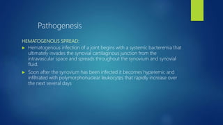
SlideShare a Scribd company logo
SEPTIC ARTHRITIS
BY DR. PONNILAVAN
DEFINITION
Acute septic
arthritis results
from bacterial
invasion of a
joint space
Hematogenous spread
Direct inoculation
from trauma or
surgery
contiguous spread from an
adjacent site of
osteomyelitis or cellulitis
RISK FACTORS
RA,
OA,
hemophilia
a prosthetic joint,
low socioeconomic status,
intravenous drug abuse,
Cancer,
alcoholism,
diabetes,
uremia,
cirrhosis.
previous intra-articular
corticosteroid injection, &
cutaneous ulcers.
CLINICAL PRESENTATION
Common in extremes of ages, can occur at any age group. Most freq in adults.
most serious sequelae from infection occur in children, especially if a hip joint is
involved & Rx has been delayed.
Lower extremity weight-bearing joints - predominantly affected (61% to 79%);
however, any joint can be involved, & multiple joint infections do occur.
It can be difficult to diagnose in neonates bcoz the inflammatory response is
blunted.
The only finding in a neonate may be infection at another site (e.g., the umbilical
catheter), irritability, failure to thrive, asymmetry of limb position, or displeasure at
being handled.
CLINICAL FACTOR ORGANISM
PATIENT AGE
Neonate Staphylococcus aureus
<2 yr Haemophilus influenzae, S. aureus
>2 yr S. aureus
Young adults (healthy, sexually active) Neisseria gonorrhoeae
Elderly adults S. aureus (50%), streptococci, gram-negative bacilli
STRUCTURAL ABNORMALITIES
Aspiration or injection S. aureus
Trauma Gram-negative bacilli, anaerobes, S. aureus
PROSTHESIS
Early infection S. epidermidis
Late infection Gram-positive cocci, anaerobes
MEDICAL CONDITIONS
Injecting drug use Atypical gram-negative bacilli (e.g., Pseudomonas species)
Rheumatoid arthritis S. aureus
Systemic lupus erythematosus,
sickle cell anemia
Salmonella species
Hemophilia S. aureus (50%), streptococci, gram-negative bacilli
Immunosuppression S. aureus, Mycobacterium species, fungi
HEMATOGENOUS SPREAD:
 Hematogenous infection of a joint begins with a systemic bacteremia that
ultimately invades the synovial cartilaginous junction from the
intravascular space and spreads throughout the synovium and synovial
fluid.
 Soon after the synovium has been infected it becomes hyperemic and
infiltrated with polymorphonuclear leukocytes that rapidly increase over
the next several days
Pathogenesis
 Destruction of the articular cartilage, which results from degradation of
ground substance, is apparent 4 to 6 days after infection.
 Complete destruction of articular cartilage occurs at approximately 4
weeks. Joint dislocation or subluxation and osteomyelitis also may occur.
IMAGING STUDIES - XRAYS
 In the first few days of infection, radiographs usually are normal; however,
they may be helpful in that they may show soft tissue swelling,
displacement of the fat pad, or joint space widening from localized edema.
As the infection progresses, joint space narrowing from the destruction of
cartilage may become evident.
 used to monitor the response to treatment
IMAGING – USG –
detect even small collections of fluid deep in the joints.
Non–echo-free effusions from clotted hemorrhagic collections are characteristic
of a septic joint
used to guide initial joint aspiration and drainage and to monitor the status of
intraarticular compartments
Radionuclide bone scans often can detect localized areas of inflammation
CT, MRI, and bone scans also may be obtained to diagnose septic arthritis;
however, these tests are not always necessary.
 If SEPTIC ARTHITIS is suspected, aspiration with a large-bore needle should be done
before antibiotic therapy is initiated.
 fluid obtained should be sent for immediate Gram staining, culture, cell counts, and
crystal analysis.
 Measuring ESR or CRP helps in following the treatment course.
 Typically, synovial leukocyte counts greater than 50,000/
 leukocyte counts < 28,000/mm3 , especially in immunocompromised patients.
 polymorphonuclear neutrophils > 90%, indicates infection.
 Cultures can be negative in up to 75% of patients with septic arthritis.
 Rx in children should be aggressive whether or not a causative organism is identified.
 Empirical antibiotic treatment is based on the patient’s age and risk factors
.
 Empirical antibiotic therapy should be given until culture and sensitivity
results are available, at which time definitive treatment is initiated .
 If no organism is isolated, empirical therapy should be continued.
TREATMENT DURATION
Infections caused by H. influenzae type b, Neisseria, or Streptococcus generally respond
rapidly to appropriate antibacterial management, and duration of therapy <2 weeks
staphylococci and gram-negative bacilli needs 4 to 6 weeks of treatment.
A longer period of therapy is required if the hip or shoulder is involved, if the patient is
immunocompromised, or if the response to treatment has been poor.
Observe for decrease in pain, swelling, temperature & for improved joint mobility.
Open surgical drainage – If response is not favorable & repeat aspiration does not show a decrease in the
synovial leukocyte count within 24 to 48 hours,.
If purulent material is deeply situated in a joint, such as the shoulder or hip, open surgical drainage should be
done. Arthroscopic drainage is a good alternative to open drainage in many instances, especially for
infections involving the knee, elbow, shoulder, or ankle.
As the infection resolves, therapy to restore normal joint function is begun, including functional splinting
initially to prevent deformity, isometric muscle strengthening, and active ROM exercises.
TARSAL JOINTS
Primary septic arthritis of the
tarsal joints is rare. An
uncontrolled infection in the
tarsal joints requires wide
surgical drainage.
ANKLE
ASPIRATION
Swelling around the ankle often makes fluctuation difficult
to locate. To avoid injuring important structures, the
needle is inserted 2.5 cm proximal and 1.3 cm anterior to
the tip of the lateral malleolus. This is just lateral to the
peroneus tertius tendon
DRAINAGE
The ankle may be drained through any of the following
approaches: anterolateral, anteromedial, posterolateral,
and posteromedial
KNEE
The knee joint is the most frequently affected.
ASPIRATION
Because the knee is a superficial joint, it can be aspirated
easily. The needle is inserted on the lateral side at the
level of the superior pole of the patella. It is advanced
through the lateral retinaculum and into the joint
DRAINAGE
In acute septic arthritis, usually anteromedial arthrotomy or arthroscopic
drainage & antibiotic Rx are adequate.
In more difficult cases, the following approaches may be used.
If the posterior compartment of the knee is distended,& a popliteal
is well established, parallel anterior incisions combined with posterolateral
and posteromedial (Henderson) incisions usually are best.
If possible, posterior drainage should be avoided because infection may
spread through the fascial planes of thigh & leg.
When fluctuation indicates a pocket of pus in posterior compartment of
joint that has not been or that cannot be drained effectively through
Henderson incisions, posterior drainage is necessary.
The posterior compartment may be divided by a median septum into medial
and lateral compartments. These may be drained effectively by the Klein or
Kelikian approach .
A posterior midline approach should not be used to drain an infected knee
because it exposes the popliteal vessels to pus and to pressure from the drain
and creates a potentially contracting scar across the joint.
ARTHROSCOPIC
DRAINAGE
Preferred treatment for acute septic arthritis of the knee in
adults.
avoids the disadvantages of needle aspiration and arthrotomy.
ADVANTAGES :
-purulent material can be removed & joint can be irrigated.
- Joint cartilage can be inspected, & loculations or adhesions can
be removed with the arthroscope.
-Partial synovectomy can be performed if necessary.
-Drains can be placed into the joint through the portal sites for
drainage or for a continuous suction drainage system.
-Arthroscopy also has the advantage of allowing much earlier
range of motion & rehabilitation of knee joint compared with
arthrotomy.
Arthroscopic irrigation of septic knee. A, Cannula is
inserted in suprapatellar pouch for outflow, and knee is
irrigated through arthroscopic sheath.
B, Small suction drain is inserted through arthroscopic
sheath.
C, Sheath is removed as drain is held in place. SEE
HIP
 Acute septic arthritis of hip is a more serious disease in children than in
adults, & severe complications are much more common in children.
 Infection begins first in metaphysis or epiphysis and is carried into the
joint.
 Septic hip places the femoral head at high risk for osteonecrosis.
 After an infected hip in an infant or child has been surgically drained, the
hip should be supported in abduction to reduce the risk of pathological
dislocation.
 B/L septic arthritis is seen more often in the hip than in other joints and
occasionally is associated with spinal infection.
ASPIRATION & DRAINAGE OF HIP
A lateral, anterior, or medial approach can be used to aspirate the hip joint.
Drainage of the hip may be accomplished through a posterior, medial,
lateral, or anterior approach.
The anterior approach is preferred in small children for several reasons: (1)
damage to the major blood supply to the femoral head is avoided, (2) the
chance of postoperative dislocation is reduced, and (3) the landmarks for the
surgical approach are much clearer in a small child.
In an adult, the posterior approach allows dependent drainage and is a
more familiar approach for most orthopaedic surgeons.
COMPLICATIONS OF ACUTE SEPTIC
ARTHRITIS OF THE HIP
 PATHOLOGICAL DISLOCATION
 OSTEOMYELITIS
 PELVIC ABSCESS
 PERSISTENT INFECTION
PATHOLOGICAL DISLOCATION
 predominantly in children.
 When dislocation is recognized before severe contracture of the soft tissue
has occurred, reduction is accomplished easily at the time of drainage and
satisfactory function may result (Fig. 22-8).
 If the femoral head has been damaged by the infection, however, skeletal
traction should be applied through the distal femur and continued until
the femoral head is at the level of the acetabulum.
 The dislocation is reduced by abduction and gentle rotation; manipulation
before the femur is displaced distally should not be attempted because the
femoral head or neck may be fractured.
 After the dislocation has been reduced, the hip is immobilized in a spica
cast until it is stable or until fibrous or bony ankylosis develops.
Appearance of hip 21 years after pathological dislocation
and treatment of septic arthritis. Joint space is slightly
narrowed, and acetabulum and femoral head are slightly
incongrous
Pathological dislocation of hip. A,
Early sequestration of epiphysis,
as shown by apparent increase
in density. B, After 16 months,
note absorption of epiphysis and
part of neck.
OSTEOMYELITIS
 When the infection is confined to the joint, prompt drainage and appropriate antibiotic
therapy should prevent osteomyelitis of the proximal femur.
 If osteomyelitis results in sequestration of the femoral head in children younger than 12
years old, however, the head may be totally reabsorbed (Fig. 22-9), or it may be replaced
by new bone after its circulation is restored.
 In older children and adults, it usually remains as an infected sequestrum and requires
excision.
 Any of the approaches described for draining the hip may be used, but the anterior
approach gives better exposure of the joint.
 In a child, osteomyelitis of the ilium may complicate acute septic arthritis of the hip or
the joint infection may be secondary to infection in the bone; in each case, the hip joint
and ilium require drainage.
 In adults, osteomyelitis of the ilium is a less common complication, but impairment of
the circulation of the femoral head may lead to pathological fracture of the neck and
sequestration of the femoral head.
PELVIC ABSCESS
 caused by suppurative infection of the iliac lymph nodes or by spread from the joint into the
sheath of the iliopsoas, which may communicate with the joint.
 The abscess is retroperitoneal and tends to gravitate along the iliopsoas muscle beneath the
inguinal ligament, eventually pointing in the medial thigh.
 In large abscesses, the pus may track proximally along the iliopsoas and point proximal to
the posterior iliac crest.
 MRI may help locate and determine the true extent of psoas involvement.
 Often this can be drained by CT-directed aspiration.
 Freiberg and Perlman advise draining pelvic abscesses as follows. When the abscess points
to the medial thigh, a Ludloff incision is made (see Fig. 1-79). By blunt dissection between
the adductor longus and brevis muscles, the abscess is found anterior or posterior to the
pectineus muscle. When the abscess points subcutaneously anterior to the pectineus, the
incision may be made directly over it, but care must be taken to avoid injuring the femoral
vessels and nerve. Drainage above the inguinal ligament is not advised because a fecal
fistula may result, and the abscess cannot be thoroughly evacuated. If the abscess points
proximal to the iliac crest posteriorly, the incision is made parallel with the crest and just
PERSISTENT INFECTION
 rare but difficult to treat.
 Usually, scarring is extensive, and draining sinuses have become established.
 Often the sinuses become blocked, causing recurring abscesses.
 Unless aggressive surgery is performed, chronic sepsis and its sequelae result.
 Girdlestone described a radical operation for chronic pyogenic infection
around the hip. In this operation, the infected area around the hip is almost
completely saucerized. In addition to resecting all of the infected bone, a mass
of muscle is resected to ensure drainage. This operation may result in a nearly
useless pseudarthrosis or ankylosis. Marked shortening of the affected
extremity results. For these reasons, this operation is a last resort. Before the
operation, sinograms should be made and appropriate antibiotic therapy
should be started. Adequate amounts of blood should be available during
surgery.
Klein et al. described a technique for the treatment of chronic sepsis of the
hip in paraplegic patients;
it consists of three separate measures to control the infection:
(1) a Girdlestone procedure,
(2) transposition of the vastus lateralis muscle into the void left by the
removal of the femoral head and neck and acetabular wall, and
(3) external fixation to prevent unrestrained motion of the femoral shaft that
might damage the transposed muscle. The external fixator spans the hip joint
with a posterior pelvic-femoral frame.
SACROILIAC JOINT
 uncommon but not rare
 Patients have buttock pain and commonly also have low back, thigh, and
abdominal pain
 Diagnosis: Routine radiographs usually are normal. the most sensitive
diagnostic study is CT
 Treatment: Most of the reported patients responded well to appropriate
antibiotic treatment. Patients who develop an abscess require open
drainage.
 Complication: Osteomyelitis of the adjacent sacrum or ilium is a common
complication
STERNOCLAVICULAR AND
ACROMIOCLAVICULAR JOINTS
 affected only when acute septic arthritis involves other joints or in heroin
addicts, in whom the causative organism now is predominantly S. aureus.
 Because these joints are subcutaneous, aspiration and surgical drainage
can be accomplished.
 Occasionally, a portion of the clavicle needs to be excised for associated
osteomyelitis.
SHOULDER
 in children it occurs as a complication of osteomyelitis of the proximal
humeral metaphysis
 in adults, it usually is associated with a debilitating disease and rarely
responds well to treatment.
 The joint should be aspirated whenever an infection is suspected, and early
surgical drainage is indicated if frank pus is obtained.
 CT or MRI can be helpful in determining if an abscess is present.
ASPIRATION
The shoulder may be aspirated anteriorly, posteriorly, or laterally.
Because the fluctuant area usually is palpable anteriorly, and the
bony landmarks can be identified more easily , the needle is
inserted here most often. The aspiration site is located half the
distance between the coracoid process and the anterolateral
edge of the acromion. The needle is directed posteriorly through
the joint capsule, and the joint is aspirated.
DRAINAGE
 anterior incision or a posterior incision, but the anterior incision is
preferable.
 In a review of adult patients treated for septic arthritis of the shoulder,
Leslie et al. found that arthrotomy yielded better results than repeat
aspirations.
ARTHROSCOPIC DRAINAGE of
shoulder
 is being seen in increasing frequency for the treatment of acute septic
arthritis of the shoulder.
 With arthroscopy, washout is done under direct vision, ensuring complete
removal of purulent material.
 Loculations and adhesions can be débrided, and a synovectomy can be
done. Drains can be placed through portal sites if needed.
 Early active mobilization may be initiated sooner than with an open
technique.
ELBOW
The elbow is best drained through a medial or lateral
approach or both.
ASPIRATION
 For elbow aspiration, the physician flexes the elbow
and inserts the needle on its posterior aspect just
lateral to the olecranon. The needle is advanced
through the skin and joint capsule, and the joint is
aspirated.
WRIST
 usually occurs only after penetrating trauma.
 Signs may be subtle, and the diagnosis is easily missed.
 Early incision and drainage should be performed to avoid the
complications of:
joint ankylosis, periarticular osteomyelitis, or suppurative flexor tenosynovitis.
ASPIRATION OF WRIST
 The most common site of aspiration is between
the first and second extensor compartments at
the radiocarpal level, immediately adjacent to the
point where the extensor pollicis longus crosses
the extensor carpi radialis longus. Other aspiration
sites are between the third and fourth extensor
compartments or between the fourth and fifth
extensor compartments
DRAINAGE
 can be drained by a medial, lateral, or posterior approach.
 Avoid opening the tendon sheaths.
OPERATIONS TO CORRECT
DEFORMITIES AFTER SEPTIC
ARTHRITISANKLE
When the ankle is fixed in equinus by soft tissue contracture, treatment by
Quengel casting or serial wedged casts or by operations such as
lengthening of the Achilles tendon with or without posterior capsulotomy
generally are effective in restoring plantigrade position of the foot.
When fixed equinus is caused by bony ankylosis, cuneiform osteotomy
through the joint is indicated.
KNEE
A flexion deformity can be corrected indirectly by a supracondylar
osteotomy that causes a compensatory deformity in the opposite direction.
This operation should be considered when the flexion deformity is not
severe but the joint is unsuitable for manipulation or soft tissue release. In
children, the osteotomy should be made well proximal to the physis.
TRANSVERSE SUPRACONDYLAR
OSTEOTOMY OF THE FEMUR
Supracondylar osteotomy for
ankylosis of knee in flexion.
Transverse osteotomy. line
indicates the femur after correction
Thompson telescoping-V osteotomy
Ankylosis of the knee in
flexion may be corrected
by the V-osteotomy
described by Thompson
pos
CUNIEFORM OSTEOTOMY
Cuneiform osteotomy based anteriorly.
Section of bone removed is indicated by
blue area
Modification of Osgood
supracondylar controlled
rotation osteotomy of
femur. A, Blue area
illustrates section of bone
to be removed. B, After
osteotomy, corrected
position is maintained by
blade plate
HIP
Harmon
reconstruction for
loss of femoral
head and neck in
child as result of
acute infectious
arthritis. Period of
growth and of
weight bearing
produces
substantial neck
and trochanter
Trochanteric
osteotomy. A, Gant
opening wedge
osteotomy fixed by
blade plate. B,
Whitman closing
wedge osteotomy. C,
Brackett ball-and-
socket osteotomy
fixed by Blount blade
plate.
Conclusion
 The principles in the management of acute septic arthritis include (1)
adequate drainage of the joint,
 (2) antibiotics to diminish the systemic effects of sepsis, and
 (3) resting the joint in a stable position. Prompt drainage and evaluation
of purulent joint fluid is crucial for preservation of articular cartilage and
for resolution of the infection.
Source- Campbell chapter 22
THANK U

More Related Content

What's hot

Septic arthritis
Septic arthritisSeptic arthritis
Septic arthritis
MOHAMMED ROSHEN
 
SIngh Index.pptx
SIngh Index.pptxSIngh Index.pptx
SIngh Index.pptx
DavinCaturputraSetia1
 
Septic arthritis
Septic arthritisSeptic arthritis
Septic arthritis
DrSagarTp
 
8. septic arthritis 30th dec 14
8. septic arthritis 30th dec 148. septic arthritis 30th dec 14
8. septic arthritis 30th dec 14
Pawan KB Agrawal
 
Septic arthritis
Septic arthritisSeptic arthritis
Septic arthritisgroup7usmkk
 
Septicarthritis
Septicarthritis Septicarthritis
Septicarthritis
M A Roshan Zameer
 
Septic arthritis in children
Septic arthritis in childrenSeptic arthritis in children
Septic arthritis in children
rangaraya medical college
 
PS SESSION : EXAMINATION OF HIP
PS SESSION : EXAMINATION OF HIPPS SESSION : EXAMINATION OF HIP
PS SESSION : EXAMINATION OF HIP
GMCA Block 4.4 @ KFU
 
Carries sicca
Carries siccaCarries sicca
Carries sicca
PratikDhabalia
 
Synovium & crystal synovitis
Synovium & crystal synovitisSynovium & crystal synovitis
Synovium & crystal synovitisorthoprince
 
Hip dislocation
Hip dislocationHip dislocation
Hip dislocation
SCGH ED CME
 
Non Union
Non UnionNon Union
Non Union
Abdullah Mamun
 
Intertrochanteric fracture
Intertrochanteric fractureIntertrochanteric fracture
Intertrochanteric fracture
MONTHER ALKHAWLANY
 
Acute and Chronic Osteomyelitis - Infection of Bone
Acute and Chronic Osteomyelitis - Infection of BoneAcute and Chronic Osteomyelitis - Infection of Bone
Acute and Chronic Osteomyelitis - Infection of Bone
Rahul Singh
 
Infected non union
Infected non unionInfected non union
Infected non union
Sagar Tomar
 
Perthes ’ disease
Perthes ’ diseasePerthes ’ disease
Perthes ’ disease
Mannan Ahmed
 
Hip dislocation class
Hip dislocation classHip dislocation class
Hip dislocation class
DrHarpreet Bhatia
 
Pilon fractures
Pilon fracturesPilon fractures
Acute osteomyelitis
Acute osteomyelitisAcute osteomyelitis
Acute osteomyelitis
hanisahwarrior
 

What's hot (20)

Septic arthritis
Septic arthritisSeptic arthritis
Septic arthritis
 
SIngh Index.pptx
SIngh Index.pptxSIngh Index.pptx
SIngh Index.pptx
 
Septic arthritis
Septic arthritisSeptic arthritis
Septic arthritis
 
8. septic arthritis 30th dec 14
8. septic arthritis 30th dec 148. septic arthritis 30th dec 14
8. septic arthritis 30th dec 14
 
Septic arthritis
Septic arthritisSeptic arthritis
Septic arthritis
 
Septicarthritis
Septicarthritis Septicarthritis
Septicarthritis
 
Septic arthritis in children
Septic arthritis in childrenSeptic arthritis in children
Septic arthritis in children
 
PS SESSION : EXAMINATION OF HIP
PS SESSION : EXAMINATION OF HIPPS SESSION : EXAMINATION OF HIP
PS SESSION : EXAMINATION OF HIP
 
Carries sicca
Carries siccaCarries sicca
Carries sicca
 
Synovium & crystal synovitis
Synovium & crystal synovitisSynovium & crystal synovitis
Synovium & crystal synovitis
 
Hip dislocation
Hip dislocationHip dislocation
Hip dislocation
 
Non Union
Non UnionNon Union
Non Union
 
Intertrochanteric fracture
Intertrochanteric fractureIntertrochanteric fracture
Intertrochanteric fracture
 
Acute and Chronic Osteomyelitis - Infection of Bone
Acute and Chronic Osteomyelitis - Infection of BoneAcute and Chronic Osteomyelitis - Infection of Bone
Acute and Chronic Osteomyelitis - Infection of Bone
 
Infected non union
Infected non unionInfected non union
Infected non union
 
Perthes ’ disease
Perthes ’ diseasePerthes ’ disease
Perthes ’ disease
 
Hip dislocation class
Hip dislocation classHip dislocation class
Hip dislocation class
 
Pilon fractures
Pilon fracturesPilon fractures
Pilon fractures
 
Non union
Non unionNon union
Non union
 
Acute osteomyelitis
Acute osteomyelitisAcute osteomyelitis
Acute osteomyelitis
 

Similar to Septic arthritis

Septic Arthritis.pptxbbbbbbbbbbbbbbbbbbbbbbbbbbbbb
Septic Arthritis.pptxbbbbbbbbbbbbbbbbbbbbbbbbbbbbbSeptic Arthritis.pptxbbbbbbbbbbbbbbbbbbbbbbbbbbbbb
Septic Arthritis.pptxbbbbbbbbbbbbbbbbbbbbbbbbbbbbb
mekuriatadesse
 
TB shoulder,elbow,wrist,short tubular bones(fingers,toes)
TB shoulder,elbow,wrist,short tubular bones(fingers,toes)TB shoulder,elbow,wrist,short tubular bones(fingers,toes)
TB shoulder,elbow,wrist,short tubular bones(fingers,toes)
Dr. Punit Gaurav
 
Acute pyogenic arthritis by dr ashutosh
Acute pyogenic arthritis by dr ashutoshAcute pyogenic arthritis by dr ashutosh
Acute pyogenic arthritis by dr ashutosh
Ashutosh Kumar
 
Paediatric musculoskeletal infections
Paediatric musculoskeletal infectionsPaediatric musculoskeletal infections
Paediatric musculoskeletal infections
RITESHJAISWAL57
 
Orthopedics 5th year, 3rd lecture (Dr. Omar Barawi)
Orthopedics 5th year, 3rd lecture (Dr. Omar Barawi)Orthopedics 5th year, 3rd lecture (Dr. Omar Barawi)
Orthopedics 5th year, 3rd lecture (Dr. Omar Barawi)
College of Medicine, Sulaymaniyah
 
Septic arthritis dr arsalan akbar
Septic arthritis dr arsalan akbarSeptic arthritis dr arsalan akbar
Septic arthritis dr arsalan akbar
SyedarsalanAkbarG
 
septicarthritis-140504005810-phpapp01.pdf
septicarthritis-140504005810-phpapp01.pdfsepticarthritis-140504005810-phpapp01.pdf
septicarthritis-140504005810-phpapp01.pdf
KeyaArere
 
SEPTIC ARTHRITIS.pptx
SEPTIC ARTHRITIS.pptxSEPTIC ARTHRITIS.pptx
SEPTIC ARTHRITIS.pptx
Ramya569989
 
Spleenectomy
Spleenectomy Spleenectomy
Spleenectomy
zohaib nadeem
 
Complications of total hip replacement final
Complications of total hip replacement finalComplications of total hip replacement final
Complications of total hip replacement final
Humayun Israr
 
spine surgical approaches along with tb spine complications
 spine surgical approaches along with tb spine complications spine surgical approaches along with tb spine complications
spine surgical approaches along with tb spine complications
Pramod Yspam
 
Osteomyelitis
OsteomyelitisOsteomyelitis
Osteomyelitis
nazirkhankmc
 
Septic arthritis dr arsalan akbar
Septic arthritis dr arsalan akbarSeptic arthritis dr arsalan akbar
Septic arthritis dr arsalan akbar
SyedarsalanAkbarG
 
Spinal infection
Spinal infectionSpinal infection
Spinal infectionorthoprince
 
Tb hip knee shoulder dactylitis
Tb hip knee shoulder dactylitisTb hip knee shoulder dactylitis
Tb hip knee shoulder dactylitis
Dr venkatesh v
 
NECROTISING FASCIITIS- The flesh eating infection
NECROTISING FASCIITIS- The flesh eating infectionNECROTISING FASCIITIS- The flesh eating infection
NECROTISING FASCIITIS- The flesh eating infection
Selvaraj Balasubramani
 
meckels, sigmoid, intussuception.pptx
meckels, sigmoid, intussuception.pptxmeckels, sigmoid, intussuception.pptx
meckels, sigmoid, intussuception.pptx
ssuser269c09
 
Complications of TAPP
Complications of TAPPComplications of TAPP
Complications of TAPP
Dr-Maryam Khan
 
Kspspot final
Kspspot finalKspspot final
Kspspot final
Kumaragoud Patil
 

Similar to Septic arthritis (20)

Septic Arthritis.pptxbbbbbbbbbbbbbbbbbbbbbbbbbbbbb
Septic Arthritis.pptxbbbbbbbbbbbbbbbbbbbbbbbbbbbbbSeptic Arthritis.pptxbbbbbbbbbbbbbbbbbbbbbbbbbbbbb
Septic Arthritis.pptxbbbbbbbbbbbbbbbbbbbbbbbbbbbbb
 
TB shoulder,elbow,wrist,short tubular bones(fingers,toes)
TB shoulder,elbow,wrist,short tubular bones(fingers,toes)TB shoulder,elbow,wrist,short tubular bones(fingers,toes)
TB shoulder,elbow,wrist,short tubular bones(fingers,toes)
 
Acute pyogenic arthritis by dr ashutosh
Acute pyogenic arthritis by dr ashutoshAcute pyogenic arthritis by dr ashutosh
Acute pyogenic arthritis by dr ashutosh
 
Paediatric musculoskeletal infections
Paediatric musculoskeletal infectionsPaediatric musculoskeletal infections
Paediatric musculoskeletal infections
 
Orthopedics 5th year, 3rd lecture (Dr. Omar Barawi)
Orthopedics 5th year, 3rd lecture (Dr. Omar Barawi)Orthopedics 5th year, 3rd lecture (Dr. Omar Barawi)
Orthopedics 5th year, 3rd lecture (Dr. Omar Barawi)
 
Septic arthritis dr arsalan akbar
Septic arthritis dr arsalan akbarSeptic arthritis dr arsalan akbar
Septic arthritis dr arsalan akbar
 
septicarthritis-140504005810-phpapp01.pdf
septicarthritis-140504005810-phpapp01.pdfsepticarthritis-140504005810-phpapp01.pdf
septicarthritis-140504005810-phpapp01.pdf
 
Osteomyelitis
OsteomyelitisOsteomyelitis
Osteomyelitis
 
SEPTIC ARTHRITIS.pptx
SEPTIC ARTHRITIS.pptxSEPTIC ARTHRITIS.pptx
SEPTIC ARTHRITIS.pptx
 
Spleenectomy
Spleenectomy Spleenectomy
Spleenectomy
 
Complications of total hip replacement final
Complications of total hip replacement finalComplications of total hip replacement final
Complications of total hip replacement final
 
spine surgical approaches along with tb spine complications
 spine surgical approaches along with tb spine complications spine surgical approaches along with tb spine complications
spine surgical approaches along with tb spine complications
 
Osteomyelitis
OsteomyelitisOsteomyelitis
Osteomyelitis
 
Septic arthritis dr arsalan akbar
Septic arthritis dr arsalan akbarSeptic arthritis dr arsalan akbar
Septic arthritis dr arsalan akbar
 
Spinal infection
Spinal infectionSpinal infection
Spinal infection
 
Tb hip knee shoulder dactylitis
Tb hip knee shoulder dactylitisTb hip knee shoulder dactylitis
Tb hip knee shoulder dactylitis
 
NECROTISING FASCIITIS- The flesh eating infection
NECROTISING FASCIITIS- The flesh eating infectionNECROTISING FASCIITIS- The flesh eating infection
NECROTISING FASCIITIS- The flesh eating infection
 
meckels, sigmoid, intussuception.pptx
meckels, sigmoid, intussuception.pptxmeckels, sigmoid, intussuception.pptx
meckels, sigmoid, intussuception.pptx
 
Complications of TAPP
Complications of TAPPComplications of TAPP
Complications of TAPP
 
Kspspot final
Kspspot finalKspspot final
Kspspot final
 

More from Ponnilavan Ponz

Cubitus varus and valgus
Cubitus varus and valgusCubitus varus and valgus
Cubitus varus and valgus
Ponnilavan Ponz
 
Rickets
RicketsRickets
Meniscal injury
Meniscal injury Meniscal injury
Meniscal injury
Ponnilavan Ponz
 
Poliomyelitis
PoliomyelitisPoliomyelitis
Poliomyelitis
Ponnilavan Ponz
 
Anatomy of cervical spine
Anatomy of cervical spineAnatomy of cervical spine
Anatomy of cervical spine
Ponnilavan Ponz
 
Congenital pseudoarthrosis of tibia
Congenital pseudoarthrosis of tibiaCongenital pseudoarthrosis of tibia
Congenital pseudoarthrosis of tibia
Ponnilavan Ponz
 
screws and plate
screws and platescrews and plate
screws and plate
Ponnilavan Ponz
 
Distal femoral fresh osteochondral allografts
Distal femoral fresh osteochondral allograftsDistal femoral fresh osteochondral allografts
Distal femoral fresh osteochondral allografts
Ponnilavan Ponz
 
External fixation
External fixation External fixation
External fixation
Ponnilavan Ponz
 
Im nail
Im nailIm nail
Krukenberg surgery
Krukenberg surgeryKrukenberg surgery
Krukenberg surgery
Ponnilavan Ponz
 
Patellofemoral disorders
Patellofemoral disordersPatellofemoral disorders
Patellofemoral disorders
Ponnilavan Ponz
 
Osteochondroma
OsteochondromaOsteochondroma
Osteochondroma
Ponnilavan Ponz
 
Dupuytren
Dupuytren   Dupuytren
Dupuytren
Ponnilavan Ponz
 
Chopart amputation
Chopart amputationChopart amputation
Chopart amputation
Ponnilavan Ponz
 
Acl reconstruction
Acl reconstructionAcl reconstruction
Acl reconstruction
Ponnilavan Ponz
 
Bladder innervation
Bladder innervationBladder innervation
Bladder innervation
Ponnilavan Ponz
 
maduramycosis
maduramycosis   maduramycosis
maduramycosis
Ponnilavan Ponz
 
Adult acquired flat foot deformity
Adult acquired flat foot deformityAdult acquired flat foot deformity
Adult acquired flat foot deformity
Ponnilavan Ponz
 

More from Ponnilavan Ponz (20)

Cubitus varus and valgus
Cubitus varus and valgusCubitus varus and valgus
Cubitus varus and valgus
 
Rickets
RicketsRickets
Rickets
 
Meniscal injury
Meniscal injury Meniscal injury
Meniscal injury
 
Poliomyelitis
PoliomyelitisPoliomyelitis
Poliomyelitis
 
Anatomy of cervical spine
Anatomy of cervical spineAnatomy of cervical spine
Anatomy of cervical spine
 
Congenital pseudoarthrosis of tibia
Congenital pseudoarthrosis of tibiaCongenital pseudoarthrosis of tibia
Congenital pseudoarthrosis of tibia
 
screws and plate
screws and platescrews and plate
screws and plate
 
Distal femoral fresh osteochondral allografts
Distal femoral fresh osteochondral allograftsDistal femoral fresh osteochondral allografts
Distal femoral fresh osteochondral allografts
 
External fixation
External fixation External fixation
External fixation
 
Im nail
Im nailIm nail
Im nail
 
Krukenberg surgery
Krukenberg surgeryKrukenberg surgery
Krukenberg surgery
 
Patellofemoral disorders
Patellofemoral disordersPatellofemoral disorders
Patellofemoral disorders
 
Avn
AvnAvn
Avn
 
Osteochondroma
OsteochondromaOsteochondroma
Osteochondroma
 
Dupuytren
Dupuytren   Dupuytren
Dupuytren
 
Chopart amputation
Chopart amputationChopart amputation
Chopart amputation
 
Acl reconstruction
Acl reconstructionAcl reconstruction
Acl reconstruction
 
Bladder innervation
Bladder innervationBladder innervation
Bladder innervation
 
maduramycosis
maduramycosis   maduramycosis
maduramycosis
 
Adult acquired flat foot deformity
Adult acquired flat foot deformityAdult acquired flat foot deformity
Adult acquired flat foot deformity
 

Recently uploaded

How libraries can support authors with open access requirements for UKRI fund...
How libraries can support authors with open access requirements for UKRI fund...How libraries can support authors with open access requirements for UKRI fund...
How libraries can support authors with open access requirements for UKRI fund...
Jisc
 
GIÁO ÁN DẠY THÊM (KẾ HOẠCH BÀI BUỔI 2) - TIẾNG ANH 8 GLOBAL SUCCESS (2 CỘT) N...
GIÁO ÁN DẠY THÊM (KẾ HOẠCH BÀI BUỔI 2) - TIẾNG ANH 8 GLOBAL SUCCESS (2 CỘT) N...GIÁO ÁN DẠY THÊM (KẾ HOẠCH BÀI BUỔI 2) - TIẾNG ANH 8 GLOBAL SUCCESS (2 CỘT) N...
GIÁO ÁN DẠY THÊM (KẾ HOẠCH BÀI BUỔI 2) - TIẾNG ANH 8 GLOBAL SUCCESS (2 CỘT) N...
Nguyen Thanh Tu Collection
 
Template Jadual Bertugas Kelas (Boleh Edit)
Template Jadual Bertugas Kelas (Boleh Edit)Template Jadual Bertugas Kelas (Boleh Edit)
Template Jadual Bertugas Kelas (Boleh Edit)
rosedainty
 
TESDA TM1 REVIEWER FOR NATIONAL ASSESSMENT WRITTEN AND ORAL QUESTIONS WITH A...
TESDA TM1 REVIEWER  FOR NATIONAL ASSESSMENT WRITTEN AND ORAL QUESTIONS WITH A...TESDA TM1 REVIEWER  FOR NATIONAL ASSESSMENT WRITTEN AND ORAL QUESTIONS WITH A...
TESDA TM1 REVIEWER FOR NATIONAL ASSESSMENT WRITTEN AND ORAL QUESTIONS WITH A...
EugeneSaldivar
 
Thesis Statement for students diagnonsed withADHD.ppt
Thesis Statement for students diagnonsed withADHD.pptThesis Statement for students diagnonsed withADHD.ppt
Thesis Statement for students diagnonsed withADHD.ppt
EverAndrsGuerraGuerr
 
Language Across the Curriculm LAC B.Ed.
Language Across the  Curriculm LAC B.Ed.Language Across the  Curriculm LAC B.Ed.
Language Across the Curriculm LAC B.Ed.
Atul Kumar Singh
 
The Art Pastor's Guide to Sabbath | Steve Thomason
The Art Pastor's Guide to Sabbath | Steve ThomasonThe Art Pastor's Guide to Sabbath | Steve Thomason
The Art Pastor's Guide to Sabbath | Steve Thomason
Steve Thomason
 
Supporting (UKRI) OA monographs at Salford.pptx
Supporting (UKRI) OA monographs at Salford.pptxSupporting (UKRI) OA monographs at Salford.pptx
Supporting (UKRI) OA monographs at Salford.pptx
Jisc
 
How to Break the cycle of negative Thoughts
How to Break the cycle of negative ThoughtsHow to Break the cycle of negative Thoughts
How to Break the cycle of negative Thoughts
Col Mukteshwar Prasad
 
Sha'Carri Richardson Presentation 202345
Sha'Carri Richardson Presentation 202345Sha'Carri Richardson Presentation 202345
Sha'Carri Richardson Presentation 202345
beazzy04
 
Synthetic Fiber Construction in lab .pptx
Synthetic Fiber Construction in lab .pptxSynthetic Fiber Construction in lab .pptx
Synthetic Fiber Construction in lab .pptx
Pavel ( NSTU)
 
Home assignment II on Spectroscopy 2024 Answers.pdf
Home assignment II on Spectroscopy 2024 Answers.pdfHome assignment II on Spectroscopy 2024 Answers.pdf
Home assignment II on Spectroscopy 2024 Answers.pdf
Tamralipta Mahavidyalaya
 
How to Create Map Views in the Odoo 17 ERP
How to Create Map Views in the Odoo 17 ERPHow to Create Map Views in the Odoo 17 ERP
How to Create Map Views in the Odoo 17 ERP
Celine George
 
Model Attribute Check Company Auto Property
Model Attribute  Check Company Auto PropertyModel Attribute  Check Company Auto Property
Model Attribute Check Company Auto Property
Celine George
 
The geography of Taylor Swift - some ideas
The geography of Taylor Swift - some ideasThe geography of Taylor Swift - some ideas
The geography of Taylor Swift - some ideas
GeoBlogs
 
special B.ed 2nd year old paper_20240531.pdf
special B.ed 2nd year old paper_20240531.pdfspecial B.ed 2nd year old paper_20240531.pdf
special B.ed 2nd year old paper_20240531.pdf
Special education needs
 
Introduction to Quality Improvement Essentials
Introduction to Quality Improvement EssentialsIntroduction to Quality Improvement Essentials
Introduction to Quality Improvement Essentials
Excellence Foundation for South Sudan
 
Welcome to TechSoup New Member Orientation and Q&A (May 2024).pdf
Welcome to TechSoup   New Member Orientation and Q&A (May 2024).pdfWelcome to TechSoup   New Member Orientation and Q&A (May 2024).pdf
Welcome to TechSoup New Member Orientation and Q&A (May 2024).pdf
TechSoup
 
How to Make a Field invisible in Odoo 17
How to Make a Field invisible in Odoo 17How to Make a Field invisible in Odoo 17
How to Make a Field invisible in Odoo 17
Celine George
 
Unit 2- Research Aptitude (UGC NET Paper I).pdf
Unit 2- Research Aptitude (UGC NET Paper I).pdfUnit 2- Research Aptitude (UGC NET Paper I).pdf
Unit 2- Research Aptitude (UGC NET Paper I).pdf
Thiyagu K
 

Recently uploaded (20)

How libraries can support authors with open access requirements for UKRI fund...
How libraries can support authors with open access requirements for UKRI fund...How libraries can support authors with open access requirements for UKRI fund...
How libraries can support authors with open access requirements for UKRI fund...
 
GIÁO ÁN DẠY THÊM (KẾ HOẠCH BÀI BUỔI 2) - TIẾNG ANH 8 GLOBAL SUCCESS (2 CỘT) N...
GIÁO ÁN DẠY THÊM (KẾ HOẠCH BÀI BUỔI 2) - TIẾNG ANH 8 GLOBAL SUCCESS (2 CỘT) N...GIÁO ÁN DẠY THÊM (KẾ HOẠCH BÀI BUỔI 2) - TIẾNG ANH 8 GLOBAL SUCCESS (2 CỘT) N...
GIÁO ÁN DẠY THÊM (KẾ HOẠCH BÀI BUỔI 2) - TIẾNG ANH 8 GLOBAL SUCCESS (2 CỘT) N...
 
Template Jadual Bertugas Kelas (Boleh Edit)
Template Jadual Bertugas Kelas (Boleh Edit)Template Jadual Bertugas Kelas (Boleh Edit)
Template Jadual Bertugas Kelas (Boleh Edit)
 
TESDA TM1 REVIEWER FOR NATIONAL ASSESSMENT WRITTEN AND ORAL QUESTIONS WITH A...
TESDA TM1 REVIEWER  FOR NATIONAL ASSESSMENT WRITTEN AND ORAL QUESTIONS WITH A...TESDA TM1 REVIEWER  FOR NATIONAL ASSESSMENT WRITTEN AND ORAL QUESTIONS WITH A...
TESDA TM1 REVIEWER FOR NATIONAL ASSESSMENT WRITTEN AND ORAL QUESTIONS WITH A...
 
Thesis Statement for students diagnonsed withADHD.ppt
Thesis Statement for students diagnonsed withADHD.pptThesis Statement for students diagnonsed withADHD.ppt
Thesis Statement for students diagnonsed withADHD.ppt
 
Language Across the Curriculm LAC B.Ed.
Language Across the  Curriculm LAC B.Ed.Language Across the  Curriculm LAC B.Ed.
Language Across the Curriculm LAC B.Ed.
 
The Art Pastor's Guide to Sabbath | Steve Thomason
The Art Pastor's Guide to Sabbath | Steve ThomasonThe Art Pastor's Guide to Sabbath | Steve Thomason
The Art Pastor's Guide to Sabbath | Steve Thomason
 
Supporting (UKRI) OA monographs at Salford.pptx
Supporting (UKRI) OA monographs at Salford.pptxSupporting (UKRI) OA monographs at Salford.pptx
Supporting (UKRI) OA monographs at Salford.pptx
 
How to Break the cycle of negative Thoughts
How to Break the cycle of negative ThoughtsHow to Break the cycle of negative Thoughts
How to Break the cycle of negative Thoughts
 
Sha'Carri Richardson Presentation 202345
Sha'Carri Richardson Presentation 202345Sha'Carri Richardson Presentation 202345
Sha'Carri Richardson Presentation 202345
 
Synthetic Fiber Construction in lab .pptx
Synthetic Fiber Construction in lab .pptxSynthetic Fiber Construction in lab .pptx
Synthetic Fiber Construction in lab .pptx
 
Home assignment II on Spectroscopy 2024 Answers.pdf
Home assignment II on Spectroscopy 2024 Answers.pdfHome assignment II on Spectroscopy 2024 Answers.pdf
Home assignment II on Spectroscopy 2024 Answers.pdf
 
How to Create Map Views in the Odoo 17 ERP
How to Create Map Views in the Odoo 17 ERPHow to Create Map Views in the Odoo 17 ERP
How to Create Map Views in the Odoo 17 ERP
 
Model Attribute Check Company Auto Property
Model Attribute  Check Company Auto PropertyModel Attribute  Check Company Auto Property
Model Attribute Check Company Auto Property
 
The geography of Taylor Swift - some ideas
The geography of Taylor Swift - some ideasThe geography of Taylor Swift - some ideas
The geography of Taylor Swift - some ideas
 
special B.ed 2nd year old paper_20240531.pdf
special B.ed 2nd year old paper_20240531.pdfspecial B.ed 2nd year old paper_20240531.pdf
special B.ed 2nd year old paper_20240531.pdf
 
Introduction to Quality Improvement Essentials
Introduction to Quality Improvement EssentialsIntroduction to Quality Improvement Essentials
Introduction to Quality Improvement Essentials
 
Welcome to TechSoup New Member Orientation and Q&A (May 2024).pdf
Welcome to TechSoup   New Member Orientation and Q&A (May 2024).pdfWelcome to TechSoup   New Member Orientation and Q&A (May 2024).pdf
Welcome to TechSoup New Member Orientation and Q&A (May 2024).pdf
 
How to Make a Field invisible in Odoo 17
How to Make a Field invisible in Odoo 17How to Make a Field invisible in Odoo 17
How to Make a Field invisible in Odoo 17
 
Unit 2- Research Aptitude (UGC NET Paper I).pdf
Unit 2- Research Aptitude (UGC NET Paper I).pdfUnit 2- Research Aptitude (UGC NET Paper I).pdf
Unit 2- Research Aptitude (UGC NET Paper I).pdf
 

Septic arthritis

  • 2. DEFINITION Acute septic arthritis results from bacterial invasion of a joint space Hematogenous spread Direct inoculation from trauma or surgery contiguous spread from an adjacent site of osteomyelitis or cellulitis
  • 3. RISK FACTORS RA, OA, hemophilia a prosthetic joint, low socioeconomic status, intravenous drug abuse, Cancer, alcoholism, diabetes, uremia, cirrhosis. previous intra-articular corticosteroid injection, & cutaneous ulcers.
  • 4. CLINICAL PRESENTATION Common in extremes of ages, can occur at any age group. Most freq in adults. most serious sequelae from infection occur in children, especially if a hip joint is involved & Rx has been delayed. Lower extremity weight-bearing joints - predominantly affected (61% to 79%); however, any joint can be involved, & multiple joint infections do occur. It can be difficult to diagnose in neonates bcoz the inflammatory response is blunted. The only finding in a neonate may be infection at another site (e.g., the umbilical catheter), irritability, failure to thrive, asymmetry of limb position, or displeasure at being handled.
  • 5. CLINICAL FACTOR ORGANISM PATIENT AGE Neonate Staphylococcus aureus <2 yr Haemophilus influenzae, S. aureus >2 yr S. aureus Young adults (healthy, sexually active) Neisseria gonorrhoeae Elderly adults S. aureus (50%), streptococci, gram-negative bacilli STRUCTURAL ABNORMALITIES Aspiration or injection S. aureus Trauma Gram-negative bacilli, anaerobes, S. aureus PROSTHESIS Early infection S. epidermidis Late infection Gram-positive cocci, anaerobes MEDICAL CONDITIONS Injecting drug use Atypical gram-negative bacilli (e.g., Pseudomonas species) Rheumatoid arthritis S. aureus Systemic lupus erythematosus, sickle cell anemia Salmonella species Hemophilia S. aureus (50%), streptococci, gram-negative bacilli Immunosuppression S. aureus, Mycobacterium species, fungi
  • 6. HEMATOGENOUS SPREAD:  Hematogenous infection of a joint begins with a systemic bacteremia that ultimately invades the synovial cartilaginous junction from the intravascular space and spreads throughout the synovium and synovial fluid.  Soon after the synovium has been infected it becomes hyperemic and infiltrated with polymorphonuclear leukocytes that rapidly increase over the next several days Pathogenesis
  • 7.  Destruction of the articular cartilage, which results from degradation of ground substance, is apparent 4 to 6 days after infection.  Complete destruction of articular cartilage occurs at approximately 4 weeks. Joint dislocation or subluxation and osteomyelitis also may occur.
  • 8. IMAGING STUDIES - XRAYS  In the first few days of infection, radiographs usually are normal; however, they may be helpful in that they may show soft tissue swelling, displacement of the fat pad, or joint space widening from localized edema. As the infection progresses, joint space narrowing from the destruction of cartilage may become evident.  used to monitor the response to treatment
  • 9. IMAGING – USG – detect even small collections of fluid deep in the joints. Non–echo-free effusions from clotted hemorrhagic collections are characteristic of a septic joint used to guide initial joint aspiration and drainage and to monitor the status of intraarticular compartments Radionuclide bone scans often can detect localized areas of inflammation CT, MRI, and bone scans also may be obtained to diagnose septic arthritis; however, these tests are not always necessary.
  • 10.  If SEPTIC ARTHITIS is suspected, aspiration with a large-bore needle should be done before antibiotic therapy is initiated.  fluid obtained should be sent for immediate Gram staining, culture, cell counts, and crystal analysis.  Measuring ESR or CRP helps in following the treatment course.  Typically, synovial leukocyte counts greater than 50,000/  leukocyte counts < 28,000/mm3 , especially in immunocompromised patients.  polymorphonuclear neutrophils > 90%, indicates infection.  Cultures can be negative in up to 75% of patients with septic arthritis.  Rx in children should be aggressive whether or not a causative organism is identified.
  • 11.  Empirical antibiotic treatment is based on the patient’s age and risk factors .  Empirical antibiotic therapy should be given until culture and sensitivity results are available, at which time definitive treatment is initiated .  If no organism is isolated, empirical therapy should be continued.
  • 12. TREATMENT DURATION Infections caused by H. influenzae type b, Neisseria, or Streptococcus generally respond rapidly to appropriate antibacterial management, and duration of therapy <2 weeks staphylococci and gram-negative bacilli needs 4 to 6 weeks of treatment. A longer period of therapy is required if the hip or shoulder is involved, if the patient is immunocompromised, or if the response to treatment has been poor.
  • 13. Observe for decrease in pain, swelling, temperature & for improved joint mobility. Open surgical drainage – If response is not favorable & repeat aspiration does not show a decrease in the synovial leukocyte count within 24 to 48 hours,. If purulent material is deeply situated in a joint, such as the shoulder or hip, open surgical drainage should be done. Arthroscopic drainage is a good alternative to open drainage in many instances, especially for infections involving the knee, elbow, shoulder, or ankle. As the infection resolves, therapy to restore normal joint function is begun, including functional splinting initially to prevent deformity, isometric muscle strengthening, and active ROM exercises.
  • 14. TARSAL JOINTS Primary septic arthritis of the tarsal joints is rare. An uncontrolled infection in the tarsal joints requires wide surgical drainage. ANKLE ASPIRATION Swelling around the ankle often makes fluctuation difficult to locate. To avoid injuring important structures, the needle is inserted 2.5 cm proximal and 1.3 cm anterior to the tip of the lateral malleolus. This is just lateral to the peroneus tertius tendon DRAINAGE The ankle may be drained through any of the following approaches: anterolateral, anteromedial, posterolateral, and posteromedial
  • 15. KNEE The knee joint is the most frequently affected. ASPIRATION Because the knee is a superficial joint, it can be aspirated easily. The needle is inserted on the lateral side at the level of the superior pole of the patella. It is advanced through the lateral retinaculum and into the joint
  • 16. DRAINAGE In acute septic arthritis, usually anteromedial arthrotomy or arthroscopic drainage & antibiotic Rx are adequate. In more difficult cases, the following approaches may be used. If the posterior compartment of the knee is distended,& a popliteal is well established, parallel anterior incisions combined with posterolateral and posteromedial (Henderson) incisions usually are best. If possible, posterior drainage should be avoided because infection may spread through the fascial planes of thigh & leg. When fluctuation indicates a pocket of pus in posterior compartment of joint that has not been or that cannot be drained effectively through Henderson incisions, posterior drainage is necessary.
  • 17. The posterior compartment may be divided by a median septum into medial and lateral compartments. These may be drained effectively by the Klein or Kelikian approach . A posterior midline approach should not be used to drain an infected knee because it exposes the popliteal vessels to pus and to pressure from the drain and creates a potentially contracting scar across the joint.
  • 18. ARTHROSCOPIC DRAINAGE Preferred treatment for acute septic arthritis of the knee in adults. avoids the disadvantages of needle aspiration and arthrotomy. ADVANTAGES : -purulent material can be removed & joint can be irrigated. - Joint cartilage can be inspected, & loculations or adhesions can be removed with the arthroscope. -Partial synovectomy can be performed if necessary. -Drains can be placed into the joint through the portal sites for drainage or for a continuous suction drainage system. -Arthroscopy also has the advantage of allowing much earlier range of motion & rehabilitation of knee joint compared with arthrotomy.
  • 19. Arthroscopic irrigation of septic knee. A, Cannula is inserted in suprapatellar pouch for outflow, and knee is irrigated through arthroscopic sheath. B, Small suction drain is inserted through arthroscopic sheath. C, Sheath is removed as drain is held in place. SEE
  • 20. HIP  Acute septic arthritis of hip is a more serious disease in children than in adults, & severe complications are much more common in children.  Infection begins first in metaphysis or epiphysis and is carried into the joint.  Septic hip places the femoral head at high risk for osteonecrosis.  After an infected hip in an infant or child has been surgically drained, the hip should be supported in abduction to reduce the risk of pathological dislocation.  B/L septic arthritis is seen more often in the hip than in other joints and occasionally is associated with spinal infection.
  • 21. ASPIRATION & DRAINAGE OF HIP A lateral, anterior, or medial approach can be used to aspirate the hip joint. Drainage of the hip may be accomplished through a posterior, medial, lateral, or anterior approach. The anterior approach is preferred in small children for several reasons: (1) damage to the major blood supply to the femoral head is avoided, (2) the chance of postoperative dislocation is reduced, and (3) the landmarks for the surgical approach are much clearer in a small child. In an adult, the posterior approach allows dependent drainage and is a more familiar approach for most orthopaedic surgeons.
  • 22. COMPLICATIONS OF ACUTE SEPTIC ARTHRITIS OF THE HIP  PATHOLOGICAL DISLOCATION  OSTEOMYELITIS  PELVIC ABSCESS  PERSISTENT INFECTION
  • 23. PATHOLOGICAL DISLOCATION  predominantly in children.  When dislocation is recognized before severe contracture of the soft tissue has occurred, reduction is accomplished easily at the time of drainage and satisfactory function may result (Fig. 22-8).  If the femoral head has been damaged by the infection, however, skeletal traction should be applied through the distal femur and continued until the femoral head is at the level of the acetabulum.  The dislocation is reduced by abduction and gentle rotation; manipulation before the femur is displaced distally should not be attempted because the femoral head or neck may be fractured.  After the dislocation has been reduced, the hip is immobilized in a spica cast until it is stable or until fibrous or bony ankylosis develops.
  • 24. Appearance of hip 21 years after pathological dislocation and treatment of septic arthritis. Joint space is slightly narrowed, and acetabulum and femoral head are slightly incongrous
  • 25. Pathological dislocation of hip. A, Early sequestration of epiphysis, as shown by apparent increase in density. B, After 16 months, note absorption of epiphysis and part of neck.
  • 26. OSTEOMYELITIS  When the infection is confined to the joint, prompt drainage and appropriate antibiotic therapy should prevent osteomyelitis of the proximal femur.  If osteomyelitis results in sequestration of the femoral head in children younger than 12 years old, however, the head may be totally reabsorbed (Fig. 22-9), or it may be replaced by new bone after its circulation is restored.  In older children and adults, it usually remains as an infected sequestrum and requires excision.  Any of the approaches described for draining the hip may be used, but the anterior approach gives better exposure of the joint.  In a child, osteomyelitis of the ilium may complicate acute septic arthritis of the hip or the joint infection may be secondary to infection in the bone; in each case, the hip joint and ilium require drainage.  In adults, osteomyelitis of the ilium is a less common complication, but impairment of the circulation of the femoral head may lead to pathological fracture of the neck and sequestration of the femoral head.
  • 27. PELVIC ABSCESS  caused by suppurative infection of the iliac lymph nodes or by spread from the joint into the sheath of the iliopsoas, which may communicate with the joint.  The abscess is retroperitoneal and tends to gravitate along the iliopsoas muscle beneath the inguinal ligament, eventually pointing in the medial thigh.  In large abscesses, the pus may track proximally along the iliopsoas and point proximal to the posterior iliac crest.  MRI may help locate and determine the true extent of psoas involvement.  Often this can be drained by CT-directed aspiration.  Freiberg and Perlman advise draining pelvic abscesses as follows. When the abscess points to the medial thigh, a Ludloff incision is made (see Fig. 1-79). By blunt dissection between the adductor longus and brevis muscles, the abscess is found anterior or posterior to the pectineus muscle. When the abscess points subcutaneously anterior to the pectineus, the incision may be made directly over it, but care must be taken to avoid injuring the femoral vessels and nerve. Drainage above the inguinal ligament is not advised because a fecal fistula may result, and the abscess cannot be thoroughly evacuated. If the abscess points proximal to the iliac crest posteriorly, the incision is made parallel with the crest and just
  • 28. PERSISTENT INFECTION  rare but difficult to treat.  Usually, scarring is extensive, and draining sinuses have become established.  Often the sinuses become blocked, causing recurring abscesses.  Unless aggressive surgery is performed, chronic sepsis and its sequelae result.  Girdlestone described a radical operation for chronic pyogenic infection around the hip. In this operation, the infected area around the hip is almost completely saucerized. In addition to resecting all of the infected bone, a mass of muscle is resected to ensure drainage. This operation may result in a nearly useless pseudarthrosis or ankylosis. Marked shortening of the affected extremity results. For these reasons, this operation is a last resort. Before the operation, sinograms should be made and appropriate antibiotic therapy should be started. Adequate amounts of blood should be available during surgery.
  • 29.
  • 30. Klein et al. described a technique for the treatment of chronic sepsis of the hip in paraplegic patients; it consists of three separate measures to control the infection: (1) a Girdlestone procedure, (2) transposition of the vastus lateralis muscle into the void left by the removal of the femoral head and neck and acetabular wall, and (3) external fixation to prevent unrestrained motion of the femoral shaft that might damage the transposed muscle. The external fixator spans the hip joint with a posterior pelvic-femoral frame.
  • 31. SACROILIAC JOINT  uncommon but not rare  Patients have buttock pain and commonly also have low back, thigh, and abdominal pain  Diagnosis: Routine radiographs usually are normal. the most sensitive diagnostic study is CT  Treatment: Most of the reported patients responded well to appropriate antibiotic treatment. Patients who develop an abscess require open drainage.  Complication: Osteomyelitis of the adjacent sacrum or ilium is a common complication
  • 32. STERNOCLAVICULAR AND ACROMIOCLAVICULAR JOINTS  affected only when acute septic arthritis involves other joints or in heroin addicts, in whom the causative organism now is predominantly S. aureus.  Because these joints are subcutaneous, aspiration and surgical drainage can be accomplished.  Occasionally, a portion of the clavicle needs to be excised for associated osteomyelitis.
  • 33. SHOULDER  in children it occurs as a complication of osteomyelitis of the proximal humeral metaphysis  in adults, it usually is associated with a debilitating disease and rarely responds well to treatment.  The joint should be aspirated whenever an infection is suspected, and early surgical drainage is indicated if frank pus is obtained.  CT or MRI can be helpful in determining if an abscess is present.
  • 34. ASPIRATION The shoulder may be aspirated anteriorly, posteriorly, or laterally. Because the fluctuant area usually is palpable anteriorly, and the bony landmarks can be identified more easily , the needle is inserted here most often. The aspiration site is located half the distance between the coracoid process and the anterolateral edge of the acromion. The needle is directed posteriorly through the joint capsule, and the joint is aspirated.
  • 35. DRAINAGE  anterior incision or a posterior incision, but the anterior incision is preferable.  In a review of adult patients treated for septic arthritis of the shoulder, Leslie et al. found that arthrotomy yielded better results than repeat aspirations.
  • 36. ARTHROSCOPIC DRAINAGE of shoulder  is being seen in increasing frequency for the treatment of acute septic arthritis of the shoulder.  With arthroscopy, washout is done under direct vision, ensuring complete removal of purulent material.  Loculations and adhesions can be débrided, and a synovectomy can be done. Drains can be placed through portal sites if needed.  Early active mobilization may be initiated sooner than with an open technique.
  • 37. ELBOW The elbow is best drained through a medial or lateral approach or both. ASPIRATION  For elbow aspiration, the physician flexes the elbow and inserts the needle on its posterior aspect just lateral to the olecranon. The needle is advanced through the skin and joint capsule, and the joint is aspirated.
  • 38. WRIST  usually occurs only after penetrating trauma.  Signs may be subtle, and the diagnosis is easily missed.  Early incision and drainage should be performed to avoid the complications of: joint ankylosis, periarticular osteomyelitis, or suppurative flexor tenosynovitis.
  • 39. ASPIRATION OF WRIST  The most common site of aspiration is between the first and second extensor compartments at the radiocarpal level, immediately adjacent to the point where the extensor pollicis longus crosses the extensor carpi radialis longus. Other aspiration sites are between the third and fourth extensor compartments or between the fourth and fifth extensor compartments
  • 40. DRAINAGE  can be drained by a medial, lateral, or posterior approach.  Avoid opening the tendon sheaths.
  • 41. OPERATIONS TO CORRECT DEFORMITIES AFTER SEPTIC ARTHRITISANKLE When the ankle is fixed in equinus by soft tissue contracture, treatment by Quengel casting or serial wedged casts or by operations such as lengthening of the Achilles tendon with or without posterior capsulotomy generally are effective in restoring plantigrade position of the foot. When fixed equinus is caused by bony ankylosis, cuneiform osteotomy through the joint is indicated.
  • 42. KNEE A flexion deformity can be corrected indirectly by a supracondylar osteotomy that causes a compensatory deformity in the opposite direction. This operation should be considered when the flexion deformity is not severe but the joint is unsuitable for manipulation or soft tissue release. In children, the osteotomy should be made well proximal to the physis.
  • 43. TRANSVERSE SUPRACONDYLAR OSTEOTOMY OF THE FEMUR Supracondylar osteotomy for ankylosis of knee in flexion. Transverse osteotomy. line indicates the femur after correction
  • 44. Thompson telescoping-V osteotomy Ankylosis of the knee in flexion may be corrected by the V-osteotomy described by Thompson pos
  • 45. CUNIEFORM OSTEOTOMY Cuneiform osteotomy based anteriorly. Section of bone removed is indicated by blue area
  • 46. Modification of Osgood supracondylar controlled rotation osteotomy of femur. A, Blue area illustrates section of bone to be removed. B, After osteotomy, corrected position is maintained by blade plate
  • 47. HIP Harmon reconstruction for loss of femoral head and neck in child as result of acute infectious arthritis. Period of growth and of weight bearing produces substantial neck and trochanter
  • 48. Trochanteric osteotomy. A, Gant opening wedge osteotomy fixed by blade plate. B, Whitman closing wedge osteotomy. C, Brackett ball-and- socket osteotomy fixed by Blount blade plate.
  • 49. Conclusion  The principles in the management of acute septic arthritis include (1) adequate drainage of the joint,  (2) antibiotics to diminish the systemic effects of sepsis, and  (3) resting the joint in a stable position. Prompt drainage and evaluation of purulent joint fluid is crucial for preservation of articular cartilage and for resolution of the infection.

Editor's Notes

  1. TECHNIQUE: Make a medial or lateral longitudinal incision 5 to 7.5 cm long. Deepen the incision to the joint capsules, and open them widely. Take appropriate material for Gram stain and cultures, and evacuate the pus by copious saline irrigation. Close the wound loosely over drains. POST OPERATIVE CARE: A posterior plaster splint is applied with the foot in neutral position and the ankle at 90 degrees. The splint is worn posterolateral approach has proved safer and more effective than any other approach.
  2. OBER / POSTERIOR APPROACH
  3. Gächter Stages of Infection Stage I Opacity of fluid, redness of the synovial membrane, possible petechial bleeding, no radiographic alterations Stage II Severe inflammation, fibrinous deposition, pus, no radiological alterations Stage III Thickening of the synovial membrane, compartment formation, no radiological alterations Stage IV Aggressive pannus with infiltration of the cartilage, undermining the cartilage, radiological signs of subchondral osteolysis, possible osseous erosions and cysts