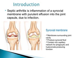
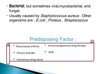
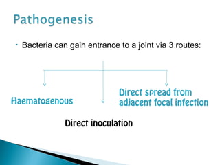
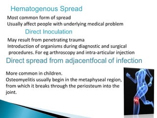
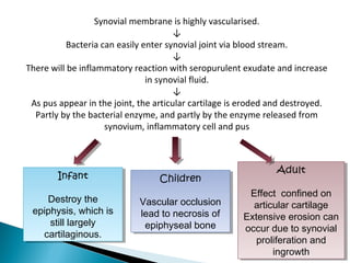
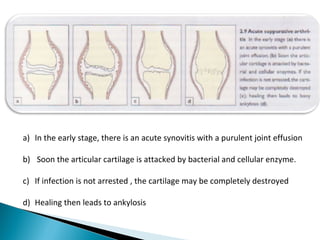
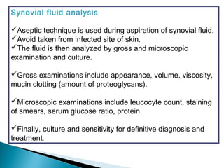
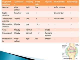
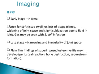
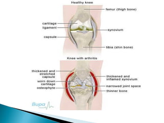
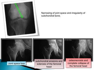
Septic arthritis of knee
Dr.ROSHANZAMEER
1st
year pg
 Introduction
 Pathogenesis
 Clinical Features
 Investigations
 Treatment
Contents
 Septic arthritis is inflammation of a synovial
membrane with purulent effusion into the joint
capsule, due to infection.
Synovial membrane
Membrane surrounding joint
cavity
Produce synovial fluid
Contain rich capillary
network for phagocytic and
hyaluronate-producing
function
 Bacterial, but sometimes viral,mycobacterial, and
fungal.
 Usually caused by Staphylococcus aureus . Other
organisms are : E.coli , Proteus , Streptococcus
Predisposing Factor :
 Rheumatoid arthritis
 Chronic disorder
 Intravenous drug abuse
 Immunosuppressive drug therapy
 AIDS
• Bacteria can gain entrance to a joint via 3 routes:
Haematogenous
Direct inoculation
Direct spread from
adjacent focal infection
Hematogenous Spread
Most common form of spread
Usually affect people with underlying medical problem
Direct Inoculation
May result from penetrating trauma
Introduction of organisms during diagnostic and surgical
procedures. For eg arthroscopy and intra-articular injection
More common in children.
Osteomyelitis usually begin in the metaphyseal region,
from which it breaks through the periosteum into the
joint.
Synovial membrane is highly vascularised.
↓
Bacteria can easily enter synovial joint via blood stream.
↓
There will be inflammatory reaction with seropurulent exudate and increase
in synovial fluid.
↓
As pus appear in the joint, the articular cartilage is eroded and destroyed.
Partly by the bacterial enzyme, and partly by the enzyme released from
synovium, inflammatory cell and pus
Infant
Destroy the
epiphysis, which is
still largely
cartilaginous.
Infant
Destroy the
epiphysis, which is
still largely
cartilaginous.
Children
Vascular occlusion
lead to necrosis of
epiphyseal bone
Children
Vascular occlusion
lead to necrosis of
epiphyseal bone
Adult
Effect confined on
articular cartilage
Extensive erosion can
occur due to synovial
proliferation and
ingrowth
Adult
Effect confined on
articular cartilage
Extensive erosion can
occur due to synovial
proliferation and
ingrowth
a) In the early stage, there is an acute synovitis with a purulent joint effusion
b) Soon the articular cartilage is attacked by bacterial and cellular enzyme.
c) If infection is not arrested , the cartilage may be completely destroyed
d) Healing then leads to ankylosis
If left untreated, it will spread to the underlying bone
and out of joint to form abscess and sinus.
Healing with:
1.Complete resolution
2.Partial loss of articular cartilage and fibrosis of joint
3.Loss of articular cartilage and bony ankylosis
4.Bony destruction and permanent deformity
Differ according to age
In new born infants
 More on septicaemia
Rather than joint pain
 Baby is irritable &
refuse to feed
 Tachycardia with fever
 Joints are warmth,
tenderness, resistance
to movement
 Umbilical cord and
inflamed IV site should be
suspicious of source of
infection
In children
o acute pain in single
large joint(esp hip)
o Pseudoparesis
o Child is ill,rapid pulse
and swingingfever
o Overlying skin looks red
& superficial joint swelling
may be obvious
o Local warmth and
marked tenderness
o All movements are
restricted by pain or spasm.
o Look for source of
infection from septic toe or
discharge ear
In adults
 Often in the superficial
joint(knee, wrist or ankle )
 Joints painful, swollen
& inflamed.
 Warmth and marked
local tenderness &
movement restricted.
 look for gonococcal
infection or drug abuse.
 Patient with
rheumatoid arthritis and
especially those on
corticosteroid may
develop “silent” joint
infection.
Physical examination:
 Lower limb  antalgic limp / cannot walk
 Upper limb  affected part is closedly
guarded
 Marked tenderness, active and passive range
of motion are limited
 Examine for synovial effusion, erythema,
heat and tenderness.
 Spasm of muscles around the joint may be
marked.
 Patient may hold the joint in a position to
reduce the intra-articular pressure to
minimize pain.
Blood
investigations
Imaging
Synovial
fluid analysis
Investigations Explaination
Full blood count Elevated white blood cell count
ESR > 40 mm/hr
CRP > 20 mg/dL
Blood culture May be positive
Synovial fluid analysis
Aseptic technique is used during aspiration of synovial fluid.
Avoid taken from infected site of skin.
The fluid is then analyzed by gross and microscopic
examination and culture.
Gross examinations include appearance, volume, viscosity,
mucin clotting (amount of proteoglycans).
Microscopic examinations include leucocyte count, staining
of smears, serum glucose ratio, protein.
Finally, culture and sensitivity for definitive diagnosis and
treatment.
Suspected
condition
Appearanc
e
Viscosity White
cells
Crystals Biochemistry Bacteriology
Normal Clear
yellow
High Few - As for plasma -
Septic
arthritis
Purulent Low + - Glucose low +
Tuberculous
arthritis
Turbid Low + - Glucose low +
Rheumatoid
arthritis
Cloudy Low + + - - -
Gout Cloudy Normal ++ Urate - -
Pseudogout Cloudy Normal + Pyropho
sphate
- -
Osteoarthrit
is
Clear
yellow
High few Often + - -
X ray
 Early Stage – Normal
Look for soft tissue swelling, loss of tissue planes,
widening of joint space and slight subluxation due to fluid in
joint. Gas may be seen with E. coli infection
 Late stage – Narrowing and irregularity of joint space
 Plain film findings of superimposed osteomyelitis may
develop (periosteal reaction, bone destruction, sequestrum
formation).
Joint space lossJoint space loss
subchondral erosions and
sclerosis of the femoral
head
subchondral erosions and
sclerosis of the femoral
head
osteonecrosis and
complete collapse of
the femoral head
osteonecrosis and
complete collapse of
the femoral head
Narrowing of joint space and irregularity of
subchondral bone.
Ultrasonography
 More reliable in revealing a joint effusion in
early cases.
 Widening of space between capsule and bone
of > 2mm indicates effusion.
 Echo-free  transient synovitis
 Positively echogenic  septic arthritis
General supportive care
-Analgesics
-IV fluids
Splintage
The joint must be rested either on a splint or in a widely split
plaster
-In neonates and infants, with hip infection the joint is held
abducted and 30 degree flexed, on traction to prevent
dislocation.
Antibiotics
Treatment is started once the blood and samples are obtained
without waiting for the detail results.
Choice of antibiotic depends on the most likely pathogen
 Surgical Drainage
 Arthroscopic debridement and copious irrigation with
normal saline – more frequently in knee joint septic
arthritis
• Bone destruction and dislocation of the joint (esp
Hip)
•Cartilage destruction
-may lead to either fibrosis or bony ankylosis
- in adult partial destruction of the joint will result
in secondary osteoarthritis
•Growth disturbance
- presenting as either localised deformity or
shortening of the bone
Campbell
Gray’s anatomy
Thank You

Septicarthritis

  • 1.
    Septic arthritis ofknee Dr.ROSHANZAMEER 1st year pg
  • 2.
     Introduction  Pathogenesis Clinical Features  Investigations  Treatment Contents
  • 3.
     Septic arthritisis inflammation of a synovial membrane with purulent effusion into the joint capsule, due to infection. Synovial membrane Membrane surrounding joint cavity Produce synovial fluid Contain rich capillary network for phagocytic and hyaluronate-producing function
  • 4.
     Bacterial, butsometimes viral,mycobacterial, and fungal.  Usually caused by Staphylococcus aureus . Other organisms are : E.coli , Proteus , Streptococcus Predisposing Factor :  Rheumatoid arthritis  Chronic disorder  Intravenous drug abuse  Immunosuppressive drug therapy  AIDS
  • 5.
    • Bacteria cangain entrance to a joint via 3 routes: Haematogenous Direct inoculation Direct spread from adjacent focal infection
  • 6.
    Hematogenous Spread Most commonform of spread Usually affect people with underlying medical problem Direct Inoculation May result from penetrating trauma Introduction of organisms during diagnostic and surgical procedures. For eg arthroscopy and intra-articular injection More common in children. Osteomyelitis usually begin in the metaphyseal region, from which it breaks through the periosteum into the joint.
  • 7.
    Synovial membrane ishighly vascularised. ↓ Bacteria can easily enter synovial joint via blood stream. ↓ There will be inflammatory reaction with seropurulent exudate and increase in synovial fluid. ↓ As pus appear in the joint, the articular cartilage is eroded and destroyed. Partly by the bacterial enzyme, and partly by the enzyme released from synovium, inflammatory cell and pus Infant Destroy the epiphysis, which is still largely cartilaginous. Infant Destroy the epiphysis, which is still largely cartilaginous. Children Vascular occlusion lead to necrosis of epiphyseal bone Children Vascular occlusion lead to necrosis of epiphyseal bone Adult Effect confined on articular cartilage Extensive erosion can occur due to synovial proliferation and ingrowth Adult Effect confined on articular cartilage Extensive erosion can occur due to synovial proliferation and ingrowth
  • 8.
    a) In theearly stage, there is an acute synovitis with a purulent joint effusion b) Soon the articular cartilage is attacked by bacterial and cellular enzyme. c) If infection is not arrested , the cartilage may be completely destroyed d) Healing then leads to ankylosis
  • 9.
    If left untreated,it will spread to the underlying bone and out of joint to form abscess and sinus. Healing with: 1.Complete resolution 2.Partial loss of articular cartilage and fibrosis of joint 3.Loss of articular cartilage and bony ankylosis 4.Bony destruction and permanent deformity
  • 10.
    Differ according toage In new born infants  More on septicaemia Rather than joint pain  Baby is irritable & refuse to feed  Tachycardia with fever  Joints are warmth, tenderness, resistance to movement  Umbilical cord and inflamed IV site should be suspicious of source of infection In children o acute pain in single large joint(esp hip) o Pseudoparesis o Child is ill,rapid pulse and swingingfever o Overlying skin looks red & superficial joint swelling may be obvious o Local warmth and marked tenderness o All movements are restricted by pain or spasm. o Look for source of infection from septic toe or discharge ear In adults  Often in the superficial joint(knee, wrist or ankle )  Joints painful, swollen & inflamed.  Warmth and marked local tenderness & movement restricted.  look for gonococcal infection or drug abuse.  Patient with rheumatoid arthritis and especially those on corticosteroid may develop “silent” joint infection.
  • 11.
    Physical examination:  Lowerlimb  antalgic limp / cannot walk  Upper limb  affected part is closedly guarded  Marked tenderness, active and passive range of motion are limited  Examine for synovial effusion, erythema, heat and tenderness.  Spasm of muscles around the joint may be marked.  Patient may hold the joint in a position to reduce the intra-articular pressure to minimize pain.
  • 12.
    Blood investigations Imaging Synovial fluid analysis Investigations Explaination Fullblood count Elevated white blood cell count ESR > 40 mm/hr CRP > 20 mg/dL Blood culture May be positive
  • 13.
    Synovial fluid analysis Aseptictechnique is used during aspiration of synovial fluid. Avoid taken from infected site of skin. The fluid is then analyzed by gross and microscopic examination and culture. Gross examinations include appearance, volume, viscosity, mucin clotting (amount of proteoglycans). Microscopic examinations include leucocyte count, staining of smears, serum glucose ratio, protein. Finally, culture and sensitivity for definitive diagnosis and treatment.
  • 14.
    Suspected condition Appearanc e Viscosity White cells Crystals BiochemistryBacteriology Normal Clear yellow High Few - As for plasma - Septic arthritis Purulent Low + - Glucose low + Tuberculous arthritis Turbid Low + - Glucose low + Rheumatoid arthritis Cloudy Low + + - - - Gout Cloudy Normal ++ Urate - - Pseudogout Cloudy Normal + Pyropho sphate - - Osteoarthrit is Clear yellow High few Often + - -
  • 15.
    X ray  EarlyStage – Normal Look for soft tissue swelling, loss of tissue planes, widening of joint space and slight subluxation due to fluid in joint. Gas may be seen with E. coli infection  Late stage – Narrowing and irregularity of joint space  Plain film findings of superimposed osteomyelitis may develop (periosteal reaction, bone destruction, sequestrum formation).
  • 17.
    Joint space lossJointspace loss subchondral erosions and sclerosis of the femoral head subchondral erosions and sclerosis of the femoral head osteonecrosis and complete collapse of the femoral head osteonecrosis and complete collapse of the femoral head Narrowing of joint space and irregularity of subchondral bone.
  • 18.
    Ultrasonography  More reliablein revealing a joint effusion in early cases.  Widening of space between capsule and bone of > 2mm indicates effusion.  Echo-free  transient synovitis  Positively echogenic  septic arthritis
  • 19.
    General supportive care -Analgesics -IVfluids Splintage The joint must be rested either on a splint or in a widely split plaster -In neonates and infants, with hip infection the joint is held abducted and 30 degree flexed, on traction to prevent dislocation. Antibiotics Treatment is started once the blood and samples are obtained without waiting for the detail results. Choice of antibiotic depends on the most likely pathogen
  • 20.
     Surgical Drainage Arthroscopic debridement and copious irrigation with normal saline – more frequently in knee joint septic arthritis
  • 21.
    • Bone destructionand dislocation of the joint (esp Hip) •Cartilage destruction -may lead to either fibrosis or bony ankylosis - in adult partial destruction of the joint will result in secondary osteoarthritis •Growth disturbance - presenting as either localised deformity or shortening of the bone
  • 22.
  • 23.