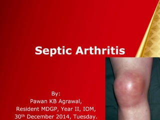
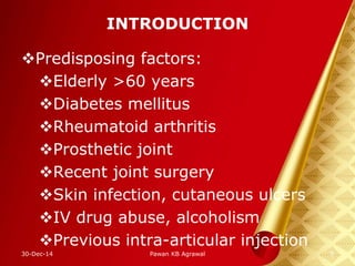
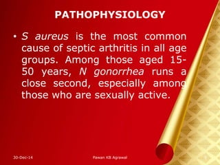
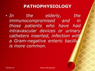
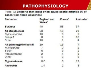
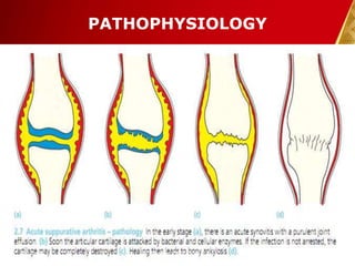
Septic Arthritis
By:
Pawan KB Agrawal,
Resident MDGP, Year II, IOM,
30th December 2014, Tuesday.
OUTLINE
• Introduction
• Pathophysiology
• Clinical features
• Diagnosis
• Differentials
• Treatment
• Complications
• Prognosis
• References
30-Dec-14 2Pawan KB Agrawal
INTRODUCTION
• Infection of joint space.
• often bacterial but could be
fungal or viral.
• rheumatologic emergency as
joint destruction occurs rapidly
and can lead to significant
morbidity and mortality.
30-Dec-14 3Pawan KB Agrawal
INTRODUCTION
• Prevalence:range from 8 to 27
%
• a 2007 systematic review that
included a total of 6242
patients with acutely painful
joints showed 653 (10 percent)
had septic arthritis1.
30-Dec-14 4Pawan KB Agrawal
INTRODUCTION
Predisposing factors:
Elderly >60 years
Diabetes mellitus
Rheumatoid arthritis
Prosthetic joint
Recent joint surgery
Skin infection, cutaneous ulcers
IV drug abuse, alcoholism
Previous intra-articular injection
30-Dec-14 5Pawan KB Agrawal
PATHOPHYSIOLOGY
• S aureus is the most common
cause of septic arthritis in all age
groups. Among those aged 15-
50 years, N gonorrhea runs a
close second, especially among
those who are sexually active.
30-Dec-14 6Pawan KB Agrawal
PATHOPHYSIOLOGY
• In the elderly, the
immunocompromised and in
those patients who have had
intravascular devices or urinary
catheters inserted, infection with
a Gram-negative enteric bacillus
is more common.
30-Dec-14 7Pawan KB Agrawal
PATHOPHYSIOLOGY
30-Dec-14 8Pawan KB Agrawal
PATHOPHYSIOLOGY
30-Dec-14 9Pawan KB Agrawal
CLINICAL FEATURES
Usually present with a single
painful swollen joint.
Low grade fever, local rise in
temperature & impaired range of
motion.
The knee is involved in more
than 50 % of cases followed by
hip, shoulder, elbow, ankle &
wrist 2.
30-Dec-14 10Pawan KB Agrawal
CLINICAL FEATURES
20 % of septic joint infections
are polyarticular3.
30-Dec-14 11Pawan KB Agrawal
CLINICAL FEATURES
Joint affected Attitude
1.Knee Flexion
2. Hip Flexion, abduction
& internal rotation.
3. Shoulder Adduction &
internal rotation.
4. Elbow Flexion & mid
pronation
5. Wrist Flexion
6. Ankle Planter flexion30-Dec-14 12Pawan KB Agrawal
DIAGNOSIS
Arthrocentesis
usually purulent with
increased count (50,000 to
150,000 cells/mm3)
The synovial fluid glucose is
often depressed and lactic acid
concentration is elevated.
30-Dec-14 13Pawan KB Agrawal
DIAGNOSIS
Arthrocentesis
usually purulent with
increased count (50,000 to
150,000 cells/mm3)
The synovial fluid glucose is
often depressed and lactic acid
concentration is elevated.
Synovial fluid culture
30-Dec-14 14Pawan KB Agrawal
DIAGNOSIS
30-Dec-14 15Pawan KB Agrawal
DIAGNOSIS
X-ray:
The earliest findings are soft
tissue swelling around the
joint and a widened joint
space from joint effusion.
Displacement of adjacent fat
pads may be present,
especially in infants and
children.
30-Dec-14 16Pawan KB Agrawal
DIAGNOSIS
30-Dec-14 17Pawan KB Agrawal
DIAGNOSIS
Later, joint-space narrowing
could be found as articular
cartilage is destroyed. Loss
of visualization of the white
cortical line over large areas
of the joint surface soon
ensues as bone destruction
begins to develop.
30-Dec-14 18Pawan KB Agrawal
DIAGNOSIS
Blood cultures are positive in
about 50 percent of cases.
Elevations of CRP are usually
present, though the sensitivity
of the ESR test in patients with
septic arthritis is inconsistent 4,5.
30-Dec-14 19Pawan KB Agrawal
DIAGNOSIS
Computed tomography (CT), or
magnetic resonance imaging
(MRI) are far more sensitive
than plain films in early septic
arthritis.
MRI:
Synovial enhancement and
the presence of a joint effusion
& perisynovial soft tissue
edema.
30-Dec-14 20Pawan KB Agrawal
DIAGNOSIS
Radionuclide bone scans:
technetium-99m
methyldiphosphonate
increase in isotope
accumulation in areas of
osteoblasts and increased
vascularity
30-Dec-14 21Pawan KB Agrawal
DIFFERENTIALS
Gout
Pseudogout
Transient synovitis
Rheumatoid arthritis
Viral arthritis
Lyme disease
30-Dec-14 22Pawan KB Agrawal
TREATMENT
Principle:
Antibiotics
Joint drainage &
Joint rest.
30-Dec-14 23Pawan KB Agrawal
TREATMENT
General support: analgesics,
antipyretics and joint splintage
for first few days.
Definitive care:
IV antibiotics for initial 1-2
wks followed by oral
antibiotics for 3-4 wks.
30-Dec-14 24Pawan KB Agrawal
TREATMENT
30-Dec-14 25Pawan KB Agrawal
TREATMENT
Concurrent systemic corticosteroids
are also supposed to shorten
duration of illness with less residual
joint damage and dysfunction7.
30-Dec-14 26Pawan KB Agrawal
TREATMENT
Joint drainage: needle
aspiration or open.
Older children with early
septic arthritis can often be
treated by repeated closed
aspiration ; however, if there
is no improvement within 48
hours, open drainage is
necessary.
30-Dec-14 27Pawan KB Agrawal
TREATMENT
30-Dec-14 Pawan KB Agrawal 28
TREATMENT
30-Dec-14 Pawan KB Agrawal 29
TREATMENT
30-Dec-14 Pawan KB Agrawal 30
TREATMENT
30-Dec-14 Pawan KB Agrawal 31
TREATMENT
30-Dec-14 Pawan KB Agrawal 32
FOLLOW UP
• Once general condition is satisfactory
and the joint is no longer painful or
warm, further damage is unlikely.
• If articular cartilage has been
preserved, gentle and gradually
increase active movements.
• If articular cartilage has been
destroyed the aim is splinting to keep
the joint immobile while ankylosis is
awaited.
30-Dec-14 Pawan KB Agrawal 33
FOLLOW UP
• If deformity is present,
subsequent osteotomy should
be planned to correct it.
30-Dec-14 Pawan KB Agrawal 34
COMPLICATIONS
Partial or complete destruction
of epiphysis.
Retarded growth
Ankylosis
Osteomyelitis
Sepsis
30-Dec-14 35Pawan KB Agrawal
PROGNOSIS
Poor outcome predictors:
Age older than 60 years
Infection of hip or shoulder
Underlying RA
Persistent positive findings.
Delay in therapy.
30-Dec-14 36Pawan KB Agrawal
PROGNOSIS
Irreversible loss of joint function
in 25-50%
Mortality ranges from 5-15%6.
30-Dec-14 37Pawan KB Agrawal
TOM SMITH ARTHRITIS
Septic arthritis of hip in infancy
Results in complete destruction
of cartilaginous femoral head.
Presentation is a child in his
preschool age with painless limp
Affected limb is shorter
X-ray shows complete absence
of head and neck of femur.
30-Dec-14 38Pawan KB Agrawal
REFERENCES
1. Margaretten ME, Kohlwes J, Moore D,
Bent S. Does this adult patient have
septic arthritis? JAMA 2007; 297:1478.
2. Goldenberg DL. Septic arthritis and other
infections of rheumatologic significance.
Rheum Dis Clin North Am 1991; 17:149.
3. Dubost JJ, Fis I, Denis P, et al.
Polyarticular septic arthritis. Medicine
(Baltimore) 1993; 72:296.
4. Ernst AA, Weiss SJ, Tracy LA, Weiss NR.
Usefulness of CRP and ESR in predicting
septic joints. South Med J 2010;
103:522.
30-Dec-14 39Pawan KB Agrawal
REFERENCES
5. Hariharan P, Kabrhel C. Sensitivity of
erythrocyte sedimentation rate and C-
reactive protein for the exclusion of
septic arthritis in emergency department
patients. J Emerg Med 2011; 40:428.
6. Kaandorp CJ, Krijnen P, Moens HJ, et al.
The outcome of bacterial arthritis: a
prospective community-based study.
Arthritis Rheum 1997; 40:884.
7. Sharff, K. A. (2013). Clinical
Management of Septic Arthritis. Curr
Rheumatol Rep .
30-Dec-14 40Pawan KB Agrawal
• THANK YOU …
30-Dec-14 41Pawan KB Agrawal

8. septic arthritis 30th dec 14

Editor's Notes

  • #7 A joint can become infected by: (1) direct invasion through a penetrating wound, intra-articular injection or arthroscopy; (2) direct spread from an adjacent bone abscess; or (3) blood spread from a distant site. Bacterial causes of septic arthritis include staphylococci (40 percent), streptococci (28 percent), gram-negative bacilli (19 percent), mycobacteria (8 per- cent), gram-negative cocci (3 percent), gram-positive bacilli (1 percent), and anaerobes (1 percent). 30
  • #8 A joint can become infected by: (1) direct invasion through a penetrating wound, intra-articular injection or arthroscopy; (2) direct spread from an adjacent bone abscess; or (3) blood spread from a distant site. Bacterial causes of septic arthritis include staphylococci (40 percent), streptococci (28 percent), gram-negative bacilli (19 percent), mycobacteria (8 per- cent), gram-negative cocci (3 percent), gram-positive bacilli (1 percent), and anaerobes (1 percent). 30
  • #9 A joint can become infected by: (1) direct invasion through a penetrating wound, intra-articular injection or arthroscopy; (2) direct spread from an adjacent bone abscess; or (3) blood spread from a distant site. Bacterial causes of septic arthritis include staphylococci (40 percent), streptococci (28 percent), gram-negative bacilli (19 percent), mycobacteria (8 per- cent), gram-negative cocci (3 percent), gram-positive bacilli (1 percent), and anaerobes (1 percent). 30
  • #10 Bacteria entering the joint initially deposit in the synovial membrane and produce an acute inflammatory cell response. Because synovial tissue has no limiting basement plate, bacterial organisms may quickly gain access to the synovial fluid, characteristically creating acute-onset, purulent joint inflammation. there is marked hyperplasia of the lining cells in the synovial membrane within seven days. In addition, inflammatory cells release cytokines and proteases that cause cartilage degradation and inhibit cartilage synthesis. Pressure necrosis from large synovial effusions may result in further cartilage and bone loss.
  • #11 Positive findings on synovial fluid cultures after 7 days of appropriate therapy Delay of 7 days or longer in instituting therapy
  • #12 Positive findings on synovial fluid cultures after 7 days of appropriate therapy Delay of 7 days or longer in instituting therapy
  • #13 Positive findings on synovial fluid cultures after 7 days of appropriate therapy Delay of 7 days or longer in instituting therapy
  • #14 The synovial fluid glucose is often depressed and lactic acid concentration is elevated; however, these tests are not sufficiently sensitive to be of widespread diagnostic utility. Ultrasonography is the most reliable method for revealing a joint effusion in early cases. Both hips should be examined for comparison. Widening of the space between capsule and bone of more than 2 mm s indicative of an effusion, which may be echo-free (perhaps a transient synovitis) or positively echogenic (more likely septic arthritis).
  • #15 The synovial fluid glucose is often depressed and lactic acid concentration is elevated; however, these tests are not sufficiently sensitive to be of widespread diagnostic utility. Ultrasonography is the most reliable method for revealing a joint effusion in early cases. Both hips should be examined for comparison. Widening of the space between capsule and bone of more than 2 mm s indicative of an effusion, which may be echo-free (perhaps a transient synovitis) or positively echogenic (more likely septic arthritis).
  • #16 The synovial fluid glucose is often depressed and lactic acid concentration is elevated; however, these tests are not sufficiently sensitive to be of widespread diagnostic utility. Ultrasonography is the most reliable method for revealing a joint effusion in early cases. Both hips should be examined for comparison. Widening of the space between capsule and bone of more than 2 mm s indicative of an effusion, which may be echo-free (perhaps a transient synovitis) or positively echogenic (more likely septic arthritis).
  • #17 The synovial fluid glucose is often depressed and lactic acid concentration is elevated; however, these tests are not sufficiently sensitive to be of widespread diagnostic utility.
  • #18 The synovial fluid glucose is often depressed and lactic acid concentration is elevated; however, these tests are not sufficiently sensitive to be of widespread diagnostic utility.
  • #19 The synovial fluid glucose is often depressed and lactic acid concentration is elevated; however, these tests are not sufficiently sensitive to be of widespread diagnostic utility.
  • #20 The synovial fluid glucose is often depressed and lactic acid concentration is elevated; however, these tests are not sufficiently sensitive to be of widespread diagnostic utility.
  • #21 The synovial fluid glucose is often depressed and lactic acid concentration is elevated; however, these tests are not sufficiently sensitive to be of widespread diagnostic utility.
  • #22 The synovial fluid glucose is often depressed and lactic acid concentration is elevated; however, these tests are not sufficiently sensitive to be of widespread diagnostic utility.
  • #33 Make a vertical incision beginning about 1 cm below the anterior superior iliac spine inferiorly.    •    Expose the sartorius muscle on the medial side and the tensor fasciae latae and vastus lateralis muscles on the lateral side. Use blunt dissection to separate these muscles.    •    Identify the lateral border of the rectus femoris, and retract this muscle medially (Fig. 17-12); this exposes the hip joint capsule.    •    Incise the capsule, evacuate the pus, and irrigate the joint with saline.    •    Leave the capsule open, but close the skin loosely over drains.    •    If wider exposure is required, extend the skin incision proximally onto the iliac crest, and subperiosteally detach the origins of the tensor fasciae latae and gluteal muscles from the ilium.    •    Protect the lateral femoral cutaneous nerve proximally and the branches of the lateral femoral circumflex artery distally, if possible.
  • #37 Positive findings on synovial fluid cultures after 7 days of appropriate therapy Delay of 7 days or longer in instituting therapy
  • #38 A joint can become infected by: (1) direct invasion through a penetrating wound, intra-articular injection or arthroscopy; (2) direct spread from an adjacent bone abscess; or (3) blood spread from a distant site.
  • #39 Positive findings on synovial fluid cultures after 7 days of appropriate therapy Delay of 7 days or longer in instituting therapy