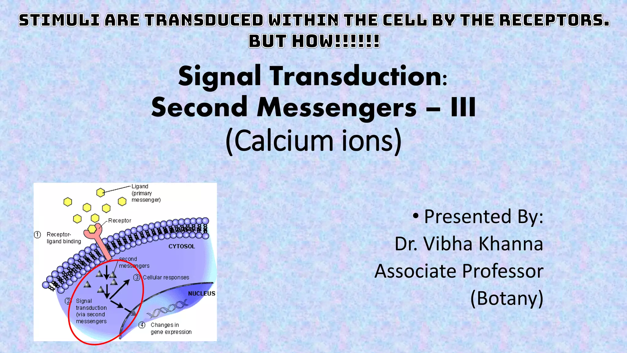
Calcium ions (Ca2+) serve as critical second messengers in a variety of developmental processes and responses to environmental stimuli in cells. The concentration of Ca2+ is regulated through various mechanisms, including active transport to maintain low levels in resting cells and the generation of calcium signatures triggered by ion channels and receptors. The signaling network involves interactions with proteins like GPCRs and RTKs, leading to the release of Ca2+ from intracellular stores and activation of downstream cellular processes.

![Calcium signatures are generated by the coordinated action of various
Ca2+ influx and efflux systems including channels, pumps, and exchangers
located at different cellular membranes.
• The cytoplasmic Ca2+ level is low in
resting cells.
• Low cytoplasmic [Ca2+] is maintained by
extrusion via
• plasma membrane Ca2+ ATPase (PMCA) and
• smooth endoplasmic reticular Ca2+ ATPase
(SERCA) transporters.
• The Na/Ca exchanger (NCX), a major
secondary regulator of [Ca2+], is
electrogenic,
• exchanging three Na ions for one Ca2+.
• Intracellular Ca2+ hyperpolarizes many
cells by activating K+ channels, and in
some cells, Cl− channels.
• This decreases CaV channel activity but
increases the driving force across active
Ca2+-permeant channels.](https://image.slidesharecdn.com/signaltransduction-secondmessengerscalciumions-210729193657/75/Second-messengers-in-Signal-transduction-Calcium-ions-5-2048.jpg)
![The Ca2+ signaling network: An Overview
• In excitatory Ca2+ signaling, plasma membrane
ion channels are triggered to open by changes
in voltage, or extra- or intracellular ligand
binding.
• Initial increases in [Ca2+] trigger more release,
primarily from ER via Ca2+-sensitive ryanodine
receptors (RyR).
• G protein-coupled receptor (GPCR) or receptor
tyrosine kinase-mediated activation of PLC
cleaves PIP2 into inositol (1,4,5) trisphosphate
(IP3) and diacylglycerol (DAG).
• IP3 is a ligand for the intracellular IP3R channel
spanning the membrane of the ER.
• GPCRs catalyze the exchange of guanosine
diphosphate (GDP) for GTP on Gα subunits,
releasing active Gα and Gβγ subunits that in
turn activate PLCβ.
• RTKs dimerize upon ligand binding, auto-
phosphorylate, and interact with other signaling
proteins to activate PLCγ.](https://image.slidesharecdn.com/signaltransduction-secondmessengerscalciumions-210729193657/75/Second-messengers-in-Signal-transduction-Calcium-ions-6-2048.jpg)


