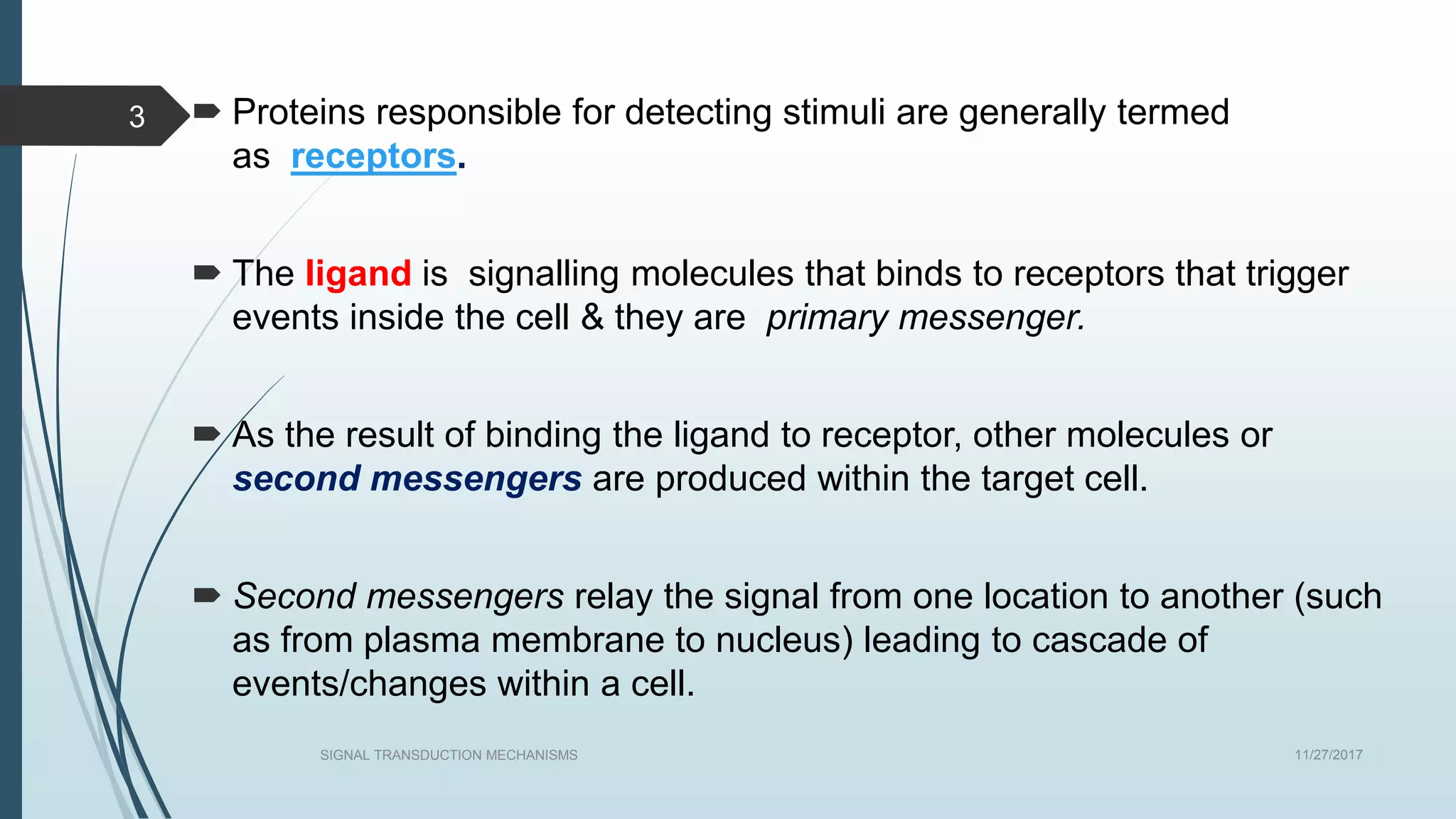
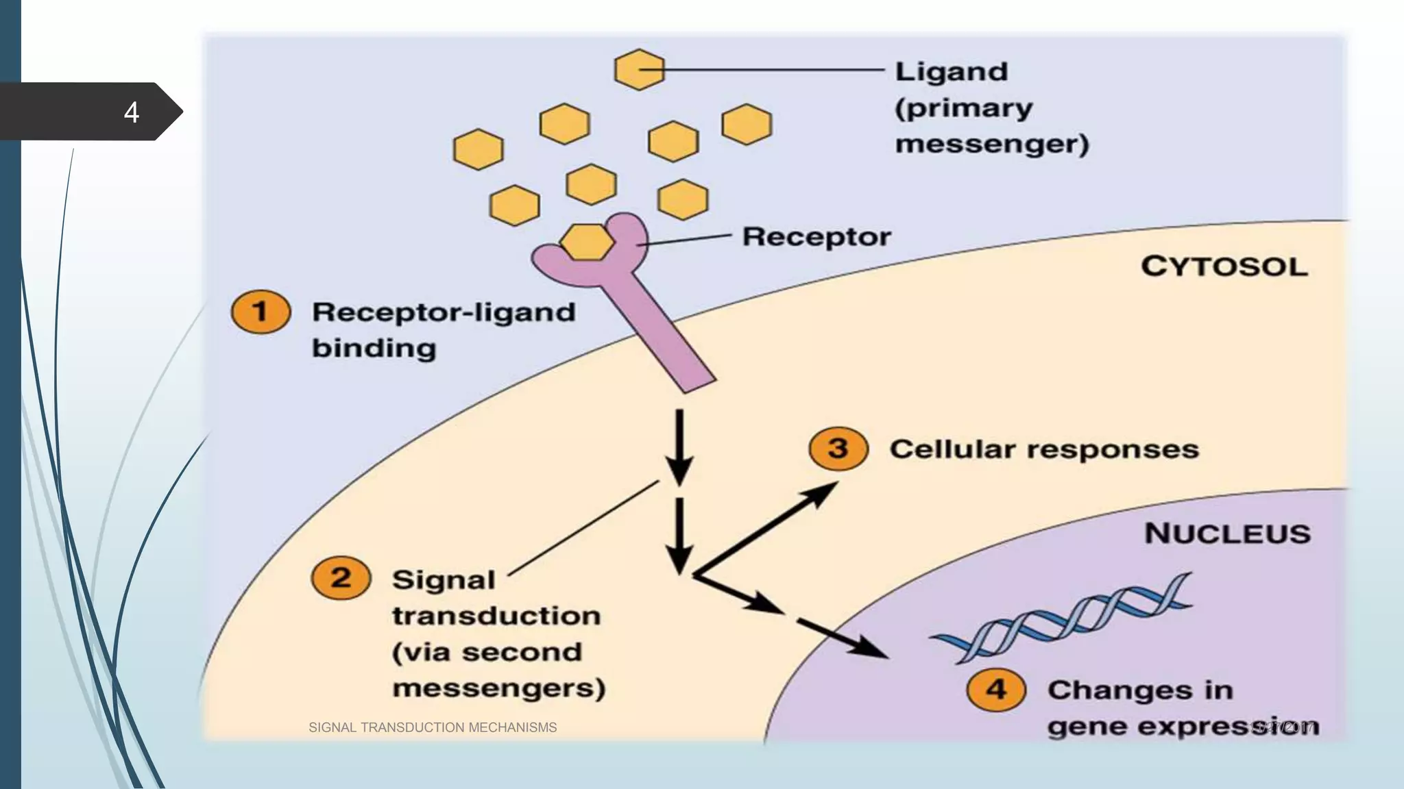
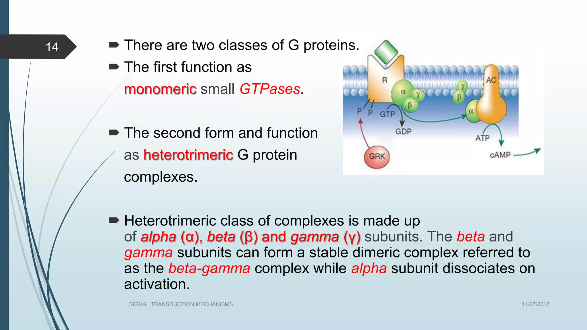
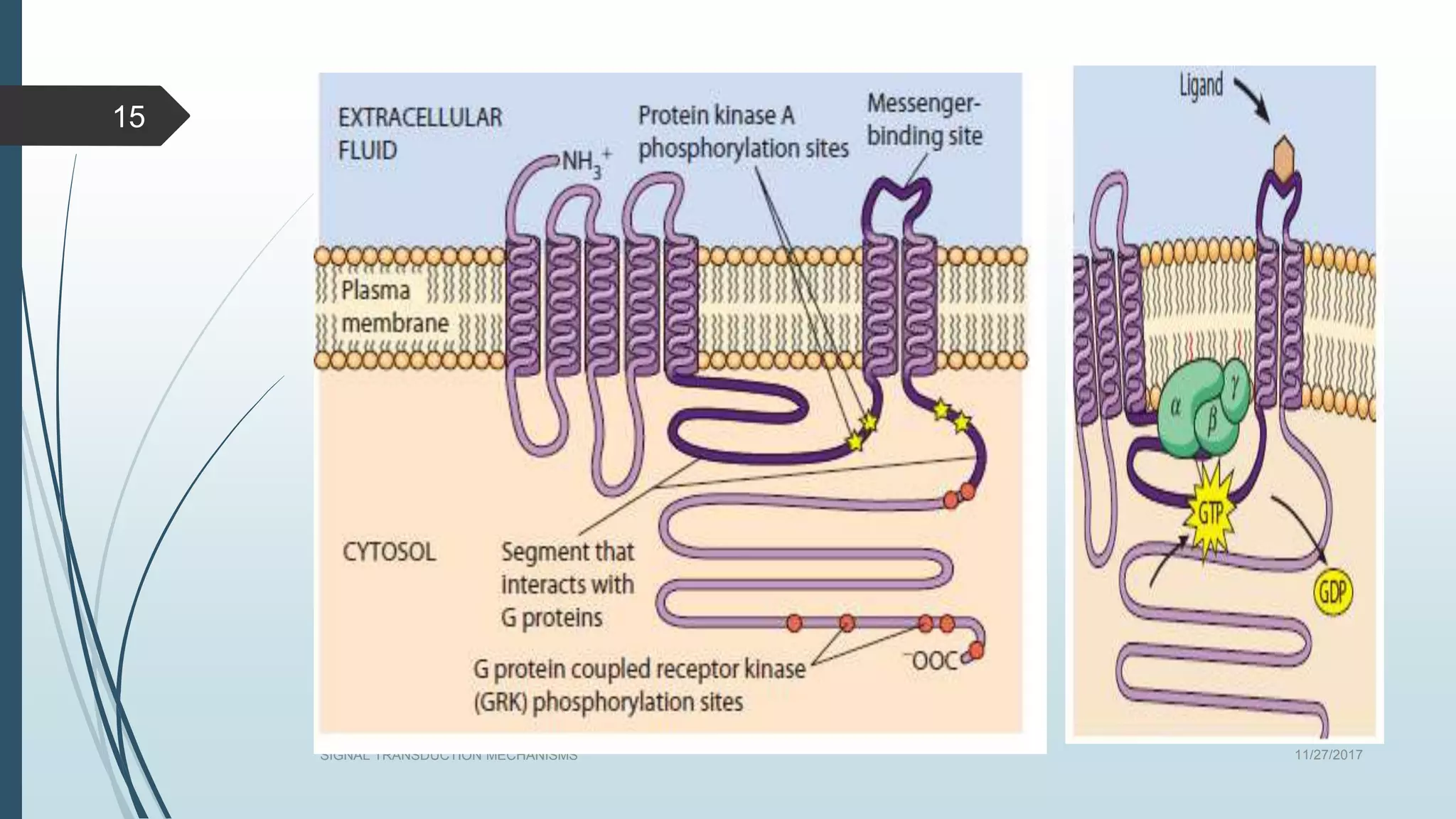
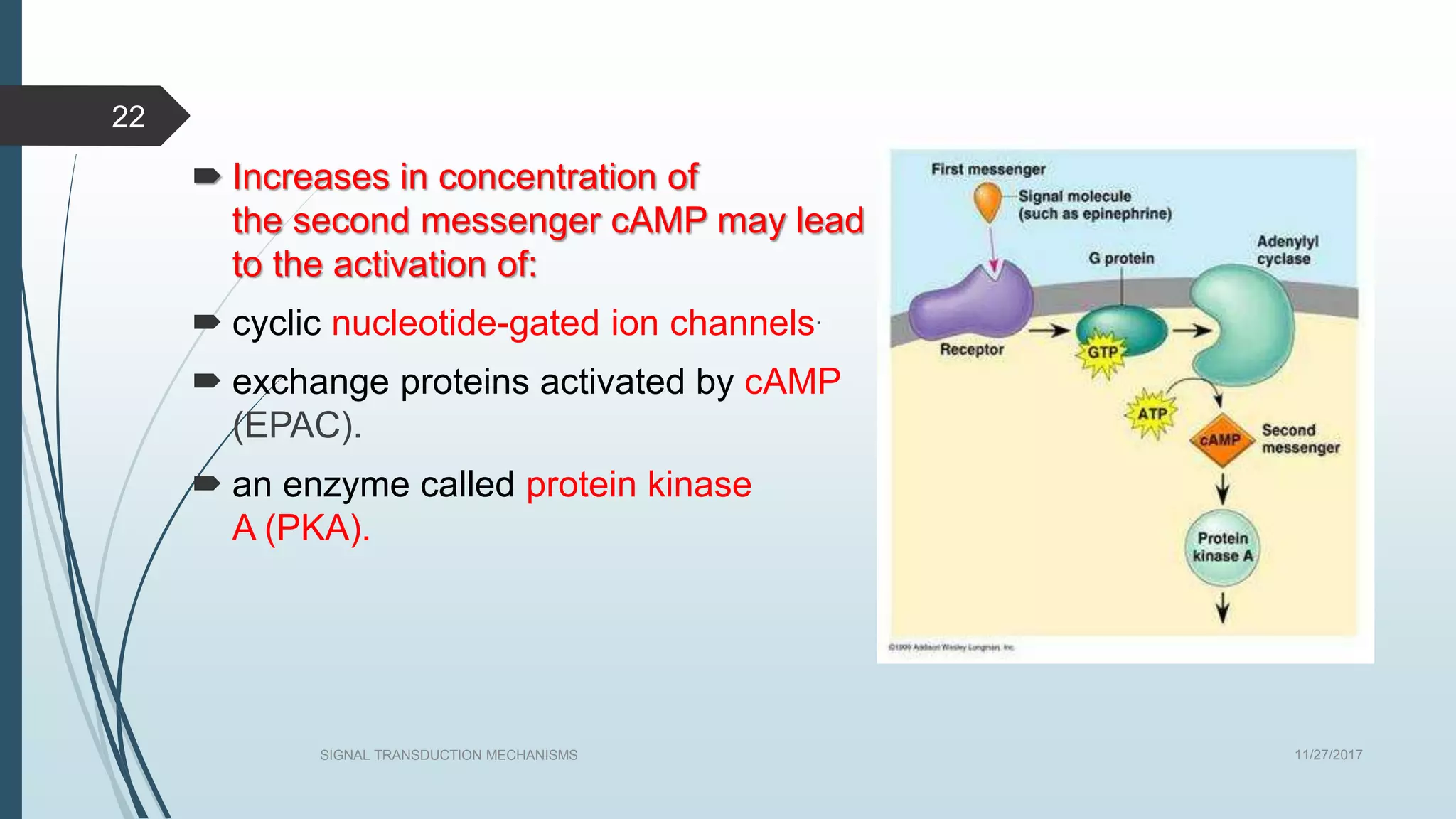
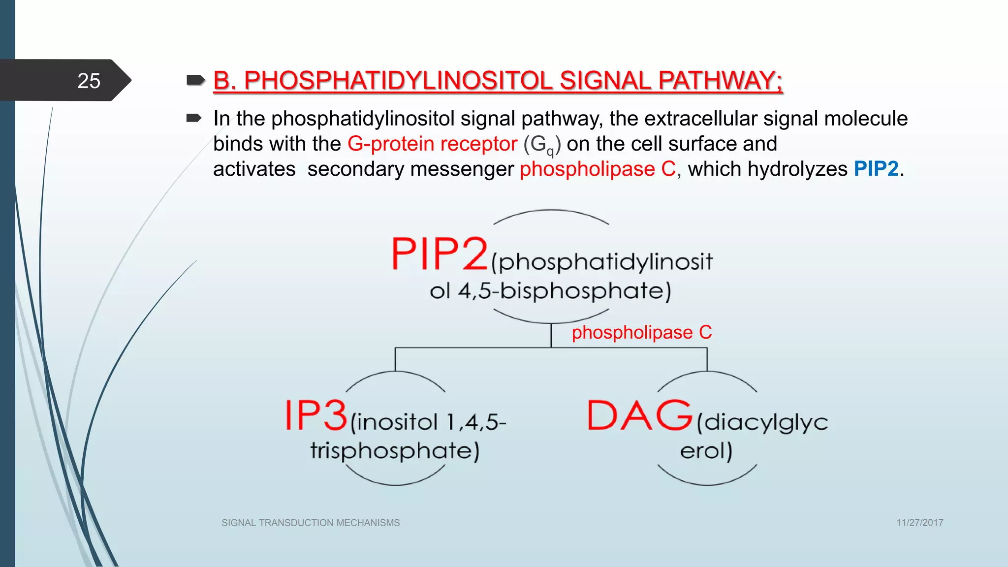
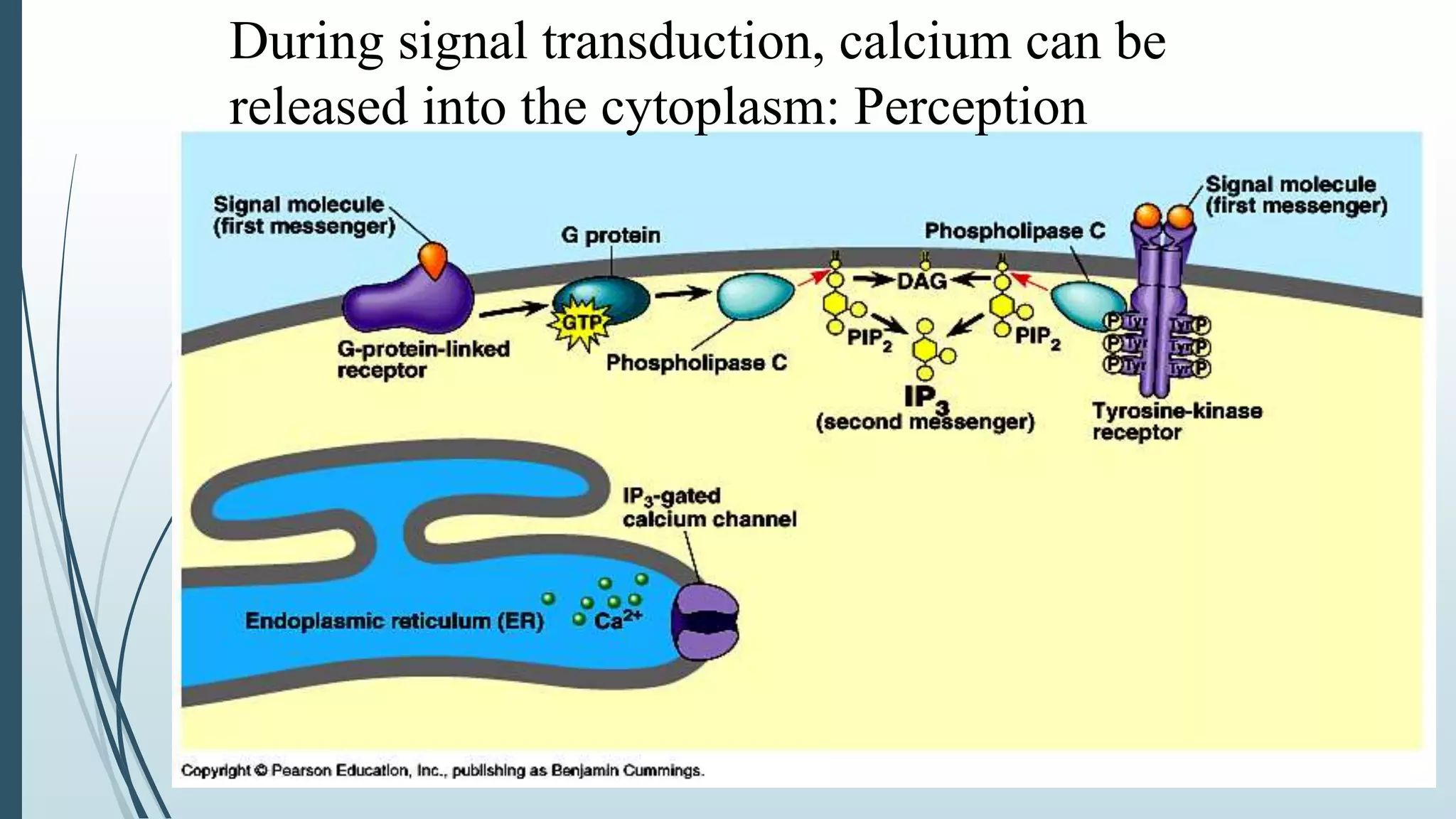
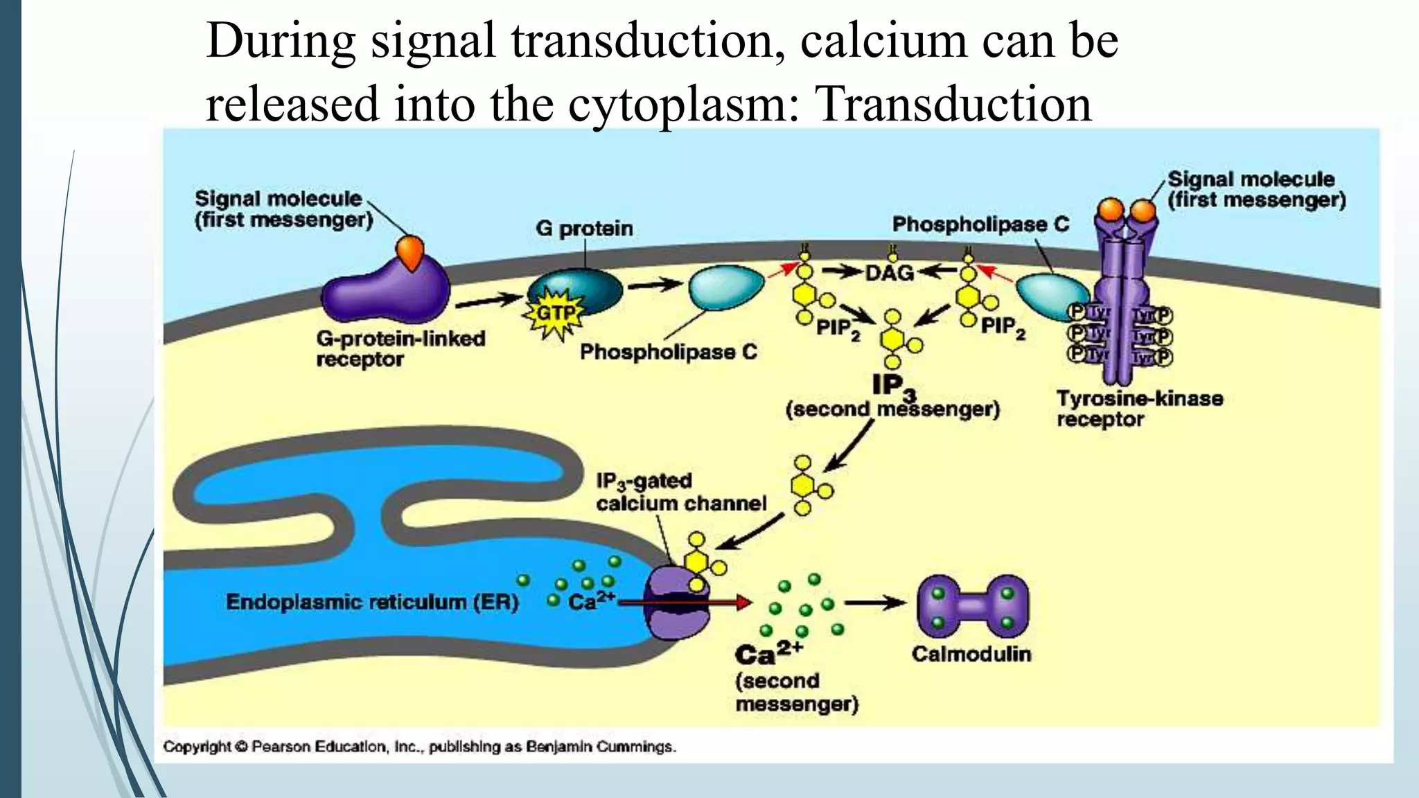
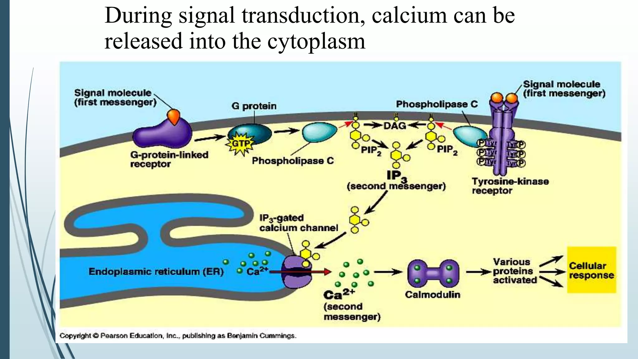
BY
S.RAJASEKHAR
TAM-16-43
MOLECULAR BIOLOGY & BIOTECHNOLOGY
SIGNAL TRANSDUCTION
Signal transduction is the process by which a chemical or physical
signal is transmitted through a cell as a series of molecular events,
most commonly protein phosphorylation
catalysed by protein kinases, which ultimately results in a cellular
response.
 Any process occurring within cells that convert one kind of
signal/stimulus into another type.
 It also known as cell signaling in which the transmission of molecular
signals from a cell's exterior to its interior.
 Signals received by cells must be transmitted effectively into the cell to
ensure an appropriate response. This step is initiated by cell-surface
receptors which triggers a biochemical chain of events inside the cell,
creating a response. 11/27/2017SIGNAL TRANSDUCTION MECHANISMS
2
 Proteins responsible for detecting stimuli are generally termed
as receptors.
 The ligand is signalling molecules that binds to receptors that trigger
events inside the cell & they are primary messenger.
 As the result of binding the ligand to receptor, other molecules or
second messengers are produced within the target cell.
 Second messengers relay the signal from one location to another (such
as from plasma membrane to nucleus) leading to cascade of
events/changes within a cell.
11/27/2017SIGNAL TRANSDUCTION MECHANISMS
3
11/27/2017SIGNAL TRANSDUCTION MECHANISMS
4
11/27/2017SIGNAL TRANSDUCTION MECHANISMS
5
In endocrine signaling, the signaling molecules are synthesized
and secreted by signaling cells (for example, those found
in endocrine glands), transported through the circulatory system
of the organism, and finally act on target cells distant
from their site of synthesis.
11/27/2017SIGNAL TRANSDUCTION MECHANISMS
6
ln paracrine signaling, the signaling molecules released by
a cell affect only those target cells in close proximity. A nerve
cell releasing a neurotransmitter (e.g., acetylcholine) that acts
on an adjacent nerve cell or on a muscle cell (inducing or inhibiting
muscle contraction) is an example of paracrine signaling.
11/27/2017SIGNAL TRANSDUCTION MECHANISMS
7
In autocrine signaling, cells respond to substances that they
themselves release. Some growth factors act in this fashion,
and cultured cells often secrete growth factors that
stimulate their own growth and proliferation.
11/27/2017SIGNAL TRANSDUCTION MECHANISMS
8
Integral membrane proteins located on the cell surface also
play an important role in signaling . In some cases, such
membrane-bound signals on one cell bind receptors on the
surface of an adjacent target cell, triggering its differentiation.
Proteins attached to the plasma membrane of one cell can interact directly with
cell-surface receptors on adjacent cells.
RECEPTORS
 Receptors can be roughly divided into two major
classes: intracellular receptors and extracellular receptors.
EXTRACELLULAR RECEPTORS
 Extracellular receptors are integral transmembrane proteins and
make up most receptors.
 They span the plasma membrane of the cell, with one part of the
receptor on the outside of the cell and the other on the inside.
 Signal transduction occurs as a result of a ligand binding to the
outside region of the receptor (the ligand does not pass through
the membrane).
11/27/2017
SIGNAL TRANSDUCTION MECHANISMS
9
Various Extracellular Receptors
 G protein-coupled receptors.
 Receptors with Kinase activity.
 Integrin receptors.
 Toll gate receptors.
 Ligand-gated ion channel receptors.
11/27/2017SIGNAL TRANSDUCTION MECHANISMS
10
 G PROTEIN–COUPLED RECEPTORS (GPCRs)
 Also known as seven-transmembrane domain receptors, 7TM
receptors, heptahelical receptors, and G protein–linked
receptors (GPLR).
 These constitute a large protein family of receptors that sense
molecules outside the cell and activate inside signal
transduction pathways and, ultimately, cellular responses.
 Coupling with G proteins, they are called seven-transmembrane
receptors because they pass through the cell membrane seven
times. 11/27/2017SIGNAL TRANSDUCTION MECHANISMS
11
11/27/2017SIGNAL TRANSDUCTION MECHANISMS
12
 G proteins, also known as guanine nucleotide-binding
proteins, are a family of proteins that act as molecular
switches inside cells, and are involved in transmitting signals
from a variety of stimuli outside a cell to its interior.
 When they are bound to GTP, they are 'on', and, when they
are bound to GDP, they are 'off'.
 G proteins belong to the larger group of enzymes
called GTPases
11/27/2017SIGNAL TRANSDUCTION MECHANISMS
13
 There are two classes of G proteins.
 The first function as
monomeric small GTPases.
 The second form and function
as heterotrimeric G protein
complexes.
 Heterotrimeric class of complexes is made up
of alpha (α), beta (β) and gamma (γ) subunits. The beta and
gamma subunits can form a stable dimeric complex referred to
as the beta-gamma complex while alpha subunit dissociates on
activation.
11/27/2017SIGNAL TRANSDUCTION MECHANISMS
14
11/27/2017SIGNAL TRANSDUCTION MECHANISMS
15
 There are two principal signal transduction pathways involving
the G protein–coupled receptors:
 A. the cAMP signal pathway and
 B. the phosphatidylinositol signal pathway.
 A. cAMP-DEPENDENT PATHWAY,
 It is also known as the adenylyl cyclase pathway.
 In a cAMP-dependent pathway, the activated Gs alpha subunit
binds to and activates an enzyme called adenylyl cyclase, which,
in turn, catalyzes the conversion of ATP into cyclic adenosine
monophosphate (cAMP).
11/27/2017SIGNAL TRANSDUCTION MECHANISMS
16
MECHANISM
 It is known that in the inactive state, the GPCR is bound to
a heterotrimeric G protein complex.
 Binding of primary messenger to the GPCR results in
a conformation change in the receptor that is transmitted to the
bound Gαsubunit of the heterotrimeric G protein.
 The activated Gα subunit exchanges GTP in place of GDP which
in turn triggers the dissociation of Gα subunit from the Gβγ dimer
and from the receptor.
11/27/2017SIGNAL TRANSDUCTION MECHANISMS
17
 CONTD..
 The dissociated Gα and Gβγ subunits interact with other
intracellular proteins to continue the signal transduction cascade.
 While the freed GPCR is able to rebind to another heterotrimeric
G protein to form a new complex that is ready to initiate another
round of signal transduction.
11/27/2017SIGNAL TRANSDUCTION MECHANISMS
18
11/27/2017SIGNAL TRANSDUCTION MECHANISMS
19
11/27/2017SIGNAL TRANSDUCTION MECHANISMS
20
11/27/2017SIGNAL TRANSDUCTION MECHANISMS
21
 In the inactive state, and subunits a represent as a complex, with GDP
bound to the subunit. When a ligand (L) binds its receptor, it binds and
activates a Gs protein.
 When a receptor is activated by ligand binding, the receptor-ligand
complex associates with the Gs protein, causing the displacement of GDP
by GTP and the dissociation of the Gsa-GTP complex.
 The GTP-Gsa complex then binds to and activates membrane-bound
adenylyl cyclase, which synthesizes cAMP.
 Activation ends when the ligand leaves the receptor, the GTP is hydrolyzed
to GDP by the GTPase activity of the Gsa subunit, and the Gsa dissociates
from adenylyl cyclase.
 Adenylyl cyclase then reverts to the inactive form, the Gsa re-associates
with the Gsbg complex, and cAMP molecules in the cytosol are hydrolyzed
to AMP by the phosphodiesterase.
11/27/2017SIGNAL TRANSDUCTION MECHANISMS
22
 Increases in concentration of
the second messenger cAMP may lead
to the activation of:
 cyclic nucleotide-gated ion channels.
 exchange proteins activated by cAMP
(EPAC).
 an enzyme called protein kinase
A (PKA).
 The PKA enzyme is also known as cAMP-dependent enzyme
because it gets activated only if cAMP is present. Once PKA is
activated, it phosphorylates a number of other proteins including:
 enzymes that convert glycogen into glucose
 transcription factors, which regulate gene expression
11/27/2017SIGNAL TRANSDUCTION MECHANISMS
23
 DEACTIVATION
 The Gs alpha subunit slowly catalyzes the hydrolysis of GTP to
GDP, which in turn deactivates the Gs protein, shutting off the
cAMP pathway.
 The pathway may also be deactivated downstream by directly
inhibiting adenylyl cyclase or dephosphorylating the proteins
phosphorylated by PKA.
 Molecules that inhibit the cAMP pathway include:
 cAMP phosphodiesterase dephosphorylates cAMP into AMP,
reducing the cAMP levels.
 Gi protein, which is a G protein that inhibits adenylyl cyclase,
reducing cAMP levels. 11/27/2017SIGNAL TRANSDUCTION MECHANISMS
24
 B. PHOSPHATIDYLINOSITOL SIGNAL PATHWAY;
 In the phosphatidylinositol signal pathway, the extracellular signal molecule
binds with the G-protein receptor (Gq) on the cell surface and
activates secondary messenger phospholipase C, which hydrolyzes PIP2.
11/27/2017SIGNAL TRANSDUCTION MECHANISMS
25
phospholipase C
11/27/2017SIGNAL TRANSDUCTION MECHANISMS
26
 IP3 binds with the IP3 receptor in the membrane of the smooth
endoplasmic reticulum and mitochondria to open
Ca2+ channels.
 DAG helps activate protein kinase C (PKC), which
phosphorylates many other proteins, changing their catalytic
activities, leading to cellular responses.
During signal transduction, calcium can be
released into the cytoplasm: Perception
During signal transduction, calcium can be
released into the cytoplasm: Transduction
During signal transduction, calcium can be
released into the cytoplasm
 The effects of Ca2+ are also remarkable:
 It cooperates with DAG in activating PKC and can activate
the CaM kinase pathway, in which calcium-modulated
protein calmodulin (CaM) binds Ca2+, undergoes a change in
conformation, and activates CaM kinase.
 The kinase then phosphorylates target enzymes, regulating their
activities. The two signal pathways are connected together by
Ca2+-CaM, which is also a regulatory subunit of adenylyl cyclase
and phosphodiesterase in the cAMP signal pathway.
11/27/2017SIGNAL TRANSDUCTION MECHANISMS
30
A wide variety of cellular effects have been linked to the
activation of protein kinase C, including the stimulation of
cell growth, the regulation of ion channels, changes
in the cytoskeleton, increases in cellular pH, and
effects on secretion of proteins and other
substances.
11/27/2017SIGNAL TRANSDUCTION MECHANISMS
31
5. INTRACELLULAR RECEPTORS
 Intracellular receptors are receptors located inside
the cell rather than on its cell membrane.
 Examples are:
 Class of nuclear receptors located in the cell
nucleus and cytoplasm
 IP3 receptor located on the endoplasmic reticulum.
11/27/2017SIGNAL TRANSDUCTION MECHANISMS
32
11/27/2017SIGNAL TRANSDUCTION MECHANISMS
33

Signal transduction mechanism

  • 1.
  • 2.
    SIGNAL TRANSDUCTION Signal transductionis the process by which a chemical or physical signal is transmitted through a cell as a series of molecular events, most commonly protein phosphorylation catalysed by protein kinases, which ultimately results in a cellular response.  Any process occurring within cells that convert one kind of signal/stimulus into another type.  It also known as cell signaling in which the transmission of molecular signals from a cell's exterior to its interior.  Signals received by cells must be transmitted effectively into the cell to ensure an appropriate response. This step is initiated by cell-surface receptors which triggers a biochemical chain of events inside the cell, creating a response. 11/27/2017SIGNAL TRANSDUCTION MECHANISMS 2
  • 3.
     Proteins responsiblefor detecting stimuli are generally termed as receptors.  The ligand is signalling molecules that binds to receptors that trigger events inside the cell & they are primary messenger.  As the result of binding the ligand to receptor, other molecules or second messengers are produced within the target cell.  Second messengers relay the signal from one location to another (such as from plasma membrane to nucleus) leading to cascade of events/changes within a cell. 11/27/2017SIGNAL TRANSDUCTION MECHANISMS 3
  • 4.
  • 5.
    11/27/2017SIGNAL TRANSDUCTION MECHANISMS 5 Inendocrine signaling, the signaling molecules are synthesized and secreted by signaling cells (for example, those found in endocrine glands), transported through the circulatory system of the organism, and finally act on target cells distant from their site of synthesis.
  • 6.
    11/27/2017SIGNAL TRANSDUCTION MECHANISMS 6 lnparacrine signaling, the signaling molecules released by a cell affect only those target cells in close proximity. A nerve cell releasing a neurotransmitter (e.g., acetylcholine) that acts on an adjacent nerve cell or on a muscle cell (inducing or inhibiting muscle contraction) is an example of paracrine signaling.
  • 7.
    11/27/2017SIGNAL TRANSDUCTION MECHANISMS 7 Inautocrine signaling, cells respond to substances that they themselves release. Some growth factors act in this fashion, and cultured cells often secrete growth factors that stimulate their own growth and proliferation.
  • 8.
    11/27/2017SIGNAL TRANSDUCTION MECHANISMS 8 Integralmembrane proteins located on the cell surface also play an important role in signaling . In some cases, such membrane-bound signals on one cell bind receptors on the surface of an adjacent target cell, triggering its differentiation. Proteins attached to the plasma membrane of one cell can interact directly with cell-surface receptors on adjacent cells.
  • 9.
    RECEPTORS  Receptors canbe roughly divided into two major classes: intracellular receptors and extracellular receptors. EXTRACELLULAR RECEPTORS  Extracellular receptors are integral transmembrane proteins and make up most receptors.  They span the plasma membrane of the cell, with one part of the receptor on the outside of the cell and the other on the inside.  Signal transduction occurs as a result of a ligand binding to the outside region of the receptor (the ligand does not pass through the membrane). 11/27/2017 SIGNAL TRANSDUCTION MECHANISMS 9
  • 10.
    Various Extracellular Receptors G protein-coupled receptors.  Receptors with Kinase activity.  Integrin receptors.  Toll gate receptors.  Ligand-gated ion channel receptors. 11/27/2017SIGNAL TRANSDUCTION MECHANISMS 10
  • 11.
     G PROTEIN–COUPLEDRECEPTORS (GPCRs)  Also known as seven-transmembrane domain receptors, 7TM receptors, heptahelical receptors, and G protein–linked receptors (GPLR).  These constitute a large protein family of receptors that sense molecules outside the cell and activate inside signal transduction pathways and, ultimately, cellular responses.  Coupling with G proteins, they are called seven-transmembrane receptors because they pass through the cell membrane seven times. 11/27/2017SIGNAL TRANSDUCTION MECHANISMS 11
  • 12.
  • 13.
     G proteins,also known as guanine nucleotide-binding proteins, are a family of proteins that act as molecular switches inside cells, and are involved in transmitting signals from a variety of stimuli outside a cell to its interior.  When they are bound to GTP, they are 'on', and, when they are bound to GDP, they are 'off'.  G proteins belong to the larger group of enzymes called GTPases 11/27/2017SIGNAL TRANSDUCTION MECHANISMS 13
  • 14.
     There aretwo classes of G proteins.  The first function as monomeric small GTPases.  The second form and function as heterotrimeric G protein complexes.  Heterotrimeric class of complexes is made up of alpha (α), beta (β) and gamma (γ) subunits. The beta and gamma subunits can form a stable dimeric complex referred to as the beta-gamma complex while alpha subunit dissociates on activation. 11/27/2017SIGNAL TRANSDUCTION MECHANISMS 14
  • 15.
  • 16.
     There aretwo principal signal transduction pathways involving the G protein–coupled receptors:  A. the cAMP signal pathway and  B. the phosphatidylinositol signal pathway.  A. cAMP-DEPENDENT PATHWAY,  It is also known as the adenylyl cyclase pathway.  In a cAMP-dependent pathway, the activated Gs alpha subunit binds to and activates an enzyme called adenylyl cyclase, which, in turn, catalyzes the conversion of ATP into cyclic adenosine monophosphate (cAMP). 11/27/2017SIGNAL TRANSDUCTION MECHANISMS 16
  • 17.
    MECHANISM  It isknown that in the inactive state, the GPCR is bound to a heterotrimeric G protein complex.  Binding of primary messenger to the GPCR results in a conformation change in the receptor that is transmitted to the bound Gαsubunit of the heterotrimeric G protein.  The activated Gα subunit exchanges GTP in place of GDP which in turn triggers the dissociation of Gα subunit from the Gβγ dimer and from the receptor. 11/27/2017SIGNAL TRANSDUCTION MECHANISMS 17
  • 18.
     CONTD..  Thedissociated Gα and Gβγ subunits interact with other intracellular proteins to continue the signal transduction cascade.  While the freed GPCR is able to rebind to another heterotrimeric G protein to form a new complex that is ready to initiate another round of signal transduction. 11/27/2017SIGNAL TRANSDUCTION MECHANISMS 18
  • 19.
  • 20.
  • 21.
    11/27/2017SIGNAL TRANSDUCTION MECHANISMS 21 In the inactive state, and subunits a represent as a complex, with GDP bound to the subunit. When a ligand (L) binds its receptor, it binds and activates a Gs protein.  When a receptor is activated by ligand binding, the receptor-ligand complex associates with the Gs protein, causing the displacement of GDP by GTP and the dissociation of the Gsa-GTP complex.  The GTP-Gsa complex then binds to and activates membrane-bound adenylyl cyclase, which synthesizes cAMP.  Activation ends when the ligand leaves the receptor, the GTP is hydrolyzed to GDP by the GTPase activity of the Gsa subunit, and the Gsa dissociates from adenylyl cyclase.  Adenylyl cyclase then reverts to the inactive form, the Gsa re-associates with the Gsbg complex, and cAMP molecules in the cytosol are hydrolyzed to AMP by the phosphodiesterase.
  • 22.
    11/27/2017SIGNAL TRANSDUCTION MECHANISMS 22 Increases in concentration of the second messenger cAMP may lead to the activation of:  cyclic nucleotide-gated ion channels.  exchange proteins activated by cAMP (EPAC).  an enzyme called protein kinase A (PKA).
  • 23.
     The PKAenzyme is also known as cAMP-dependent enzyme because it gets activated only if cAMP is present. Once PKA is activated, it phosphorylates a number of other proteins including:  enzymes that convert glycogen into glucose  transcription factors, which regulate gene expression 11/27/2017SIGNAL TRANSDUCTION MECHANISMS 23
  • 24.
     DEACTIVATION  TheGs alpha subunit slowly catalyzes the hydrolysis of GTP to GDP, which in turn deactivates the Gs protein, shutting off the cAMP pathway.  The pathway may also be deactivated downstream by directly inhibiting adenylyl cyclase or dephosphorylating the proteins phosphorylated by PKA.  Molecules that inhibit the cAMP pathway include:  cAMP phosphodiesterase dephosphorylates cAMP into AMP, reducing the cAMP levels.  Gi protein, which is a G protein that inhibits adenylyl cyclase, reducing cAMP levels. 11/27/2017SIGNAL TRANSDUCTION MECHANISMS 24
  • 25.
     B. PHOSPHATIDYLINOSITOLSIGNAL PATHWAY;  In the phosphatidylinositol signal pathway, the extracellular signal molecule binds with the G-protein receptor (Gq) on the cell surface and activates secondary messenger phospholipase C, which hydrolyzes PIP2. 11/27/2017SIGNAL TRANSDUCTION MECHANISMS 25 phospholipase C
  • 26.
    11/27/2017SIGNAL TRANSDUCTION MECHANISMS 26 IP3 binds with the IP3 receptor in the membrane of the smooth endoplasmic reticulum and mitochondria to open Ca2+ channels.  DAG helps activate protein kinase C (PKC), which phosphorylates many other proteins, changing their catalytic activities, leading to cellular responses.
  • 27.
    During signal transduction,calcium can be released into the cytoplasm: Perception
  • 28.
    During signal transduction,calcium can be released into the cytoplasm: Transduction
  • 29.
    During signal transduction,calcium can be released into the cytoplasm
  • 30.
     The effectsof Ca2+ are also remarkable:  It cooperates with DAG in activating PKC and can activate the CaM kinase pathway, in which calcium-modulated protein calmodulin (CaM) binds Ca2+, undergoes a change in conformation, and activates CaM kinase.  The kinase then phosphorylates target enzymes, regulating their activities. The two signal pathways are connected together by Ca2+-CaM, which is also a regulatory subunit of adenylyl cyclase and phosphodiesterase in the cAMP signal pathway. 11/27/2017SIGNAL TRANSDUCTION MECHANISMS 30
  • 31.
    A wide varietyof cellular effects have been linked to the activation of protein kinase C, including the stimulation of cell growth, the regulation of ion channels, changes in the cytoskeleton, increases in cellular pH, and effects on secretion of proteins and other substances. 11/27/2017SIGNAL TRANSDUCTION MECHANISMS 31
  • 32.
    5. INTRACELLULAR RECEPTORS Intracellular receptors are receptors located inside the cell rather than on its cell membrane.  Examples are:  Class of nuclear receptors located in the cell nucleus and cytoplasm  IP3 receptor located on the endoplasmic reticulum. 11/27/2017SIGNAL TRANSDUCTION MECHANISMS 32
  • 33.