Download as PDF, PPTX

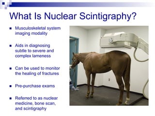


Nuclear scintigraphy uses radioactive isotopes injected into the horse's bloodstream to identify areas of bone with increased metabolic activity. The isotopes accumulate in actively healing bone and damaged soft tissues, emitting gamma rays detected by a camera to create images. It is a sensitive way to diagnose subtle or complex lameness issues, monitor fracture healing, and examine areas like the pelvis or back inaccessible to other modalities. The horse is hospitalized after the scan for 48 hours as the isotope decays to ensure handler safety. Scintigraphy highlights injury locations but may require other tests to characterize findings fully.


![[5]Isotope_Scan_Surgical_Diseases](https://cdn.slidesharecdn.com/ss_thumbnails/1664464-thumbnail.jpg?width=640&height=640&fit=bounds)


