Downloaded 244 times


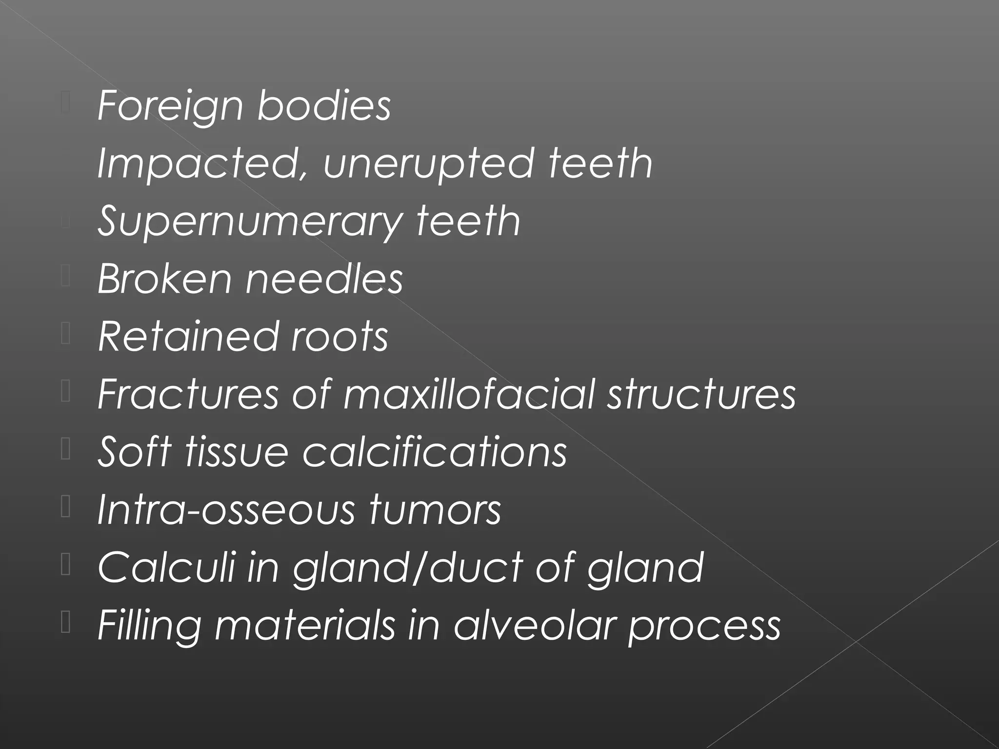
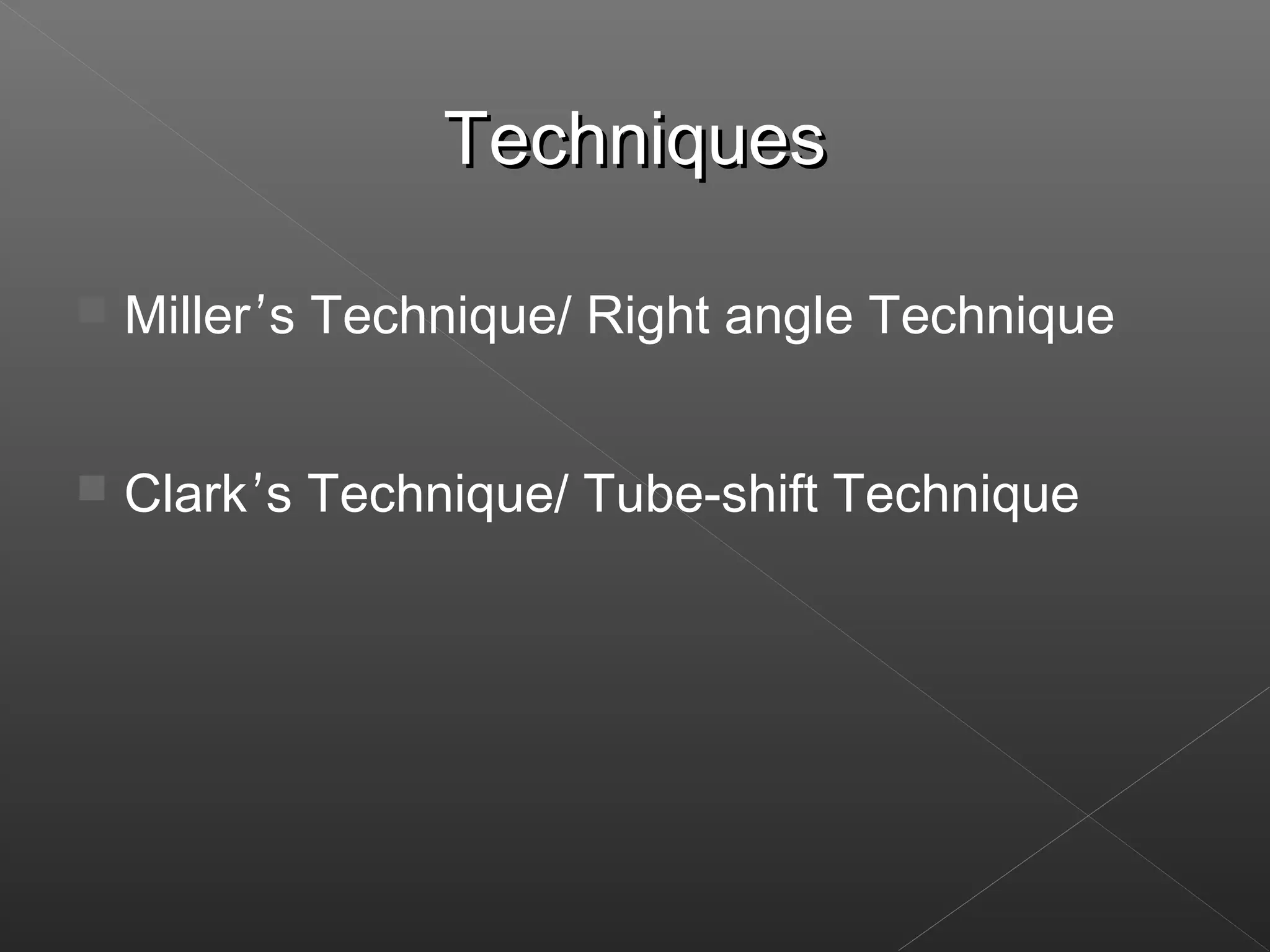
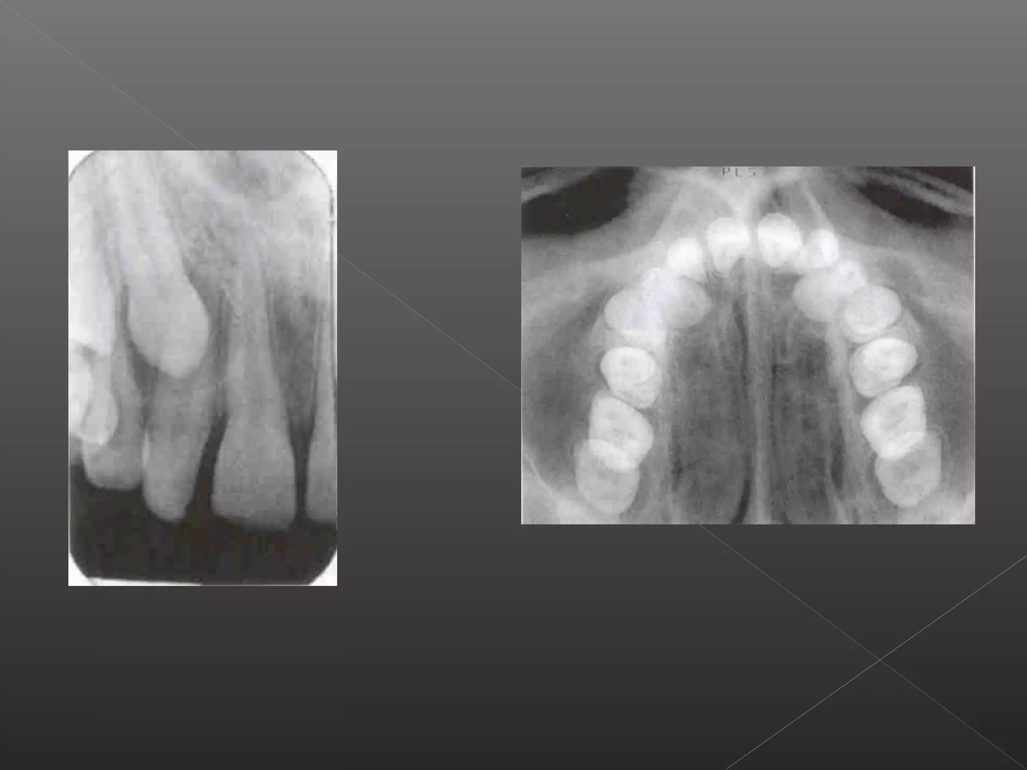
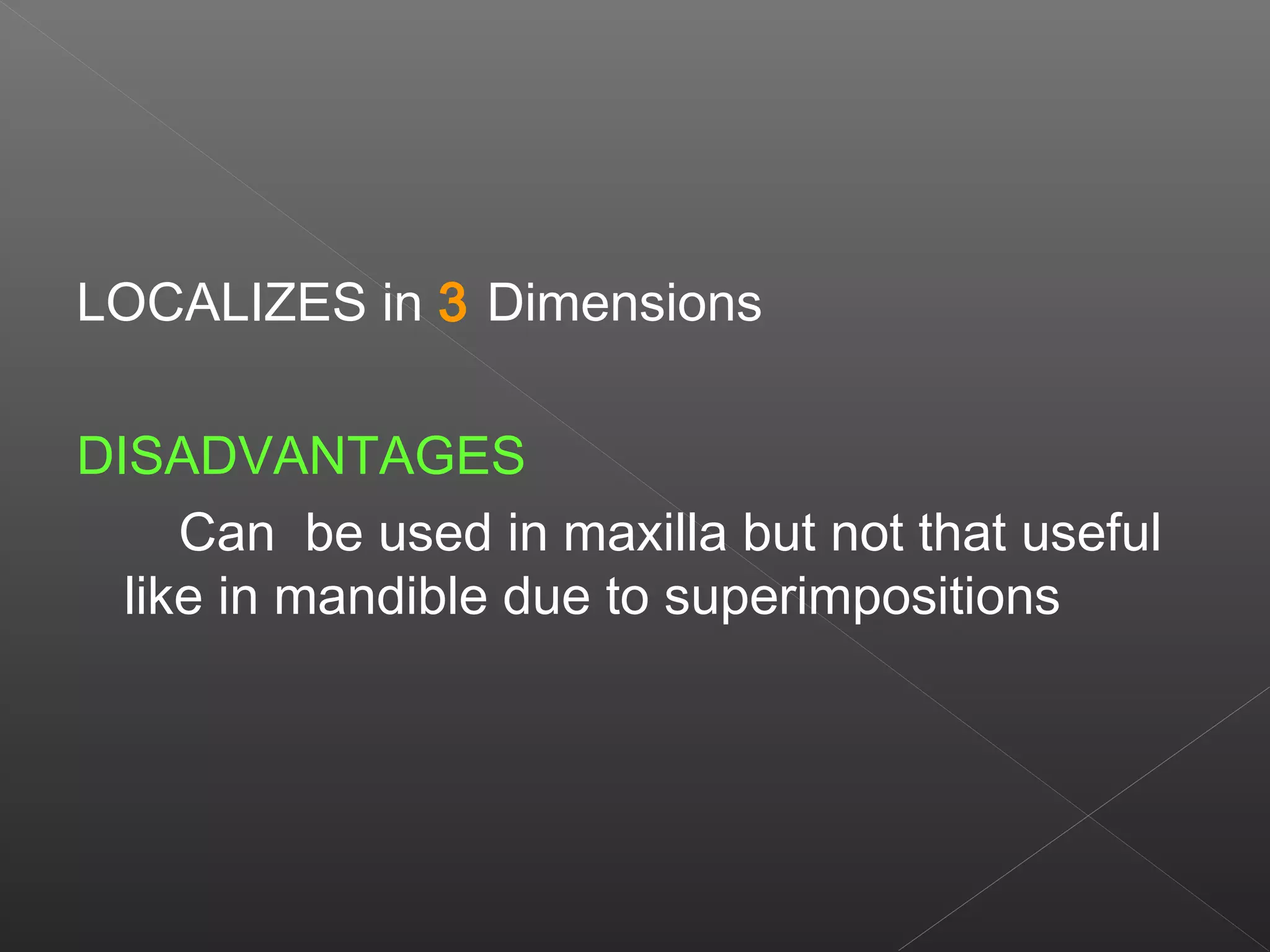
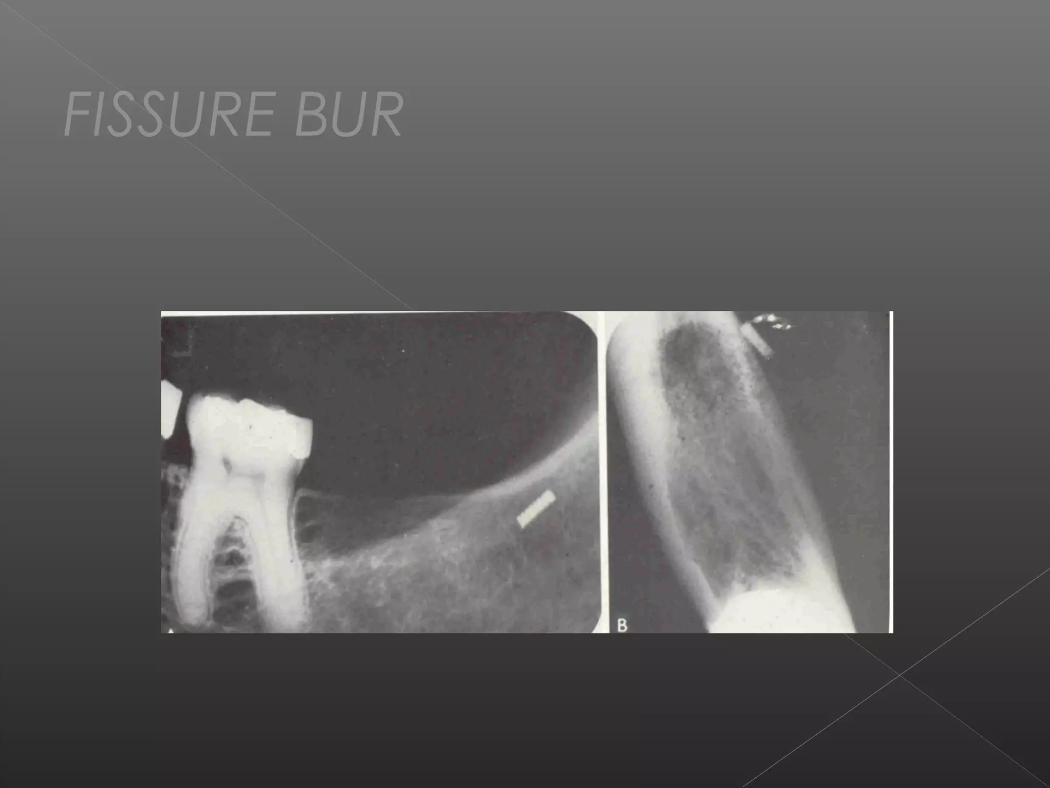
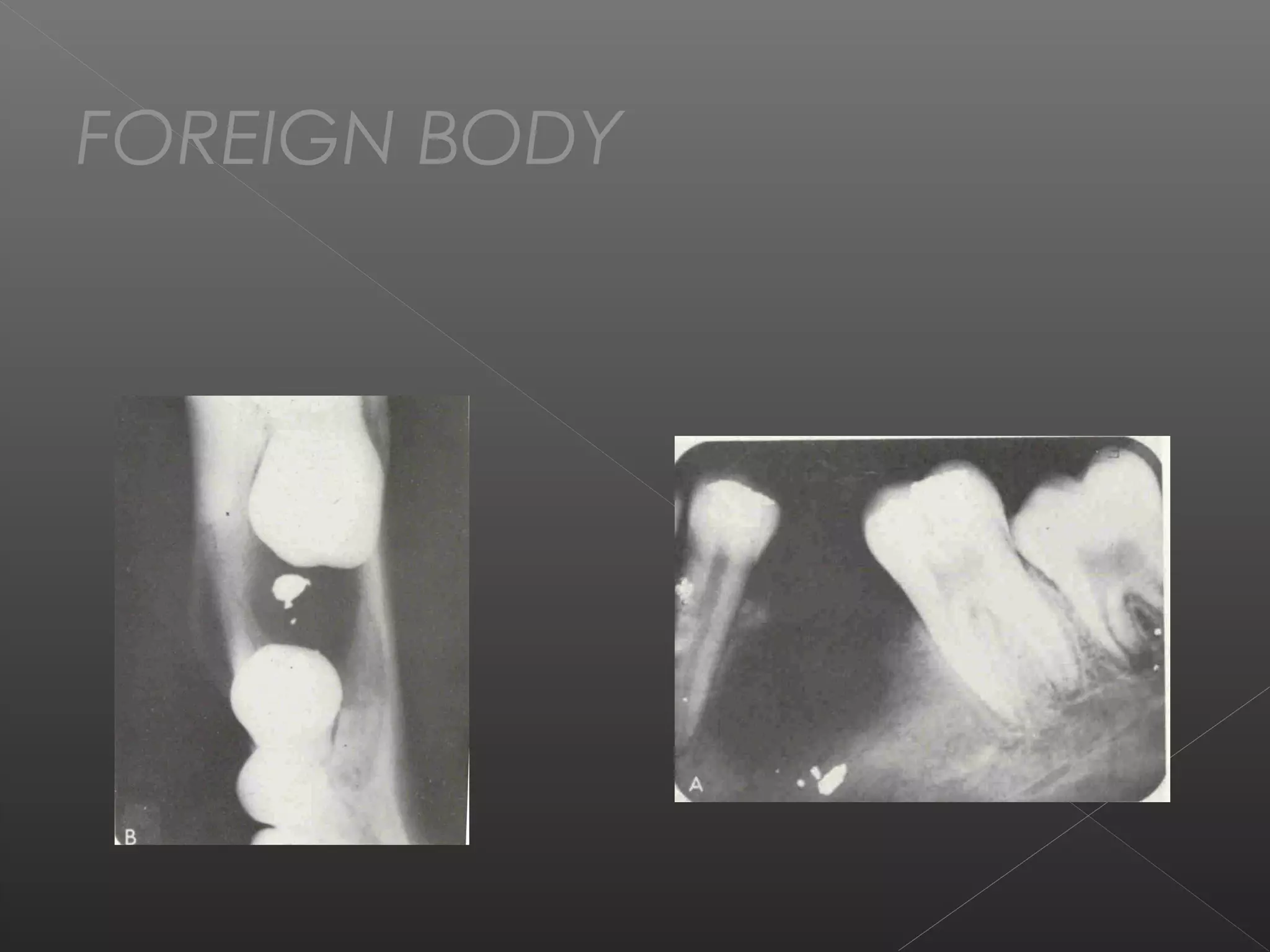
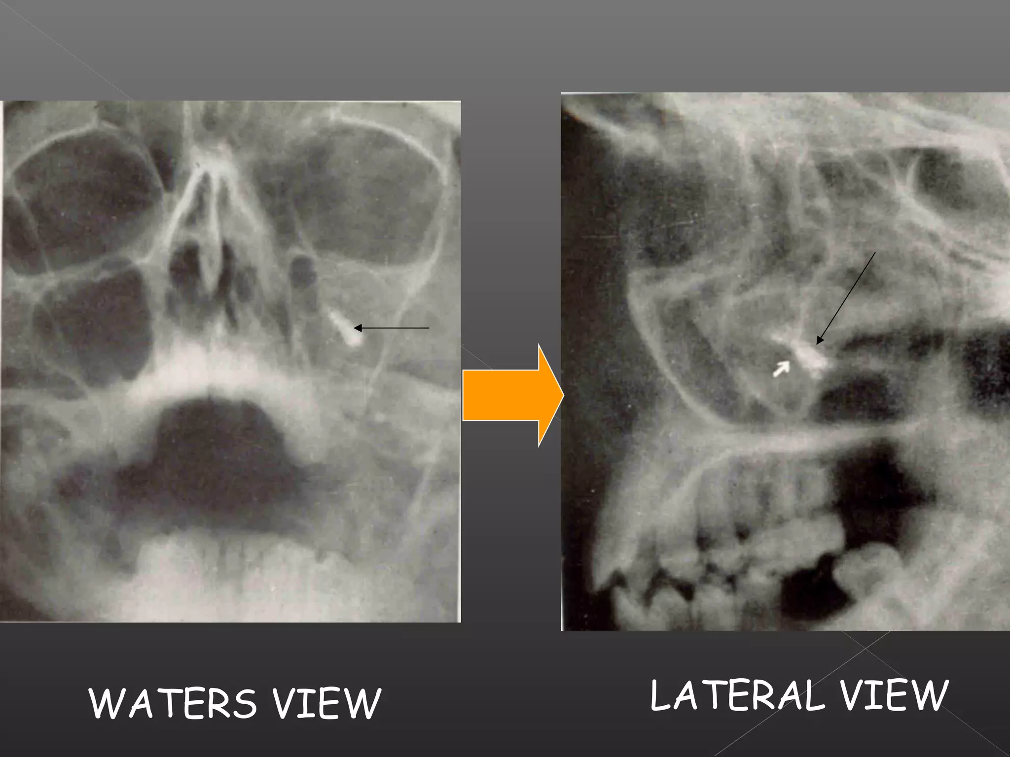
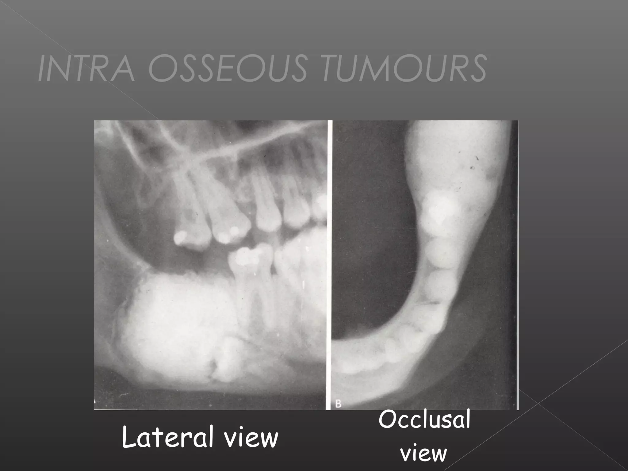
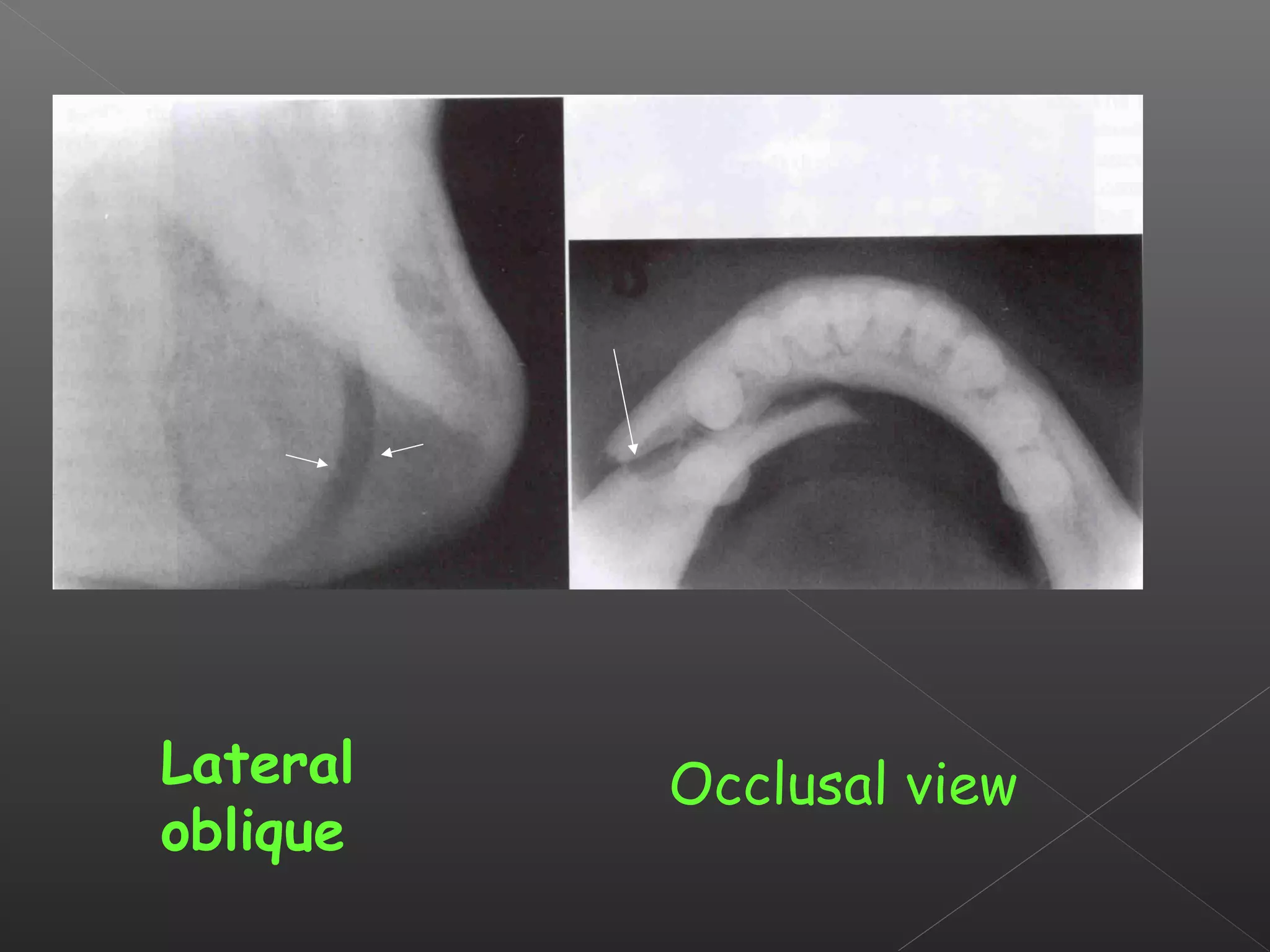
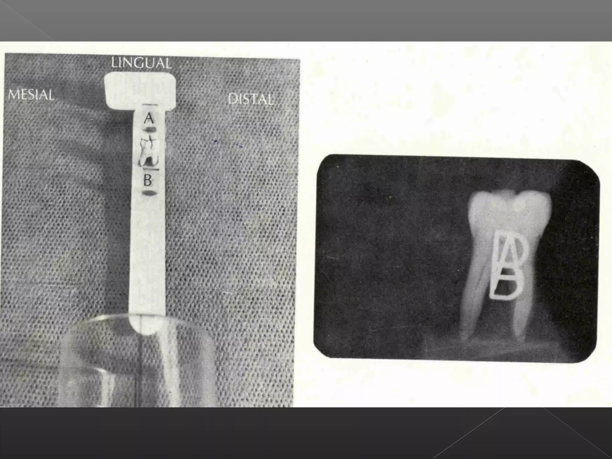
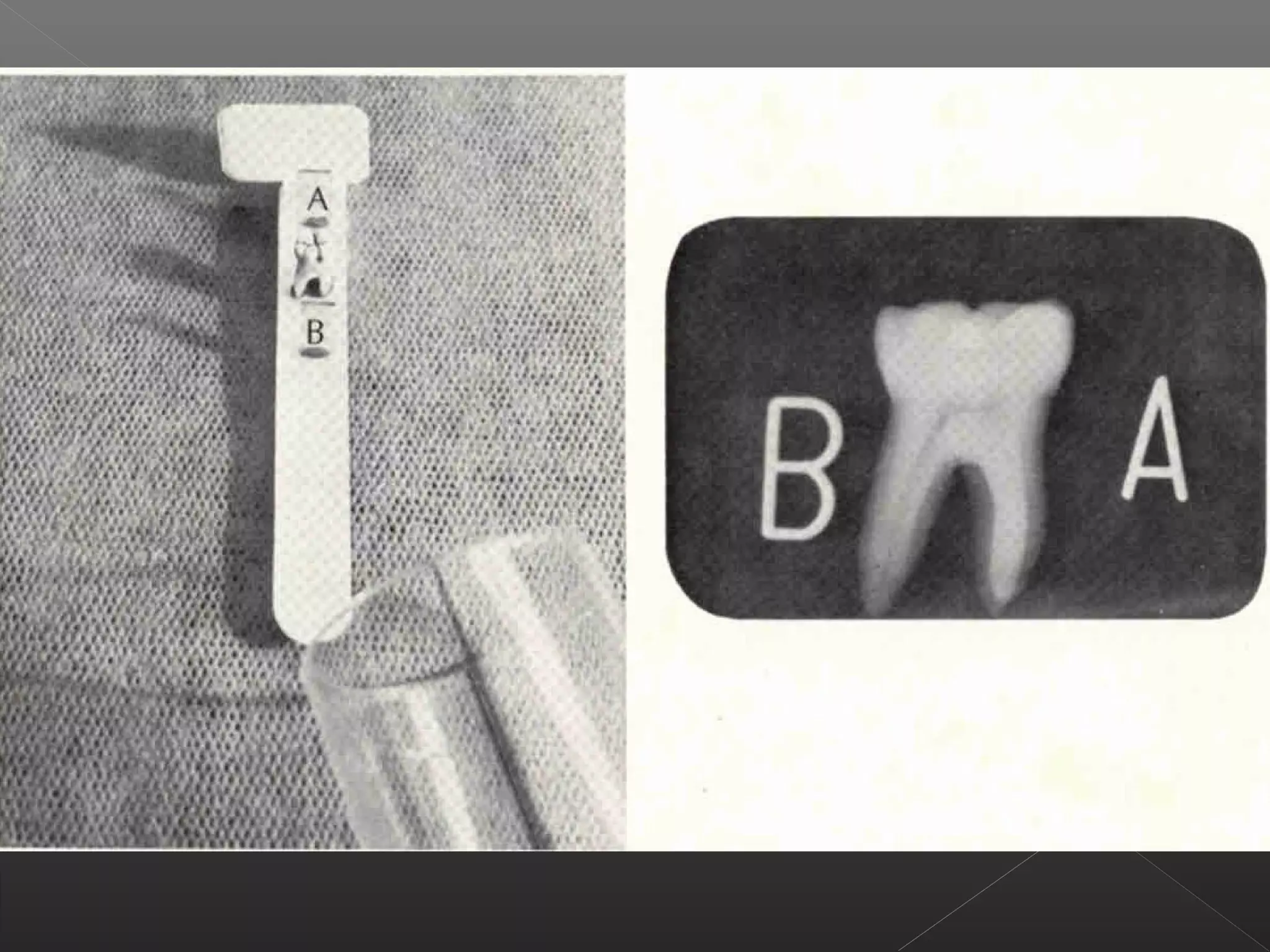
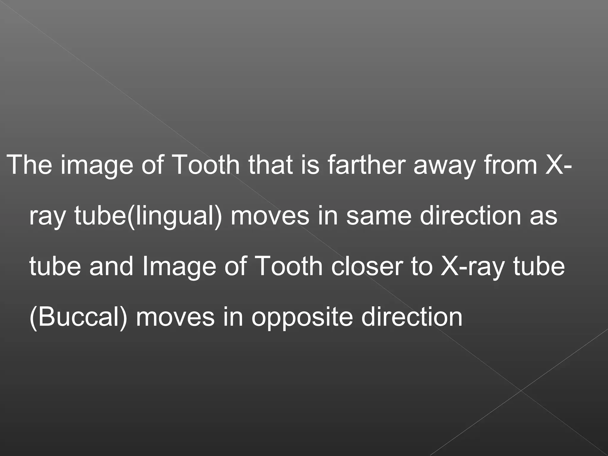

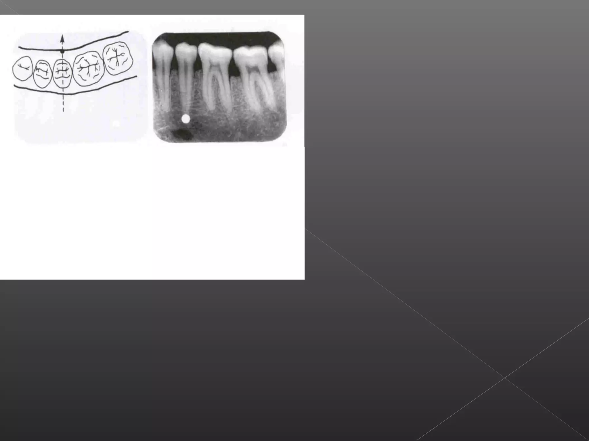
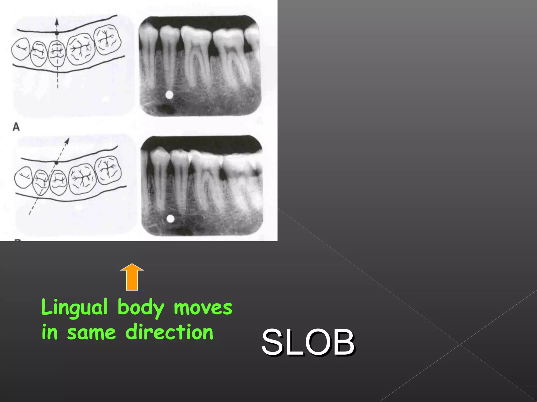
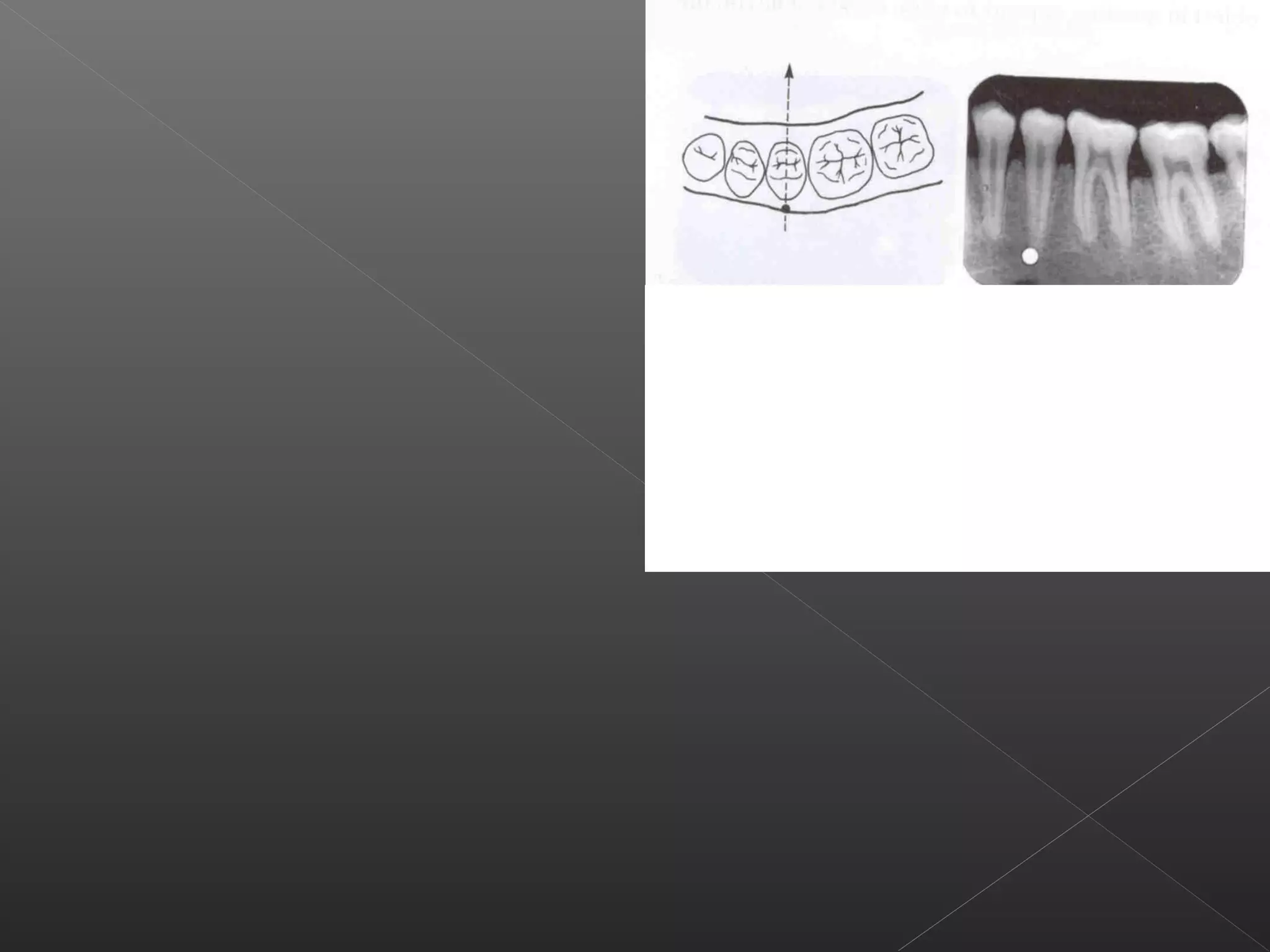
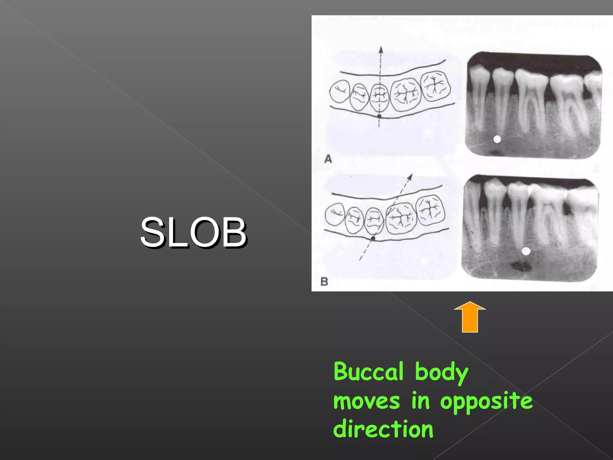


This document discusses techniques for localizing objects using radiography. It describes common reasons for needing to localize foreign bodies or other objects like unerupted teeth, fractures, or tumors. Two main techniques are described: Miller's technique which uses two radiographs at right angles, and Clark's tube-shift technique which analyzes how an object's image shifts when the projection angle is changed. The advantages and disadvantages of each technique are provided.