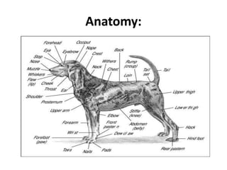
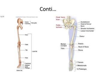
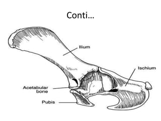
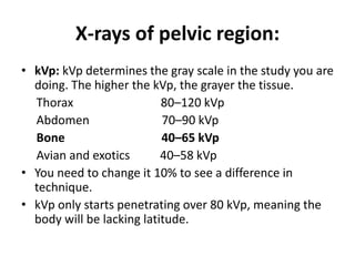
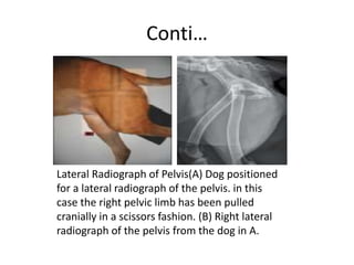
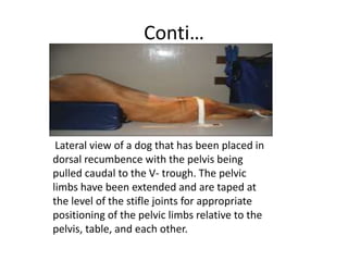
X-rays of the pelvic limb can help diagnose bone and joint abnormalities in dogs. Key views include lateral, ventrodorsal, and oblique images of the pelvis, femur, tibia, fibula, and tarsus. Proper positioning and technical settings like kVp, mA, and exposure time are needed to obtain quality radiographic images. Abnormal findings may include fractures, osteoarthritis, tumors, and ligament injuries. Pelvic x-rays can assist with surgical planning and post-operative evaluation.


![ONFH[AVN HIP] -TRIPLE REGIME -A NOVAL SURGICAL CONCEPT .pptx](https://cdn.slidesharecdn.com/ss_thumbnails/onfhavnhip2026koaconcalicutdrgokuldevdrmashraf-260210064517-213ec005-thumbnail.jpg?width=640&height=640&fit=bounds)


