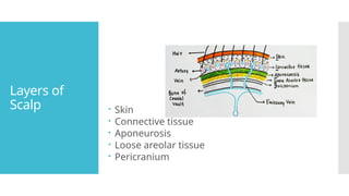
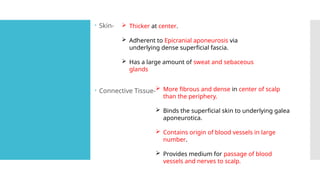
SCALP
ABDULLAH, ABHIJITH, ABHIRAMI, ADHITHYA, ADILA,
ADITHI
Extent
 Anterior- superciliary arches
 Posterior- Superior nuchal line
 Lateral- Superior temporal line
Layers of
Scalp  Skin
 Connective tissue
 Aponeurosis
 Loose areolar tissue
 Pericranium
 Skin-
 Connective Tissue-
 Thicker at center.
 Adherent to Epicranial aponeurosis via
underlying dense superficial fascia.
 Has a large amount of sweat and sebaceous
glands
 More fibrous and dense in center of scalp
than the periphery.
 Binds the superficial skin to underlying galea
aponeurotica.
 Contains origin of blood vessels in large
number.
 Provides medium for passage of blood
vessels and nerves to scalp.
 Aponeurosis-  It is a tough fibrous sheath of connective tissue
underlying the superficial fascia.
 It covers the scalp forming third layer of scalp.
 It forms the last layer of surgical layers of scalp.
 It is freely movable on pericranium.
 Anteriorly it receives insertion of frontalis muscle
 Posteriorly it receives the insertion of occipitalis
muscle.
 Posteriorly it is attached to superior nuchal lines; on
either side to superior temporal lines where it sends
down an expansion which is attached to the
zygomatic arch.
 It provides pathway for passage of emissary veins
which links the extracranial and Dural venous
sinuses.
• Loose areolar tissue-
Made up of loose areolar tissue
 Natural plane of cleavage during craniotomy
Traversed by emissary veins
Emissary veins connects veins of scalp with intracranial dural
venous sinuses
 Pericranium-
Formed by periosteum of bones of vault of skull
Loosely attached to bones,can be easily stripped
Firmly attached to sutural membranes at sutures,which inturn
attached to endocranium
Applied
Aspects
SURGICAL LAYERS OF SCALP
 First three layers of scalp are firmly adherent to each other
and is inseparable.
 These form the scalp proper and is known as the surgical
layers of scalp.
DANGEROUS AREA OF SCALP
 Loose areolar tissue layer is the dangerous layer of scalp.
 Blood or pus can easily collect in this region and can
travel readily through emissary veins into intracranial
dural venous sinus.
Applied
aspects
BLACK EYE
 The swelling caused due to blood accumulation following
a blow on head cannot pass into occipital or temple
region due to bony attachment of occipitofrontalis.
 But blood can track down through eyelids where there is
no bony attachments
 This cause black discoloration of skin around eye due to
hematoma.
Blood Supply
Arterial Supply
On each side, supplied by 5 arteries.
• In front of auricle.
i. Supratrochlear – Branch of ophthalmic
artery from ICA
ii. Supraorbital – Branch of ophthalmic
artery from ICA
iii. Superficial Temporal – Branch of ECA
• Behind the auricle.
i. Posterior Auricular Artery – Branch of
ECA
ii. Occipital Artery – Branch of ECA
Venous Drainage
On each side, drained by 5 veins
1. Supratrochlear + Supraorbital Veins - Join at medial
angle of eye to form the Angular vein, which
continues down as Facial vein
2. Superficial Temporal Vein - Enter parotid gland
where it join maxillary vein to form retromandibular
vein. Anterior division unites with facial vein to form
common facial vein, which drains into Internal jugular
vein
3. Posterior Auricular Vein - unite with posterior
division of retromandibular vein to form External
jugular vein, which drains into subclavian vein
4. Occipital Vein - Terminates in the suboccipital venous
plexus
• Emissary Veins
The veins of scalp communicate with intracranial dural venous sinus through emissary veins.
On each side of midline,
1. Parietal emissary veins - pass through parietal foramen and communicates with superior
sagittal sinus
2. Mastoid emissary vein- pass through mastoid foramen and communicates with sigmoid sinus
Nerve
Supply
IN FRONT OF AURICLE BEHIND THE AURICLE
SENSORY From TRIGEMINAL NERVE
● Supratroachlear
(Opthalmic)
● Supraorbital
(Opthalmic)
● Zygomaticotemporal
(Maxillary)
● Auricotemporal
(Mandibular)
From CERVICAL NERVE
● Great auricular (C2 ,C3)
● Lesser occipital(C2)
● Greater occipital(C2)
● Third occipital(C3)
MOTOR ● Temporal branch of the
facial nerve
● Posterior auricular branch of the
facial nerve
Applied
aspects
SAFETY VALVE HEMATOMA
#Blood from intracranial haemorrhage communicates with sub
aponeurotic space of scalp through fracture lines .
#The signs of cerebral compression does not develop until the sub
aponeurotic space is fully filled with blood.
#So collection of blood in fourth layer is called safety valve hematoma .
CEPHALHYDROCELE
#CSF collects in sub aponeurotic space.
CEPHALHEMATOMA
#Sub periosteal collection of blood.
#Hematoma is surrounded by sutural lines ,so take up the shape of related
bones .
APPLIED
ASPECTS
CAPUT SUCCEDANEUM
#Sub cutaneous oedema over the presenting part of head
at delivery.
#Takes place during passage of head through birth canal
due to interference of venous return.
# Usually occurs over the occiput and crosses the suture
lines .
#Scalp feels soft and margins are partly defined.
#Oedema subsides in few days.

scalp anatomy -layers and applied aspects

  • 1.
    SCALP ABDULLAH, ABHIJITH, ABHIRAMI,ADHITHYA, ADILA, ADITHI
  • 2.
    Extent  Anterior- superciliaryarches  Posterior- Superior nuchal line  Lateral- Superior temporal line
  • 3.
    Layers of Scalp Skin  Connective tissue  Aponeurosis  Loose areolar tissue  Pericranium
  • 4.
     Skin-  ConnectiveTissue-  Thicker at center.  Adherent to Epicranial aponeurosis via underlying dense superficial fascia.  Has a large amount of sweat and sebaceous glands  More fibrous and dense in center of scalp than the periphery.  Binds the superficial skin to underlying galea aponeurotica.  Contains origin of blood vessels in large number.  Provides medium for passage of blood vessels and nerves to scalp.
  • 5.
     Aponeurosis- It is a tough fibrous sheath of connective tissue underlying the superficial fascia.  It covers the scalp forming third layer of scalp.  It forms the last layer of surgical layers of scalp.  It is freely movable on pericranium.  Anteriorly it receives insertion of frontalis muscle  Posteriorly it receives the insertion of occipitalis muscle.  Posteriorly it is attached to superior nuchal lines; on either side to superior temporal lines where it sends down an expansion which is attached to the zygomatic arch.  It provides pathway for passage of emissary veins which links the extracranial and Dural venous sinuses.
  • 6.
    • Loose areolartissue- Made up of loose areolar tissue  Natural plane of cleavage during craniotomy Traversed by emissary veins Emissary veins connects veins of scalp with intracranial dural venous sinuses  Pericranium- Formed by periosteum of bones of vault of skull Loosely attached to bones,can be easily stripped Firmly attached to sutural membranes at sutures,which inturn attached to endocranium
  • 7.
    Applied Aspects SURGICAL LAYERS OFSCALP  First three layers of scalp are firmly adherent to each other and is inseparable.  These form the scalp proper and is known as the surgical layers of scalp. DANGEROUS AREA OF SCALP  Loose areolar tissue layer is the dangerous layer of scalp.  Blood or pus can easily collect in this region and can travel readily through emissary veins into intracranial dural venous sinus.
  • 8.
    Applied aspects BLACK EYE  Theswelling caused due to blood accumulation following a blow on head cannot pass into occipital or temple region due to bony attachment of occipitofrontalis.  But blood can track down through eyelids where there is no bony attachments  This cause black discoloration of skin around eye due to hematoma.
  • 9.
    Blood Supply Arterial Supply Oneach side, supplied by 5 arteries. • In front of auricle. i. Supratrochlear – Branch of ophthalmic artery from ICA ii. Supraorbital – Branch of ophthalmic artery from ICA iii. Superficial Temporal – Branch of ECA • Behind the auricle. i. Posterior Auricular Artery – Branch of ECA ii. Occipital Artery – Branch of ECA
  • 10.
    Venous Drainage On eachside, drained by 5 veins 1. Supratrochlear + Supraorbital Veins - Join at medial angle of eye to form the Angular vein, which continues down as Facial vein 2. Superficial Temporal Vein - Enter parotid gland where it join maxillary vein to form retromandibular vein. Anterior division unites with facial vein to form common facial vein, which drains into Internal jugular vein 3. Posterior Auricular Vein - unite with posterior division of retromandibular vein to form External jugular vein, which drains into subclavian vein 4. Occipital Vein - Terminates in the suboccipital venous plexus
  • 11.
    • Emissary Veins Theveins of scalp communicate with intracranial dural venous sinus through emissary veins. On each side of midline, 1. Parietal emissary veins - pass through parietal foramen and communicates with superior sagittal sinus 2. Mastoid emissary vein- pass through mastoid foramen and communicates with sigmoid sinus
  • 12.
    Nerve Supply IN FRONT OFAURICLE BEHIND THE AURICLE SENSORY From TRIGEMINAL NERVE ● Supratroachlear (Opthalmic) ● Supraorbital (Opthalmic) ● Zygomaticotemporal (Maxillary) ● Auricotemporal (Mandibular) From CERVICAL NERVE ● Great auricular (C2 ,C3) ● Lesser occipital(C2) ● Greater occipital(C2) ● Third occipital(C3) MOTOR ● Temporal branch of the facial nerve ● Posterior auricular branch of the facial nerve
  • 14.
    Applied aspects SAFETY VALVE HEMATOMA #Bloodfrom intracranial haemorrhage communicates with sub aponeurotic space of scalp through fracture lines . #The signs of cerebral compression does not develop until the sub aponeurotic space is fully filled with blood. #So collection of blood in fourth layer is called safety valve hematoma . CEPHALHYDROCELE #CSF collects in sub aponeurotic space. CEPHALHEMATOMA #Sub periosteal collection of blood. #Hematoma is surrounded by sutural lines ,so take up the shape of related bones .
  • 15.
    APPLIED ASPECTS CAPUT SUCCEDANEUM #Sub cutaneousoedema over the presenting part of head at delivery. #Takes place during passage of head through birth canal due to interference of venous return. # Usually occurs over the occiput and crosses the suture lines . #Scalp feels soft and margins are partly defined. #Oedema subsides in few days.