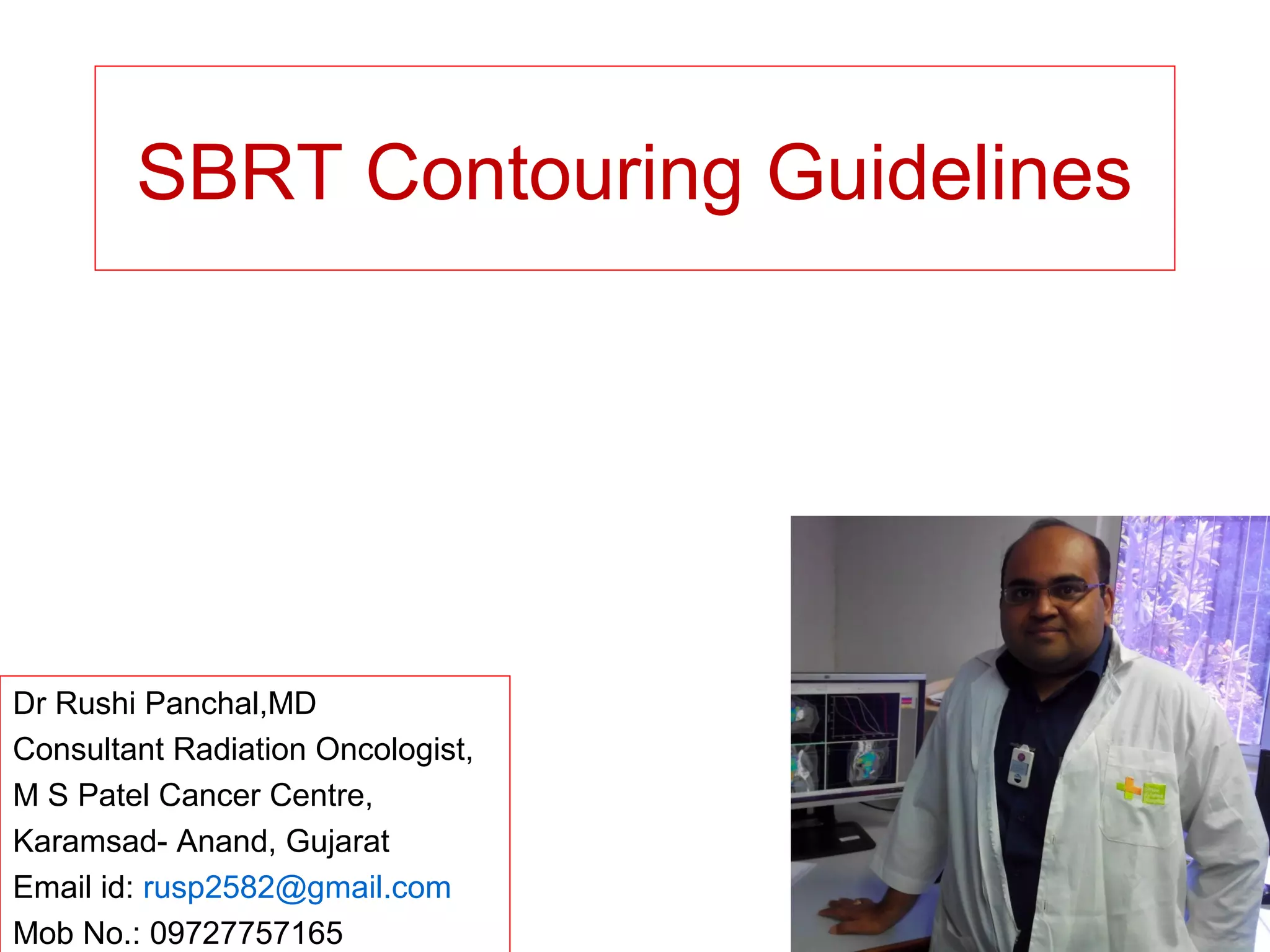
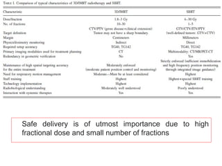
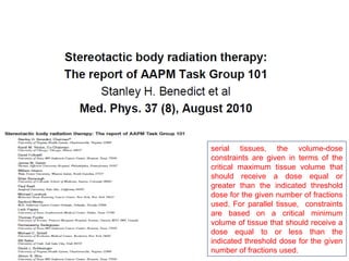
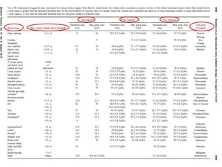
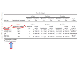
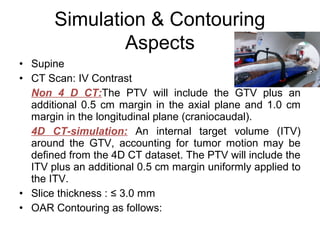
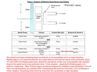
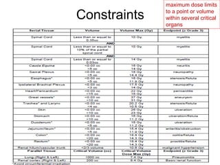
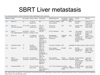
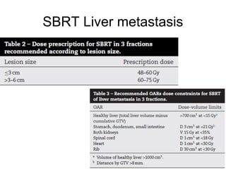
This document provides contouring and treatment planning guidelines for stereotactic body radiation therapy (SBRT). It discusses indications, contraindications, simulation, target volume delineation, organ at risk contouring, dose prescription, and plan evaluation for SBRT treatment of lung, spine, liver, and other cancers. Key considerations include ensuring accurate tumor targeting given organ motion, minimizing dose to nearby organs at risk, and prescribing ablative doses in a small number of fractions to achieve tumor control.
Hypofractionation in early breast cancer is no more a research scholars topic. Multiple studies with robust data have proven its utility. It may hold an important role in many countries with constrained resources. This is a short presentation incorporating important completed and ongoing trials. Feel free to use this.
Accelerated partial breast irradiation is an alternative to whole breast irradiation in carcinoma breast patients Post breast conserving surgery with equivalent outcome, less duration & less burden on the patient.
This is a made easy summary of ICRU 89 guidelines for gynecological brachytherapy. Extra practical questions for MD/DNB Radiotherapy exams are also attached.
EBCTCG METAANALYSIS
INDICATION OF POST OP RADIOTHERAPY
Immobilization devices
Conventional planning
Alignment of the Tangential Beam with the Chest Wall Contour
Doses To Heart & Lung By Tangential Fields
Title: Sense of Taste
Presenter: Dr. Faiza, Assistant Professor of Physiology
Qualifications:
MBBS (Best Graduate, AIMC Lahore)
FCPS Physiology
ICMT, CHPE, DHPE (STMU)
MPH (GC University, Faisalabad)
MBA (Virtual University of Pakistan)
Learning Objectives:
Describe the structure and function of taste buds.
Describe the relationship between the taste threshold and taste index of common substances.
Explain the chemical basis and signal transduction of taste perception for each type of primary taste sensation.
Recognize different abnormalities of taste perception and their causes.
Key Topics:
Significance of Taste Sensation:
Differentiation between pleasant and harmful food
Influence on behavior
Selection of food based on metabolic needs
Receptors of Taste:
Taste buds on the tongue
Influence of sense of smell, texture of food, and pain stimulation (e.g., by pepper)
Primary and Secondary Taste Sensations:
Primary taste sensations: Sweet, Sour, Salty, Bitter, Umami
Chemical basis and signal transduction mechanisms for each taste
Taste Threshold and Index:
Taste threshold values for Sweet (sucrose), Salty (NaCl), Sour (HCl), and Bitter (Quinine)
Taste index relationship: Inversely proportional to taste threshold
Taste Blindness:
Inability to taste certain substances, particularly thiourea compounds
Example: Phenylthiocarbamide
Structure and Function of Taste Buds:
Composition: Epithelial cells, Sustentacular/Supporting cells, Taste cells, Basal cells
Features: Taste pores, Taste hairs/microvilli, and Taste nerve fibers
Location of Taste Buds:
Found in papillae of the tongue (Fungiform, Circumvallate, Foliate)
Also present on the palate, tonsillar pillars, epiglottis, and proximal esophagus
Mechanism of Taste Stimulation:
Interaction of taste substances with receptors on microvilli
Signal transduction pathways for Umami, Sweet, Bitter, Sour, and Salty tastes
Taste Sensitivity and Adaptation:
Decrease in sensitivity with age
Rapid adaptation of taste sensation
Role of Saliva in Taste:
Dissolution of tastants to reach receptors
Washing away the stimulus
Taste Preferences and Aversions:
Mechanisms behind taste preference and aversion
Influence of receptors and neural pathways
Impact of Sensory Nerve Damage:
Degeneration of taste buds if the sensory nerve fiber is cut
Abnormalities of Taste Detection:
Conditions: Ageusia, Hypogeusia, Dysgeusia (parageusia)
Causes: Nerve damage, neurological disorders, infections, poor oral hygiene, adverse drug effects, deficiencies, aging, tobacco use, altered neurotransmitter levels
Neurotransmitters and Taste Threshold:
Effects of serotonin (5-HT) and norepinephrine (NE) on taste sensitivity
Supertasters:
25% of the population with heightened sensitivity to taste, especially bitterness
Increased number of fungiform papillae
Couples presenting to the infertility clinic- Do they really have infertility...Sujoy Dasgupta
Dr Sujoy Dasgupta presented the study on "Couples presenting to the infertility clinic- Do they really have infertility? – The unexplored stories of non-consummation" in the 13th Congress of the Asia Pacific Initiative on Reproduction (ASPIRE 2024) at Manila on 24 May, 2024.
Knee anatomy and clinical tests 2024.pdfvimalpl1234
This includes all relevant anatomy and clinical tests compiled from standard textbooks, Campbell,netter etc..It is comprehensive and best suited for orthopaedicians and orthopaedic residents.
Recomendações da OMS sobre cuidados maternos e neonatais para uma experiência pós-natal positiva.
Em consonância com os ODS – Objetivos do Desenvolvimento Sustentável e a Estratégia Global para a Saúde das Mulheres, Crianças e Adolescentes, e aplicando uma abordagem baseada nos direitos humanos, os esforços de cuidados pós-natais devem expandir-se para além da cobertura e da simples sobrevivência, de modo a incluir cuidados de qualidade.
Estas diretrizes visam melhorar a qualidade dos cuidados pós-natais essenciais e de rotina prestados às mulheres e aos recém-nascidos, com o objetivo final de melhorar a saúde e o bem-estar materno e neonatal.
Uma “experiência pós-natal positiva” é um resultado importante para todas as mulheres que dão à luz e para os seus recém-nascidos, estabelecendo as bases para a melhoria da saúde e do bem-estar a curto e longo prazo. Uma experiência pós-natal positiva é definida como aquela em que as mulheres, pessoas que gestam, os recém-nascidos, os casais, os pais, os cuidadores e as famílias recebem informação consistente, garantia e apoio de profissionais de saúde motivados; e onde um sistema de saúde flexível e com recursos reconheça as necessidades das mulheres e dos bebês e respeite o seu contexto cultural.
Estas diretrizes consolidadas apresentam algumas recomendações novas e já bem fundamentadas sobre cuidados pós-natais de rotina para mulheres e neonatos que recebem cuidados no pós-parto em unidades de saúde ou na comunidade, independentemente dos recursos disponíveis.
É fornecido um conjunto abrangente de recomendações para cuidados durante o período puerperal, com ênfase nos cuidados essenciais que todas as mulheres e recém-nascidos devem receber, e com a devida atenção à qualidade dos cuidados; isto é, a entrega e a experiência do cuidado recebido. Estas diretrizes atualizam e ampliam as recomendações da OMS de 2014 sobre cuidados pós-natais da mãe e do recém-nascido e complementam as atuais diretrizes da OMS sobre a gestão de complicações pós-natais.
O estabelecimento da amamentação e o manejo das principais intercorrências é contemplada.
Recomendamos muito.
Vamos discutir essas recomendações no nosso curso de pós-graduação em Aleitamento no Instituto Ciclos.
Esta publicação só está disponível em inglês até o momento.
Prof. Marcus Renato de Carvalho
www.agostodourado.com
- Video recording of this lecture in English language: https://youtu.be/lK81BzxMqdo
- Video recording of this lecture in Arabic language: https://youtu.be/Ve4P0COk9OI
- Link to download the book free: https://nephrotube.blogspot.com/p/nephrotube-nephrology-books.html
- Link to NephroTube website: www.NephroTube.com
- Link to NephroTube social media accounts: https://nephrotube.blogspot.com/p/join-nephrotube-on-social-media.html
Lung Cancer: Artificial Intelligence, Synergetics, Complex System Analysis, S...Oleg Kshivets
RESULTS: Overall life span (LS) was 2252.1±1742.5 days and cumulative 5-year survival (5YS) reached 73.2%, 10 years – 64.8%, 20 years – 42.5%. 513 LCP lived more than 5 years (LS=3124.6±1525.6 days), 148 LCP – more than 10 years (LS=5054.4±1504.1 days).199 LCP died because of LC (LS=562.7±374.5 days). 5YS of LCP after bi/lobectomies was significantly superior in comparison with LCP after pneumonectomies (78.1% vs.63.7%, P=0.00001 by log-rank test). AT significantly improved 5YS (66.3% vs. 34.8%) (P=0.00000 by log-rank test) only for LCP with N1-2. Cox modeling displayed that 5YS of LCP significantly depended on: phase transition (PT) early-invasive LC in terms of synergetics, PT N0—N12, cell ratio factors (ratio between cancer cells- CC and blood cells subpopulations), G1-3, histology, glucose, AT, blood cell circuit, prothrombin index, heparin tolerance, recalcification time (P=0.000-0.038). Neural networks, genetic algorithm selection and bootstrap simulation revealed relationships between 5YS and PT early-invasive LC (rank=1), PT N0—N12 (rank=2), thrombocytes/CC (3), erythrocytes/CC (4), eosinophils/CC (5), healthy cells/CC (6), lymphocytes/CC (7), segmented neutrophils/CC (8), stick neutrophils/CC (9), monocytes/CC (10); leucocytes/CC (11). Correct prediction of 5YS was 100% by neural networks computing (area under ROC curve=1.0; error=0.0).
CONCLUSIONS: 5YS of LCP after radical procedures significantly depended on: 1) PT early-invasive cancer; 2) PT N0--N12; 3) cell ratio factors; 4) blood cell circuit; 5) biochemical factors; 6) hemostasis system; 7) AT; 8) LC characteristics; 9) LC cell dynamics; 10) surgery type: lobectomy/pneumonectomy; 11) anthropometric data. Optimal diagnosis and treatment strategies for LC are: 1) screening and early detection of LC; 2) availability of experienced thoracic surgeons because of complexity of radical procedures; 3) aggressive en block surgery and adequate lymph node dissection for completeness; 4) precise prediction; 5) adjuvant chemoimmunoradiotherapy for LCP with unfavorable prognosis.
263778731218 Abortion Clinic /Pills In Harare ,sisternakatoto
263778731218 Abortion Clinic /Pills In Harare ,ABORTION WOMEN’S CLINIC +27730423979 IN women clinic we believe that every woman should be able to make choices in her pregnancy. Our job is to provide compassionate care, safety,affordable and confidential services. That’s why we have won the trust from all generations of women all over the world. we use non surgical method(Abortion pills) to terminate…Dr.LISA +27730423979women Clinic is committed to providing the highest quality of obstetrical and gynecological care to women of all ages. Our dedicated staff aim to treat each patient and her health concerns with compassion and respect.Our dedicated group ABORTION WOMEN’S CLINIC +27730423979 IN women clinic we believe that every woman should be able to make choices in her pregnancy. Our job is to provide compassionate care, safety,affordable and confidential services. That’s why we have won the trust from all generations of women all over the world. we use non surgical method(Abortion pills) to terminate…Dr.LISA +27730423979women Clinic is committed to providing the highest quality of obstetrical and gynecological care to women of all ages. Our dedicated staff aim to treat each patient and her health concerns with compassion and respect.Our dedicated group of receptionists, nurses, and physicians have worked together as a teamof receptionists, nurses, and physicians have worked together as a team wwww.lisywomensclinic.co.za/
Flu Vaccine Alert in Bangalore Karnatakaaddon Scans
As flu season approaches, health officials in Bangalore, Karnataka, are urging residents to get their flu vaccinations. The seasonal flu, while common, can lead to severe health complications, particularly for vulnerable populations such as young children, the elderly, and those with underlying health conditions.
Dr. Vidisha Kumari, a leading epidemiologist in Bangalore, emphasizes the importance of getting vaccinated. "The flu vaccine is our best defense against the influenza virus. It not only protects individuals but also helps prevent the spread of the virus in our communities," he says.
This year, the flu season is expected to coincide with a potential increase in other respiratory illnesses. The Karnataka Health Department has launched an awareness campaign highlighting the significance of flu vaccinations. They have set up multiple vaccination centers across Bangalore, making it convenient for residents to receive their shots.
To encourage widespread vaccination, the government is also collaborating with local schools, workplaces, and community centers to facilitate vaccination drives. Special attention is being given to ensuring that the vaccine is accessible to all, including marginalized communities who may have limited access to healthcare.
Residents are reminded that the flu vaccine is safe and effective. Common side effects are mild and may include soreness at the injection site, mild fever, or muscle aches. These side effects are generally short-lived and far less severe than the flu itself.
Healthcare providers are also stressing the importance of continuing COVID-19 precautions. Wearing masks, practicing good hand hygiene, and maintaining social distancing are still crucial, especially in crowded places.
Protect yourself and your loved ones by getting vaccinated. Together, we can help keep Bangalore healthy and safe this flu season. For more information on vaccination centers and schedules, residents can visit the Karnataka Health Department’s official website or follow their social media pages.
Stay informed, stay safe, and get your flu shot today!
Explore natural remedies for syphilis treatment in Singapore. Discover alternative therapies, herbal remedies, and lifestyle changes that may complement conventional treatments. Learn about holistic approaches to managing syphilis symptoms and supporting overall health.
New Directions in Targeted Therapeutic Approaches for Older Adults With Mantl...i3 Health
i3 Health is pleased to make the speaker slides from this activity available for use as a non-accredited self-study or teaching resource.
This slide deck presented by Dr. Kami Maddocks, Professor-Clinical in the Division of Hematology and
Associate Division Director for Ambulatory Operations
The Ohio State University Comprehensive Cancer Center, will provide insight into new directions in targeted therapeutic approaches for older adults with mantle cell lymphoma.
STATEMENT OF NEED
Mantle cell lymphoma (MCL) is a rare, aggressive B-cell non-Hodgkin lymphoma (NHL) accounting for 5% to 7% of all lymphomas. Its prognosis ranges from indolent disease that does not require treatment for years to very aggressive disease, which is associated with poor survival (Silkenstedt et al, 2021). Typically, MCL is diagnosed at advanced stage and in older patients who cannot tolerate intensive therapy (NCCN, 2022). Although recent advances have slightly increased remission rates, recurrence and relapse remain very common, leading to a median overall survival between 3 and 6 years (LLS, 2021). Though there are several effective options, progress is still needed towards establishing an accepted frontline approach for MCL (Castellino et al, 2022). Treatment selection and management of MCL are complicated by the heterogeneity of prognosis, advanced age and comorbidities of patients, and lack of an established standard approach for treatment, making it vital that clinicians be familiar with the latest research and advances in this area. In this activity chaired by Michael Wang, MD, Professor in the Department of Lymphoma & Myeloma at MD Anderson Cancer Center, expert faculty will discuss prognostic factors informing treatment, the promising results of recent trials in new therapeutic approaches, and the implications of treatment resistance in therapeutic selection for MCL.
Target Audience
Hematology/oncology fellows, attending faculty, and other health care professionals involved in the treatment of patients with mantle cell lymphoma (MCL).
Learning Objectives
1.) Identify clinical and biological prognostic factors that can guide treatment decision making for older adults with MCL
2.) Evaluate emerging data on targeted therapeutic approaches for treatment-naive and relapsed/refractory MCL and their applicability to older adults
3.) Assess mechanisms of resistance to targeted therapies for MCL and their implications for treatment selection
How to Give Better Lectures: Some Tips for Doctors
SBRT Contouring Guidelines
1. SBRT Contouring Guidelines
Dr Rushi Panchal,MD
Consultant Radiation Oncologist,
M S Patel Cancer Centre,
Karamsad- Anand, Gujarat
Email id: rusp2582@gmail.com
Mob No.: 09727757165
2. Safe delivery is of utmost importance due to high
fractional dose and small number of fractions
3. Unlike intracranial radiosurgery, where the PTV is immobile within the skull and therefore
relatively easy to fix in relation to the radiation source, at extra-cranial sites there are the
problems of internal organ motion and external patient movement
4.
5. serial tissues, the volume-dose
constraints are given in terms of the
critical maximum tissue volume that
should receive a dose equal or
greater than the indicated threshold
dose for the given number of fractions
used. For parallel tissue, constraints
are based on a critical minimum
volume of tissue that should receive a
dose equal to or less than the
indicated threshold dose for the given
number of fractions used.
6.
7.
8. SBRT
• Primary cancer
• Oligometastatic disease” is defined as a state in which metastases
are limited in number and site and characterized by unusual cancer
biology and behaviour(stable tumour state somewhere between
purely localised and widely metastatic) In this setting local ablative
therapy could have a potential curative role.
• in an analysis of over 5000 patients with lung metastases, survival
after complete surgical resection was 36% at 5 years and 22% at 15
years, Pastorino UB et al. J Thorac Cardiovasc Surg
1997;113:37–49.
10. Indication
• Patients with T1, T2 (≤ 5 cm), T3 (≤ 5 cm), N0, M0 non-
small cell lung cancer(squamous cell carcinoma, adenocarcinoma, large
cell carcinoma, large cell neuroendocrine, and non-small cell carcinoma not otherwise
specified) patients with T3 tumors must have chest wall
primary tumors only.
• Oligometastasis of lung from primary elsewhere.
the 2-year OS was 60.3% for patients with
1–3 metastases compared with 21.9% for
patients with4–5 metastases
12. Simulation & Contouring
Aspects
However, due to respiratory
motion, standard free-breathing
computed tomography both distorts
and generally underestimates the
tumour volume therefore, GTV on
a single free-breathing computed
tomography scan is both an
inaccurate representation of the
tumour dimensions and of the
mean tumour position relative to
other organs
18. • Supine
• CT Scan: IV Contrast
Non 4 D CT:The PTV will include the GTV plus an
additional 0.5 cm margin in the axial plane and 1.0 cm
margin in the longitudinal plane (craniocaudal).
4D CT-simulation: An internal target volume (ITV)
around the GTV, accounting for tumor motion may be
defined from the 4D CT dataset. The PTV will include the
ITV plus an additional 0.5 cm margin uniformly applied to
the ITV.
• Slice thickness : ≤ 3.0 mm
• OAR Contouring as follows:
Simulation & Contouring
Aspects
25. • As per TG 101 for 1/3/5# protocol
• 4 fraction protocol as follows:
Constraints
26. Premedication
• Although not mandatory, it is recommended that patients
receive corticosteroid premedication (e.g.,
Dexamethasone, 4 mg, p.o. in a single dose, or
equivalent) 15-60 minutes before each SBRT treatment
for the intended purpose of modulating immediate
pulmonary inflammatory effects.
• Analgesic premedication to avoid general discomfort
during long treatment durations also is recommended
when appropriate
28. Indication
• Patients with localized spine metastasis from the C1 to
L5 levels (a solitary spine metastasis; 2 separate spine
levels; or up to 3 separate sites); each of the separate
sites must have a maximal involvement of 2 contiguous
vertebral bodies. (e.g. C5, T5-6, and T12)?
• ≥5 NRPS(Numerical Rating Pain Scale) of at least one of
the planned sites for spine radiosurgery
• Zubrod Performance Status : 0-2
• Patients with epidural compression are eligible provided
that there is a ≥ 3 mm gap between the spinal cord and
the edge of the epidural lesion.
• A paraspinal mass (≤ 5 cm in greatest dimension),
contiguous with the spine metastasis
29. There can be multiple small metastatic lesions
shown in other vertebral bodies as shown in
diagram (4) above. The metastatic lesion of
each spine should be less than 20% of the
vertebral body as opposed to the diffuse
vertebral involvement. These small lesions are
often seen in the MRI even when bone scan or
PET was negative. Most of these lesions are not
clinically required to be treated and are
therefore not included in the target volume. Only
the painful spine (pain score≥ 5) is to be
treated .
30. Contraindication
• myeloma or lymphoma
• Non-ambulatory patients
• spinal instability due a compression fracture
• > 50% loss of vertebral body height
• Frank spinal cord compression or displacement or
epidural compression within 3 mm of the spinal cord
• Rapid neurologic decline
• Bony retropulsion causing neurologic abnormality
• Prior radiation to Index Spine
• Patients for whom an MRI of the spine is medically
contraindicated
31.
32. Points to remember
• Patients can have other visceral metastasis, and radioresistant
tumors (including soft tissue sarcomas, melanomas, and renal cell
carcinomas) are eligible
• MRI (contrast is not required but strongly recommended) of the
involved spine to determine the extent of the spine involvement; an
MRI is required as it is superior to a CT scan in delineating the
spinal cord as well as identifying an epidural or paraspinal soft
tissue component.MRI can be used as the required MRI for
treatment planning.
• Patients with mild to moderate neurological signs are eligible. These
neurological signs include radiculopathy, dermatomal sensory
change, and muscle strength of involved extremity 4/5 (lower
extremity for ambulation or upper extremity for raising arms and/or
arm function).
• Imaging study(bone scan, PET, CT scan, or MRI)
33. Simulation & Contouring
Aspects
• Supine
• Immobilized with vacuum bag, alpha cradle, or
stereotactic frames, for cervical spine or cervicothoracic
junctional areas, a rigid head and neck immobilization
• CT-MRI Fusion T1 PC, T2
For soft tissue tumor & Cord
• <3mm slice thickness
35. Target Volume delineation
In any circumstance, when there is an epidural or paraspinal soft tissue tumor component, the visible
epidural or paraspinal tumors are included in the target volume
37. Target Volume delineation
Anterior VB with no epidural extension
Left pedicle, posterolateral VB, and neural
foramen. Involvement of the ventral and left lateral
epidural space, mild spinal canal compromise, and
abutment of the spinal cord
Collapse deformity, ventral epidural disease,
moderate spinal canal compromise, mild spinal
cord displacement, extension to the bilateral
neural
foramina, and paraspinal extension
Pedicle and posterior elements, mild ventral and
right lateral epidural disease narrowing of the right
neural foramina
Centered in the spinous process and extending to
the bilateral lamina, bilateral posterior paraspinal
musculature, and bilateral dorsal epidural space
extension with mild spinal canal compromise
38. Target Volume delineation
Expansile mass in right-sided VB and right posterior
elements with mild right ventral, lateral, and dorsal
epidural disease Involvement of the right neural
foramina
Posterior VB lesion extending into the left neural
foramen with mild spinal canal compromise and left
ventral and lateral epidural extension
Lesion in posterior VB
Diffuse marrow replacement including left pedicle and
articular facets, ventral epidural extension, left lateral
recess extension, and left neural foramen
involvement
Lesion with mild superior and inferior endplate
infractions resulting in mild loss of VB height. Mild
anterior paraspinal extension. Patient underwent
T5 kyphoplasty
40. Dose Prescription & Plan
evaluation
• 16-18Gy/1# or 24Gy/3#
• >/=90% coverage of the target volume by the prescribed
dose( up to 80% is minor deviation)
• 80-90% isodose line is used as prescription line
• Dose inhomogeneity can exist within the target volume
• Outside of the target : >/= to 105% Dose to less than or
equal to 2.0 cc (3cc is major violation)
• >/= to 105% dose to a region within 1.0 cm(1.5 cm is
major violation) from the edge of the target volume
• excludes all doses of greater than or equal to
110%(115% is major violation) of the prescription dose
outside of the target volume.
41. Any spinal cord dose exceeding these constraints is not acceptable and is a major deviation.
Radiosurgery is not recommended for any cases that do not meet the spinal cord constraints. Each
CT slice within the Radiosurgery plan should be checked to screen any unacceptably high radiation
dose to the spinal cord at any particular slice. In this situation, stopping the Radiosurgery or to
perform re-planning. Other Critical organs should be analyzed for radiation dose distribution if any of
them are transected by any radiation field. Exceeding these limits by more than 2.5% constitutes a
minor violation. Exceeding these limits by more than 5% constitutes a major deviation
45. Indication for HCC SBRT
• Unsuitable for resection or transplant or radiofrequency
ablation (RFA)
• Unsuitable for TACE or refractory to TACE
• Barcelona Clinic Liver Cancer Stage (BCLC)
Intermediate (B) or Advanced (C)
• Often used in 1-3 lesions, could be considered for larger
lesions or more extensive disease if there is sufficient
uninvolved liver and liver radiation tolerance can be
respected.
• Child Pugh A/ Early Child Pugh B.
46.
47. Contraindication for HCC SBRT
• Any one hepatocellular carcinoma > 15 cm
• Total maximal sum of hepatocellular carcinoma > 20 cm
• More than 5 discrete intrahepatic parenchymal foci of HCC
• Direct tumor extension into the stomach, duodenum, small bowel or
large bowel
• Measureable common or main branch biliary duct involvement with
HCC
• Extrahepatic metastases or malignant nodes (that enhance with
typical features of HCC) > 2.0 cm, in sum of maximal diameters
(e.g. presence of one 2.4 cm metastatic lymph node or two 1.2 cm
lung lesions). Note that benign non-enhancing periportal
lymphadenopathy is not unusual in the presence of hepatitis and is
permitted, even if the sum of enlarged nodes is > 2.0 cm
• Hepatic insufficiency resulting in clinical jaundice, encephalopathy
and/or variceal bleed.
48. Age is not considered in selection criteria, as even elderly and fragile patients
can safely undergo SBRT. This non-invasive and well-tolerated therapy is a
good option for patients unsuitable for surgery.
The intrinsic radio-sensitivity of tumours is not an issue ,as SBRT can be used
regardless of histopathology thanks to the use of ablative doses, with similar
local control rates in radioresistant and radiosensitive primary tumour.
49. Prerequisite
• For all patients, the following criteria calculated from baseline CT or
MR scans should be met:
• Liver volume minus intrahepatic GTV > 700 cc.
• Intrahepatic tumor GTV/liver volume ratio <80%.
• Minimal distance from GTV to stomach, duodenum, small or large
bowel > 1 cm.
50. Simulation & Contouring
Aspects HCC
• Supine
• Custom immobilization is recommended (e.g. With vacuum
immobilization, patient positioning boards, knee cushions, and/or
breath hold immobilization with active breathing control).
53. • The primary tumor(s) and any tumor vascular thrombi must be treated
• The Gross Tumor Volume (GTV) is defined as all parenchymal and vascular
HCC visualized on contrast enhanced CT and/or MRI, most often best seen on
arterial phase (as hyperintensity) and/or venous or delayed phase (as hypointensity
relative to liver). Vascular HCC thrombi (GTVv) most often are best seen on venous
phase imaging as hypointensity relative to the contrast in the vessel, Vascular HCC
may be combined with parenchymal HCC (labeled as GTVp or GTVpv) if they are to
be treated to the same dose.
• Treatment of non-tumor extrahepatic vascular thrombi, RFA cavities and prior TACE
sites is not recommended. no prophylactic nodal irradiation is allowed.
• It is expected that there will be no expansion from GTV to CTV for the majority
of cases. However, CTV expansions to include regions at high risk for microscopic
disease, including non-tumor vascular (v) thrombi (CTVv), prior TACE (t) sites (CTVt),
or adjacent RFA (r) (or other ablation) sites (CTVr) are permitted.
• PTV will provide a margin around each CTV to compensate for set-up and
internal organ motion
• minimum PTV margin of 4 mm around each CTV is required in all directions (for
example if active breathing control is used with excellent reproducibility). The
maximum permitted PTV margin is 20 mm, expected to be used uncommonly. PTV
margins ≤ 10 mm are a goal.
Target Volume delineation for
HCC
54. OAR Contouring for HCC
• At minimum, these structures are required to be contoured at the
level of the PTV and over any region received > 10 Gy.
• An upper abdominal/liver atlas, posted at the ITC website, may be
used as a guide for contouring.
• all portions of the duodenum are recommended to be contoured
55.
56. Dose Prescription & Plan
evaluation for HCC
• 27.5 Gy - 50 Gy in 5 fractions. The prescription dose may be 50 Gy,
45 Gy, 40 Gy, 35 Gy, 30 Gy or 27.5 Gy in 5 fractions, based on
normal tissue constraints.
• The dose to multiple PTVs may be different.
• The goal is to use the highest allowable prescription dose to the
primary target, while respecting normal tissue constraints. The
minimal planned prescription dose to PTVs is 27.5 Gy.
• The prescription isodose should encompass 95% of PTV. The dose
to multiple PTVs within the same patient may vary, with each
specific covering isodose planned to encompass 95% of each PTV.
The highest allowable doses to the target volumes that
maintain normal tissue constraints should be used. A goal is
that 100% of the CTV is encompassed by the prescription dose.
57. Dose Prescription & Plan
evaluation for HCC
• Dose prescription: Is based on the volume of normal tissues
irradiated (correlated with mean liver dose), as well as proximity of
stomach, duodenum, small and large bowel (GI luminal structures)
to the target volumes, as normal tissue constraints must be
maintained.
• In the absence of adjacent GI luminal structures that may limit dose,
the PTV dose prescription should be as high as possible based on
mean liver dose (MLD, defined as the mean dose to the liver minus
all GTVs)
• Vascular tumor thrombosis (e.g., portal vein thrombosis) dose
should be the same as the HCC prescription dose. However, lower
doses are acceptable if required to maintain normal tissue limits,
58. Dose Prescription & Plan
evaluation for HCC
• Maximum dose within PTV = 150%. If multiple PTVs exist, 150% of
the maximal PTV prescription dose is permitted for all PTVs
• Maximum dose outside PTV = 120% of the maximal PTV
prescription.
• Efforts should be made to keep the 30Gy isodose as conformal as
possible.
• Different isodoses may cover different PTVs. If multiple PTVs, the
MLD should be evaluated with the prescription dose corresponding
to the highest dose level that any PTV is treated.
• Reducing the maximal dose to all luminal gastrointestinal
normal tissues should be a planning priority to reduce the risk
of gastrointestinal toxicity.
59. Dose Prescription & Plan
evaluation for HCC
3 × 15–25 Gy for patients with tumors <3 cm in size and adequate liver
reserve (CP-A5)
5 × 10–12 Gy for patients with tumors between 3 and 5 cm or inadequate liver
reserve (CP-A 6)
10 × 5–5.5 Gy for patients with tumors >5 cm in size or CP-B scores
65. Indication
• Gleason scores 2-6; Clinical stage T1-2a; PSA < 10
ng/mL (PSA should not be obtained within 10 days after
prostate biopsy)
66. Contraindication
• Evidence of distant metastases
• Regional lymph node involvement
• Previous radical surgery (prostatectomy), cryosurgery, or
HIFU for prostate cancer
• Previous pelvic irradiation, prostate Brachytherapy
67. Simulation & Contouring
Aspects
• Supine with Elekta Body fix / Vacuum Cushion + knee
Rest.
• Planning CT Scan with Axial cuts of 2.5 mm or less will
be acquired throughout the pelvis and prostate from the
top of the iliac crests Superior to the perineum inferior
with full bladder and empty rectum.
• MRI images is not must.
• Oral, IV, urethral, and bladder contrast are allowed but
not required.
70. Dose Prescription & Plan
evaluation
• 36.25 Gy/5# 7.25Gy/#. The 5 treatments will be scheduled to be
delivered twice a week over approximately 15-17 days. A minimum
of 72 hours and a maximum of 96 hours should separate each
treatment. No more than 2 fractions will be delivered per week. The
total duration of treatment will be no shorter than 15 days and no
longer than 17 days.
• prescription dose should cover a minimum of 95% of the PTV.
• The minimum dose within the PTV to a point that is 0.03 cc in size
must be ≥95% of the prescribed dose.
• The maximum dose within the PTV is 7% above the prescribed dose
for a point that is 0.03 cc in size.