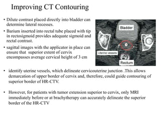
This is a made easy summary of ICRU 89 guidelines for gynecological brachytherapy. Extra practical questions for MD/DNB Radiotherapy exams are also attached.
EBCTCG METAANALYSIS
INDICATION OF POST OP RADIOTHERAPY
Immobilization devices
Conventional planning
Alignment of the Tangential Beam with the Chest Wall Contour
Doses To Heart & Lung By Tangential Fields
Hypofractionation in early breast cancer is no more a research scholars topic. Multiple studies with robust data have proven its utility. It may hold an important role in many countries with constrained resources. This is a short presentation incorporating important completed and ongoing trials. Feel free to use this.
Techniques for Inguinal/Groin IrradiationAjeet Gandhi
Inguinal radiotherapy delivery is many a times a complex dosimetric uncertainty and we need to judiciously choose the technique for best patient outcome
A review of advances in Brachytherapy treatment planning and delivery in last decade or so, with main focus on brachytherapy for Prostate cancer, Breast cancer and Cervical cancer
the role of brachytherapy in oral cavity carcinoma.
physics of brachytherapy
radiobiology of brachytherapy
clinical application in tongue, buccal mucosa cancer
Energy Absorption in Radiobiology
Ionization vs. Excitation
Ionizing Versus Non-ionizing Radiation
Absorption Mechanisms
Ionization by alpha particle, Xray & neutron
Cancer of Right Breast with Single Liver Metastasis - Simultaneous Treatment ...Kanhu Charan
Cancer of Right Breast with Single Liver Metastasis - Simultaneous
Treatment of Chest Wall with Radiotherapy for Carcinoma Breast and
SBRT for Liver Lesion - Procedural Details of the Complex Procedure
Management of cacrinoma cervix: Techniques of radiotherapy (2D conventional, 3D Conformal radiotherapy (3DCRT) and IMRT with a review of various contouring guidelines.
Cancer of Right Breast with Single-Liver MetastasisSimultaneous Treatment of ...Kanhu Charan
Cancer of Right Breast with Single-Liver Metastasis Simultaneous Treatment of Chest Wall with Radiotherapy for Carcinoma Breast and SBRT for Liver Lesion: Procedural
Details of the Complex Procedure
Physical Models For Time Dose & FractionationIsha Jaiswal
Physical Models For Time Dose & Fractionation
Strandqvist Plot
Cohen’s Formula
Fowler Concepts
NSD Model
TDF model
Target Theory
L Q model
BED calculation of different fractionation regimen
Introduction
Time dose & fractionation
Therapeutic index
Four R’s Of Radiobiology
Radiation response
Survival Curves Of Early & Late Responding Cells
Various fractionation schedules
Clinical trials of altered fractionation
General management
Management of low grade gliomas: overview
Pilocytic astrocytoma
non pilocytic/diffuse infiltrating gliomas
Management of high grade gliomas: overview
Anaplastic gliomas
Glioblastoma multiformae
describes relationship between radiation dose and the fraction of cells that “survive” that dose
model of cell killing
target model
linear quadratic model
Knee anatomy and clinical tests 2024.pdfvimalpl1234
This includes all relevant anatomy and clinical tests compiled from standard textbooks, Campbell,netter etc..It is comprehensive and best suited for orthopaedicians and orthopaedic residents.
Acute scrotum is a general term referring to an emergency condition affecting the contents or the wall of the scrotum.
There are a number of conditions that present acutely, predominantly with pain and/or swelling
A careful and detailed history and examination, and in some cases, investigations allow differentiation between these diagnoses. A prompt diagnosis is essential as the patient may require urgent surgical intervention
Testicular torsion refers to twisting of the spermatic cord, causing ischaemia of the testicle.
Testicular torsion results from inadequate fixation of the testis to the tunica vaginalis producing ischemia from reduced arterial inflow and venous outflow obstruction.
The prevalence of testicular torsion in adult patients hospitalized with acute scrotal pain is approximately 25 to 50 percent
Recomendações da OMS sobre cuidados maternos e neonatais para uma experiência pós-natal positiva.
Em consonância com os ODS – Objetivos do Desenvolvimento Sustentável e a Estratégia Global para a Saúde das Mulheres, Crianças e Adolescentes, e aplicando uma abordagem baseada nos direitos humanos, os esforços de cuidados pós-natais devem expandir-se para além da cobertura e da simples sobrevivência, de modo a incluir cuidados de qualidade.
Estas diretrizes visam melhorar a qualidade dos cuidados pós-natais essenciais e de rotina prestados às mulheres e aos recém-nascidos, com o objetivo final de melhorar a saúde e o bem-estar materno e neonatal.
Uma “experiência pós-natal positiva” é um resultado importante para todas as mulheres que dão à luz e para os seus recém-nascidos, estabelecendo as bases para a melhoria da saúde e do bem-estar a curto e longo prazo. Uma experiência pós-natal positiva é definida como aquela em que as mulheres, pessoas que gestam, os recém-nascidos, os casais, os pais, os cuidadores e as famílias recebem informação consistente, garantia e apoio de profissionais de saúde motivados; e onde um sistema de saúde flexível e com recursos reconheça as necessidades das mulheres e dos bebês e respeite o seu contexto cultural.
Estas diretrizes consolidadas apresentam algumas recomendações novas e já bem fundamentadas sobre cuidados pós-natais de rotina para mulheres e neonatos que recebem cuidados no pós-parto em unidades de saúde ou na comunidade, independentemente dos recursos disponíveis.
É fornecido um conjunto abrangente de recomendações para cuidados durante o período puerperal, com ênfase nos cuidados essenciais que todas as mulheres e recém-nascidos devem receber, e com a devida atenção à qualidade dos cuidados; isto é, a entrega e a experiência do cuidado recebido. Estas diretrizes atualizam e ampliam as recomendações da OMS de 2014 sobre cuidados pós-natais da mãe e do recém-nascido e complementam as atuais diretrizes da OMS sobre a gestão de complicações pós-natais.
O estabelecimento da amamentação e o manejo das principais intercorrências é contemplada.
Recomendamos muito.
Vamos discutir essas recomendações no nosso curso de pós-graduação em Aleitamento no Instituto Ciclos.
Esta publicação só está disponível em inglês até o momento.
Prof. Marcus Renato de Carvalho
www.agostodourado.com
NVBDCP.pptx Nation vector borne disease control programSapna Thakur
NVBDCP was launched in 2003-2004 . Vector-Borne Disease: Disease that results from an infection transmitted to humans and other animals by blood-feeding arthropods, such as mosquitoes, ticks, and fleas. Examples of vector-borne diseases include Dengue fever, West Nile Virus, Lyme disease, and malaria.
The prostate is an exocrine gland of the male mammalian reproductive system
It is a walnut-sized gland that forms part of the male reproductive system and is located in front of the rectum and just below the urinary bladder
Function is to store and secrete a clear, slightly alkaline fluid that constitutes 10-30% of the volume of the seminal fluid that along with the spermatozoa, constitutes semen
A healthy human prostate measures (4cm-vertical, by 3cm-horizontal, 2cm ant-post ).
It surrounds the urethra just below the urinary bladder. It has anterior, median, posterior and two lateral lobes
It’s work is regulated by androgens which are responsible for male sex characteristics
Generalised disease of the prostate due to hormonal derangement which leads to non malignant enlargement of the gland (increase in the number of epithelial cells and stromal tissue)to cause compression of the urethra leading to symptoms (LUTS
These lecture slides, by Dr Sidra Arshad, offer a quick overview of physiological basis of a normal electrocardiogram.
Learning objectives:
1. Define an electrocardiogram (ECG) and electrocardiography
2. Describe how dipoles generated by the heart produce the waveforms of the ECG
3. Describe the components of a normal electrocardiogram of a typical bipolar leads (limb II)
4. Differentiate between intervals and segments
5. Enlist some common indications for obtaining an ECG
Study Resources:
1. Chapter 11, Guyton and Hall Textbook of Medical Physiology, 14th edition
2. Chapter 9, Human Physiology - From Cells to Systems, Lauralee Sherwood, 9th edition
3. Chapter 29, Ganong’s Review of Medical Physiology, 26th edition
4. Electrocardiogram, StatPearls - https://www.ncbi.nlm.nih.gov/books/NBK549803/
5. ECG in Medical Practice by ABM Abdullah, 4th edition
6. ECG Basics, http://www.nataliescasebook.com/tag/e-c-g-basics
Couples presenting to the infertility clinic- Do they really have infertility...Sujoy Dasgupta
Dr Sujoy Dasgupta presented the study on "Couples presenting to the infertility clinic- Do they really have infertility? – The unexplored stories of non-consummation" in the 13th Congress of the Asia Pacific Initiative on Reproduction (ASPIRE 2024) at Manila on 24 May, 2024.
TEST BANK for Operations Management, 14th Edition by William J. Stevenson, Ve...kevinkariuki227
TEST BANK for Operations Management, 14th Edition by William J. Stevenson, Verified Chapters 1 - 19, Complete Newest Version.pdf
Ethanol (CH3CH2OH), or beverage alcohol, is a two-carbon alcohol
that is rapidly distributed in the body and brain. Ethanol alters many
neurochemical systems and has rewarding and addictive properties. It
is the oldest recreational drug and likely contributes to more morbidity,
mortality, and public health costs than all illicit drugs combined. The
5th edition of the Diagnostic and Statistical Manual of Mental Disorders
(DSM-5) integrates alcohol abuse and alcohol dependence into a single
disorder called alcohol use disorder (AUD), with mild, moderate,
and severe subclassifications (American Psychiatric Association, 2013).
In the DSM-5, all types of substance abuse and dependence have been
combined into a single substance use disorder (SUD) on a continuum
from mild to severe. A diagnosis of AUD requires that at least two of
the 11 DSM-5 behaviors be present within a 12-month period (mild
AUD: 2–3 criteria; moderate AUD: 4–5 criteria; severe AUD: 6–11 criteria).
The four main behavioral effects of AUD are impaired control over
drinking, negative social consequences, risky use, and altered physiological
effects (tolerance, withdrawal). This chapter presents an overview
of the prevalence and harmful consequences of AUD in the U.S.,
the systemic nature of the disease, neurocircuitry and stages of AUD,
comorbidities, fetal alcohol spectrum disorders, genetic risk factors, and
pharmacotherapies for AUD.
Tom Selleck Health: A Comprehensive Look at the Iconic Actor’s Wellness Journeygreendigital
Tom Selleck, an enduring figure in Hollywood. has captivated audiences for decades with his rugged charm, iconic moustache. and memorable roles in television and film. From his breakout role as Thomas Magnum in Magnum P.I. to his current portrayal of Frank Reagan in Blue Bloods. Selleck's career has spanned over 50 years. But beyond his professional achievements. fans have often been curious about Tom Selleck Health. especially as he has aged in the public eye.
Follow us on: Pinterest
Introduction
Many have been interested in Tom Selleck health. not only because of his enduring presence on screen but also because of the challenges. and lifestyle choices he has faced and made over the years. This article delves into the various aspects of Tom Selleck health. exploring his fitness regimen, diet, mental health. and the challenges he has encountered as he ages. We'll look at how he maintains his well-being. the health issues he has faced, and his approach to ageing .
Early Life and Career
Childhood and Athletic Beginnings
Tom Selleck was born on January 29, 1945, in Detroit, Michigan, and grew up in Sherman Oaks, California. From an early age, he was involved in sports, particularly basketball. which played a significant role in his physical development. His athletic pursuits continued into college. where he attended the University of Southern California (USC) on a basketball scholarship. This early involvement in sports laid a strong foundation for his physical health and disciplined lifestyle.
Transition to Acting
Selleck's transition from an athlete to an actor came with its physical demands. His first significant role in "Magnum P.I." required him to perform various stunts and maintain a fit appearance. This role, which he played from 1980 to 1988. necessitated a rigorous fitness routine to meet the show's demands. setting the stage for his long-term commitment to health and wellness.
Fitness Regimen
Workout Routine
Tom Selleck health and fitness regimen has evolved. adapting to his changing roles and age. During his "Magnum, P.I." days. Selleck's workouts were intense and focused on building and maintaining muscle mass. His routine included weightlifting, cardiovascular exercises. and specific training for the stunts he performed on the show.
Selleck adjusted his fitness routine as he aged to suit his body's needs. Today, his workouts focus on maintaining flexibility, strength, and cardiovascular health. He incorporates low-impact exercises such as swimming, walking, and light weightlifting. This balanced approach helps him stay fit without putting undue strain on his joints and muscles.
Importance of Flexibility and Mobility
In recent years, Selleck has emphasized the importance of flexibility and mobility in his fitness regimen. Understanding the natural decline in muscle mass and joint flexibility with age. he includes stretching and yoga in his routine. These practices help prevent injuries, improve posture, and maintain mobilit
Report Back from SGO 2024: What’s the Latest in Cervical Cancer?bkling
Are you curious about what’s new in cervical cancer research or unsure what the findings mean? Join Dr. Emily Ko, a gynecologic oncologist at Penn Medicine, to learn about the latest updates from the Society of Gynecologic Oncology (SGO) 2024 Annual Meeting on Women’s Cancer. Dr. Ko will discuss what the research presented at the conference means for you and answer your questions about the new developments.
Explore natural remedies for syphilis treatment in Singapore. Discover alternative therapies, herbal remedies, and lifestyle changes that may complement conventional treatments. Learn about holistic approaches to managing syphilis symptoms and supporting overall health.
Are There Any Natural Remedies To Treat Syphilis.pdf
image guided brachytherapy carcinoma cervix
1. Brachytherapy In Carcinoma Cervix:
Part II
Made by: Dr. isha jaiswal
Guided by: Dr. Sandip Barik
Date: 28th July 2015
2. Contents
• Advances in gynecological brachytherapy
• ABS recommendations for HDR BT
• Image guided brachytherapy
• Steps in IGBT
• Imaging
• Limitations of (2 D)radiographic imaging
• Volumetric imaging
• GEC-ESTRO recommendations for cervical cancer BT
3. ADVANCES IN GYNAECOLOGICAL BRACHYTHERAPY
• Remote after loading HDR BT
• Applicator development: Intracavitary (IC), Interstitial (IS) & IC+IS
• In corporation of Newer Imaging Modalities: CT, MR, PET, etc.
• Advances in Treatment Planning Systems
• IGBT: Volume Based Brachytherapy
4. • The American Brachytherapy Society (ABS) Gynaecologic Cervical
Cancer Task group has developed general criteria for the management of
cervical cancer, designed to guide Radiation Oncologists and assist in
making decisions regarding therapy
5. Gynaecologic Brachytherapy*
General Inclusion Criteria:
Stage IA1-IA2
1B1:
IB2 - IVA :radically with concurrent chemo radiation followed by BT
Stage IVB cervical cancer :may be palliative treated with BT with or without EBRT
Exclusion Criteria:
Absolute contraindications to radical treatment
Prior pelvic radiotherapy with brachytherapy
Life expectancy < 6 month
The use of (IMRT) or 3D CRT is not a substitute for brachytherapy.
*American Brachytherapy society cervical cancer task group
6. All treatment, including EBRT & BT must be completed within 56 days
from initiation.
HDR-BT commences after 45Gy with up to 2 #/week during the conclusion
of EBRT and during the parametrial boost portion of treatment.
BT may be initiated earlier(but no earlier than approximately 20 Gy), if the
physician determines that the applicator placed at this time point is adequate.
Chemotherapy is not typically given on the days of HDR-BT
Timing of BT
American Brachytherapy society cervical cancer task group
7. Pre-implant evaluation
Applicator selection & insertion
Imaging
Delineation
Prescription
Treatment planning
Target dose specification
Treatment delivery
Steps In IGBT
American Brachytherapy society cervical cancer task group
8. patient should have a detailed gynecologic examination
assess the anatomy, residual tumor & decide brachytherapy applicator best suited
appropriate medical evaluations and a pre procedure anesthesia assessment
instructions on fasting, bowel preparation, and preoperative testing, including
laboratory studies, should be provided
Pre-implant evaluation
9. Variety of applicators
tandem and ovoids
tandem and ring
tandem and cylinder,
tandem and ovoid or ring with guides for interstitial needles
10. APPLICATOR SELECTION
1-Intracavitary applicators
Patients with an intact uterus should have a tandem placed;
2-Interstitial application
recommended for
extensive cervical lesions
extension to the lateral parametria or pelvic sidewall
lower vaginal extension,
a narrow vaginal apex
os not negotiable
poorly fitting intracavitary applicators
3-Both
if patient had a prior supracervical hysterectomy, a short tandem with interstitial
implant can be used
for patients with large, bulky tumors, to enhance coverage of cervix and reduce the
dose to the organs at risk (OAR)
11. 1-Tandem & ovoid
• most widely used applicator
• largest ovoid that can be placed snugly into the lateral fornices should be
used however oversized ovoids can result in displacement of the applicator
down the vagina.
• Fletcher-Suit ovoids provide a wider surface area on the cervix as
compared to other applicators
2-Tandem and ring applicator :
• Indication:
for patients who have shallow vaginal fornices.
• Disadvantage:
slightly narrower distribution than ovoid
result in a higher vaginal dose.
12. 3-Tandem and cylinder:
• Indications:
cases with upper vaginal stenosis (inability to place ovoids or a ring)
cases with superficial disease involving the lower vagina
• Disadvantage:
dose distribution significantly different
higher doses to the bladder and rectum, lower dose to the parametrium
4-Tandem and ring or ovoids with short interstitial needles
• for patients with large, bulky tumors, to cover the depth of the cervix and
reduce the dose to the organs at risk (OAR)
• The main part of delivered dose results from loading in intrauterine
applicators, whereas needles are used to shape, fine tune, and enlarge the
treated volume.
• improve lateral coverage by an additional 10 mm when compared with
Intracavitary applicators alone
13. Tandem and ovoid, tandem and ring or tandem and cylinders for
intracavitary applications, inserted free hand
Hollow interstitial needles inserted either freehand or with
template or ultrasound guidance .
APPLICATOR INSERTION
14. Imaging after applicator insertion
X ray
USG
CT
MRI
When available, the ABS recommends the use of cross-sectional
imaging, such as magnetic resonance imaging
(MRI) or computed tomography (CT), to obtain measurements
of tumor size, volume, and extent of disease
16. For determination of target
• point based dosimetry
• point A may overestimate or underestimate the tumor dose based on
3D imaging*
• no optimization:
• tumor coverage relies on tumor volume at time of BT, larger tumors
requiring greater optimization to be adequately covered by the prescribed
isodose line
• Kim et al** found that dose to point A was significantly lower than
the D90 for HR-CTV calculated using 3D image-based optimization
• dose escalation not possible
• *Kim RY, Pareek P. Radiography-based treatment planning compared with computed tomography (CT)-based treatment planning for
intracavitary brachytherapy in cancer of the cervix: analysis of dose-volume histograms. Brachytherapy 2003;2:200–206.
• **Kim H, Beriwal S, Houser C, et al. Dosimetric analysis of 3D image-guided HDR brachytherapy planning for the treatment of
cervical cancer: is point A-based dose prescription still valid in image-guided brachytherapy? Med Dosim 2011;36:166–170.
17. AIM
to analyze dosimetric outcome of 3D IGBT &compare dose coverage of HRCTV to
traditional Point A dose.
• N=32patients (stage IA2-IIIB cervical cancer) treated with IGBT
• dose: 5.0-6.0 Gy/# ×5 fractions.
• delineation of CTV as per GYN GEC/ESTRO guidelines.
• D90 for HRCTV was 80-85 Gy,
• D2cc of bladder, rectum, and sigmoid was limited to 85 Gy, 75 Gy &75 Gy.
• RESULTS
• The mean D90 for HRCTV was 83.2 ± 4.3 Gy SD significantly higher (p
<0.0001) than mean value of Point A dose (78.6 ± 4.4 Gy).
• The dose levels of the OARs were within acceptable limits
• Dose to Point A was found to be significantly lower than the D90 for HRCTV
• Image-based 3D brachytherapy provides adequate dose coverage to HRCTV,
with acceptable dose to OARs in most patients.
18. • ICRU bladder point:
• Foley Bulb in the trigone of bladder with7 cc of dilute contrast is used
• only report point estimates.
• wide range of anatomic variations in bladder points along he length of implant
• doses may be different at bladder base & neck, multiple points have to be taken
• ICRU point may underestimate maximum doses to the OAR, in particular
for the bladder
• ICRU bladder volume point does not represent the hottest part of the bladder that
usually falls about 2 cm superior. highest dose often is about 2-4 times the dose
at the bulb
For determination of OAR
Bladder Point
19. Rectal point
ICRU rectal point:
• rectal markers is used which tend to lie on posterior wall of rectum while the
anterior wall is at greater risk.
• Stiff markers can move rectum, flimsy ones are difficult to push deep.
• ICRU rectal point doesn’t usually represent the maximum rectal does, which,
again often is 2-4 cm cephalad.
• maximum does is up to 3 times the ICRU point
None of this localizes the superior bowel - an organ very much at risk.
20. Ultrasound
Can be very useful
during tandem insertion
Localizing the cervical cannel when obscured by large tumor,
detecting a retroverted uterus before tandem insertion.
Determines uterine width & height
US does not define the target volume as clearly as MRI
Transrectal US may assist with interstitial brachytherapy when other
imaging modalities are not available
21. VOLUMETRIC 3 D IMAGING
• Aim to
1. localize the source positions.
2. Localize the target.
3. Localize the organs at risk.
4. Determine the relationships between all the above
22. CT SCAN
Plain CT scan is obtained after applicator insertion with 3-5mm cuts
Advantages:
• verifies proper placement of applicator
• reasonable estimate of the location of uterus
• fairly good for visualizing bladder and rectum.
• analyses 3D BT dose distribution
• depicts changes in the OAR related to tumor shrinkage & filling status.
• 3D dose calculations & optimisation possible
• OAR dosimetry based on CT is similar to that based on MRI when optimized
similarly*
• long experience in treatment planning for EBRT
• readily available in radiotherapy departments
• *Eskander RN, Scanderbeg D, Saenz CC, et al. Comparison of computed tomography and magnetic
resonance imaging in cervical cancer brachytherapy target and normal tissue contouring. Int J Gynecol
Cancer 2010;20:47–53
23. Problems with CT treatment planning
• produce artifact with metallic applicators
• expensive
• GTV not identified
• overestimate tumor contours compared to MRI (although additional width
contoured on CT may not be of detriment)
• fails to provide differentiation between the uterus, cervix, pariuterine tissues
so CT-based contouring guidelines recommend delineating entire cervix and uterus.
not provide sufficient detail of tumor if selected dose escalation is required,
• contouring sigmoid difficult due to lack of contrast
• contrast placed in OAR may cause artifact.in contouring wall of organ.
• requires moving patient after application from CT to treatment room: can
produce motion artifact that nullifies the increased accuracy of IGBT
*Viswanathan AN et al. Computed tomography versus magnetic resonance imaging-based contouring in cervical cancer
brachytherapy: results of a prospective trial and preliminary guidelines for standardized contours. Int J Radiat Oncol
Biol Phys2007;68:491–498
24. MRIADVANTAGES
• multiplaner imaging
• excellent soft tissue contrast
• better visualisation of tumor & parametrium. involvement
• differentiate between uterus, cervix, tumor, other pelvic tissues & OAR
• particularly useful in patients with advanced or deeply infiltrating tumors.
• specific signal intensities allow for distinct separation on T1- and T2 WI
cervix (low T1, low T2), parametrium (high T1, high T2), tumor (low T1,
high T2)
• regression of cervical tumors can be documented so dose escalation possible
• organ wall may be more clearly visualized for contouring OAR.
DISADVANTAGES
• requires special applicators. non-ferromagnetic, metal or plastic/graphite
• very expensive
• can produce motion artifact
25. The criteria for an adequate implant
(regardless of imaging modality used)
• The tandem should bisect the ovoids on an AP and lateral image.
• On a lateral image, the ovoids should not be displaced inferiorly from the flange
(cervical stop) and should be as symmetrical as possible (should overlap one
another).
• The tandem should be approximately one-half to one third the distance between the
symphysis and the sacral promontory, approximately equidistant between a
contrast-filled bladder and rectum-sigmoid.
• The superior tip of the tandem should be located below the sacral promontory
within the pelvis
• Radio-opaque packing will be visible on radiographic images and should be placed
anterior and posterior to the ovoids, with no packing visible superior to the ovoids.
27. DELINEATION
Advances in image guidance for applicator insertion and treatment planning have
resulted in 3D tissue contouring guidelines (GEC-ESTRO)
After insertion of applicators, the target volumes and normal-tissue structures are
delineated on images in TPS.
The delineation is to be performed at time of each BT application.
The delineation process is based on clinical examination at diagnosis and at BT and on a
set of sectional images (preferably MRI T2 weighted) taken at diagnosis and at BT with
applicator in place.
29. Gross tumor volume: GTV
Gross tumour volume (diagnosis) (GTVD)
includes macroscopic tumour extension at diagnosis as detected by
clinical examination (visualisation and palpation) and as visualised on MRI:
Gross tumour volume (BT) (GTVB1, GTVB2, GTVB3,.)
includes macroscopic tumour extension at time of BT as detected by clinical examination
and as visualised on MRI:
In patients treated with upfront BT or with BT alone, GTVB is identical with GTVD.
30. Clinical target volume: CTV
,
HR-CTV :with macroscopic ds.
includes GTV + whole cervix + presumed
extracervical tumour extension+ residual ds
Pathologic residual tissue(s) as defined by
palpable indurations and/or grey zones in
parametria, uterine corpus, vagina or rectum and
bladder are included in HR-CTV.
No safety margin are added.
IR-CTV :represent significant microscopic ds
includes HR-CTV +different safety margins are
added (minimal 5 to 15 mm)
LR-CTV :including potential microscopic spread treated
by surgery and/or EBRT
31. IR-CTV includes HRCTV
different safety margins are added according to potential spread
5 mm AP limited by bladder or rectum
10 mm cranially into uterine corpus
10 mm caudally below the cervical os into the vagina.
10 mm laterally into both parametria, usually representing internal third of the
parametrium
+ 5 mm if endocervical tumour in BT only
+ 5 mm laterally if lateral macroscopic tumour in BT only
IRCTV: for limited disease (tumour size<4cm)
Schematic diagram for limited disease, with GTV, high risk CTV and intermediate risk CTV :coronal and transversal view
32. IR-CTV: for extensive disease
• IR CTV is based on macroscopic tumour at diagnosis (GTVD) which
is superimposed on HR-CTV at time of BT (ie GTVBT + Cervix +
extracervical tissue)taking original anatomical tumour spread as
reference
• Margins are added depending on the regression in initial tumor extent
present at diagnosis
Schematic representation for HR and IR CTV lateral parametrial limits for extensive ds
33. IR-CTV extensive disease :safety margins
Complete remission
IRCTV=HR CTV(red) + GTVdiagnosis(blue dot)
no safety margin
Partial response: IRCTV include
HRCTV ie GTV + cervix+ extracervical residual ds (e.g. parametria)
+GTVdiagnosis(blue dot)
+safety margin of minimum 10 mm added into the direction of
potential spread (parametria, vagina, uterus)
In case of stable disease
IRCTV include HRCT
+GTVdiagnosis (blue dot)
+safety margin of 10 mm is added to the initial tumour extension at diagnosis
34. Planning target volume: PTV
• It is assumed that no extra margins are needed neither for patient
related uncertainties (e.g. organ movement) nor for set up
uncertainties.
• Therefore, the PTV is identical to the CTV
•Contouring of OARS:
• rectum : began 1 cm above the anus, ended at the sigmoid flexure,
and covered the outer wall of the organ.
• sigmoid :begin at the level of the rectosigmoid flexure and ended
at the anterior crossing of the sigmoid by the pubic symphysis.
• bladder contour included the outer wall of the bladder and ended at
the beginning of the urethra.
35. 2007
Purpose: To compare contours and DVH of tumor & OAR with CT vs. MRI
in cervical cancer BT
A standardized approach to contouring on CT (CTStd) was developed,
implemented and compared with the MRI contours
primary endpoint :assess the feasibility of using these CT-standardized
(CTStd) contours to approximate MRI-based treatment parameters.
secondary endpoint: to determine whether CT and MRI provide dosimetrically
similar results for the organs at risk (OARs)
36. 10 patients (Stage IIA–IIIB)
underwent pelvic EBRT+ CT f/b tandem and ring BT
Planning CT and MRI were performed and contouring carried out separately,no contrast
used in study.
MRI contoured in accordance with the GEC-ESTRO recommendations
The CT and MRI volumes were fused
CT contours of the HR-CTVCT and IR-CTVCT were adapted based on the GEC-ESTRO
recommendations for MRI.
The GTV could not be defined on CT. Also difficulty delineating the superior border of
cervix and lateral border of parametria (if involved) and accurate delineation of the OARs
CT Vs MRI CONTOURING
Vishwanathan et al. IJROBP 2007
37. Volumetric and DVH values
A two-sided t test comparing mean values of height, thickness,& volume showed no
significant differences among three.
However, the tumor width was significantly different for HR-CTVCTStd and
compared with MRI values.
difference in width resulted in statistically significant differences in D100 and D90.
38. the tumor width was significantly different for IR-CTVCTStd compared with corresponding
MRI values resulting in statistically significant differences in D100, and D90
No statistically significant differences in the dose to 0.1 cm3, 1 cm3, and 2 cm3 for the OARs
were noted
39. RESULTS
CT significantly overestimated the
width of the tumor and altered the
D90, D100
OAR DVH were the same
MRI remains standard for contouring
CT contouring results can be improved by
contrast-enhanced imaging
integration of information obtained from clinical examination
multiplaner imaging
MRI immediately before brachytherapy
40. Improving CT Contouring
• Dilute contrast placed directly into bladder can
determine lateral recesses.
• Barium inserted into rectal tube placed with tip
in rectosigmoid provides adequate sigmoid and
rectal contrast.
• sagittal images with the applicator in place can
ensure that superior extent of cervix
encompasses average cervical height of 3 cm
• identify uterine vessels, which delineate cervicouterine junction .This allows
demarcation of upper border of cervix and, therefore, could guide contouring of
superior border of HR-CTV.
• However, for patients with tumor extension superior to cervix, only MRI
immediately before or at brachytherapy can accurately delineate the superior
border of the HR-CTV
41. TREATMENT PRESCRIPTION
It should at least include the following items:
• target
• target dose, dose rate
• dose per fraction
• fractionation plan.
Complete description of brachytherapy technique radionuclide; source type (wire,
stepping source); source strength; applicator type; type of afterloading (manual or
remote); description of additional interstitial needles if any
The treatment plan, including:
• a. The dose distribution to the target and
• b. The critical organs and their dose limits.
42. Treatment planning
Treatment planning and dosimetry should be performed every time applicators
are inserted to assess doses to the target and normal tissues
• Planning : TPS
• Target dose specification
• OAR specification
• Catheter reconstruction:
• Loading pattern
• dose specification & optimisation method, if applied
44. 3D dose-volume parameters for BT of cervical carcinoma
Defined dose volume parameters for target & OAR
Cumulative dose volume histograms (DVH) are recommended for evaluation
45. Target dose specification for GTV,HRCTV & IRCTV with their uses
& limitation
• Minimum target dose(D100) & D90 :minimum dose delivered to 100% &
90% of target
• Uses:
• D100 and D90: both highly recommended for reporting
• can easily be calculated from DVH and converted to EQD2 doses which
makes them suitable for plan comparison of all dose rate techniques.
• D100: limitation: extremely sensitive to inaccuracies in contouring &
dose calculation .Due to the steep dose gradient, small spikes in the
contour cause large deviations in D100.
• D90 is less sensitive to these influences and is therefore considered to be a
more stable parameter
46. • V100 :volume receiving 100 % of prescribed dose describes
• Uses:
• V100 asses dose coverage to whole target & is 100 % when entire target
covered by prescribed dose
• Limitation:
• V100 is based on prescribed physical dose, only relevant within a
specific dose rate and fractionation, cannot be used for
intercomparison purposes, should be applied solely for intra-patient
plan comparison
• The intercomparison problem is avoided when BED are used, e.g. V(60
GyEQD2),V(85 GyEQD2).
• For fractionated treatment, however, this parameter is only usable for
evaluation after last fraction, as it uses summed doses of all #.
47. Dose volume parameters for OAR
• Typical adverse effects from BT such as ulceration, necrosis and fistulas occur
mainly in limited volumes adjacent to applicator irradiated with high doses
(>80 Gy),
• whole organ side effects like overall organ inflammation, fibrosis or
telangiectasia occur mainly after whole organ irradiation with intermediate or
high doses (60–70CGy). within short time periods
• When assessing late effects from BT , small organ (wall) volumes irradiated to
a high dose is of major interest.
• As there is rapid dose fall-off near the sources, adjacent small organ (wall)
volumes, dose assessment has to refer to one (or more) defined dose point(s)
in these limited volumes.
48. OAR specification with volumetric imaging
recommendations suggest three quantities to characterize the dose
distribution for OAR. .
minimum dose in the most irradiated tissue volume adjacent to the
applicator (0.1, 1 & 2 cc) is recommended for recording
D2 cc can be useful during dose planning and for evaluating toxicities
D0.1 cc is indicative of the maximum dose.
49. RECOMMENDED DOSE PRESCRIPTION
TARGET
For HDR: cumulative dose from EBRT +BT
HR-CTV dose: EQD2=85-90Gy
The IR-CTV dose: EQD2=60 Gy.
Target coverage D90 should be equal to 100 % prescribed dose
OAR
D2cc bladder ≤90 Gy EQD2
D2cc rectum & sigmoid ≤70-75Gy EQD2
50. Radiotherapy and Oncology (2010)
In IGBT :geometry of the applicator is extracted from patient 3D images and
introduced into TPS
Due to the steep dose gradients, reconstruction errors can lead to major dose
deviations
applicator commissioning and reconstruction methods must be implemented in
order to minimise errors.
Applicator commissioning verifies location of source positions in relation to
applicator
for optimal visualisation of applicator. Para-transverse imaging with small slice
thickness (≤5 mm) is recommended
contouring and reconstruction should be performed in same image series in order
to avoid fusion uncertainties.
Under well-controlled circumstances reconstruction uncertainties are in general
smaller than other brachytherapy uncertainties.
51. • MR imaging criteria have to be fulfilled.
• Technical requirements, patient preparation, as well as image acquisition
protocols have to be tailored to the needs of BT
• pelvic MRI scanning to be performed prior to radiotherapy (‘‘Pre-RT-MRI
examination’’) and at time of BT (‘‘BT MRI examination’’) with one MR imager.
• Multiplanar (transversal, sagittal, coronal and oblique image orientation) T2-
weighted images obtained with pelvic surface coils are considered as the
golden standard for visualisation of the tumour & OAR.
Radiotherapy and Oncology (2012)