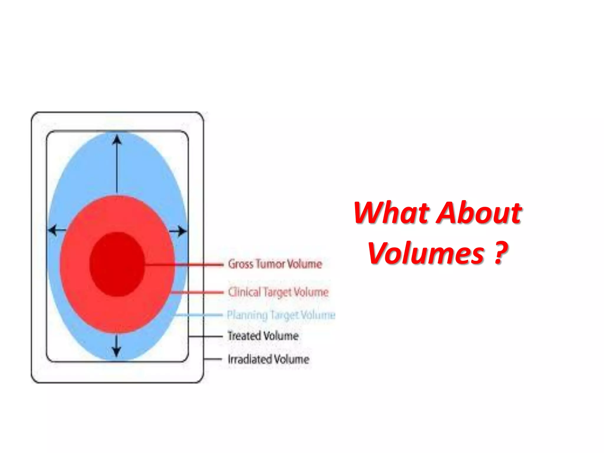
Dr. Heba Gomaa
Assistant Consultant Radiation Oncology
I HAVE SEEN MY
DEATH !!!
What We Are
Searching for !?
Why IMRT ?
 Multiple fields, including oblique and noncoplanar
Fields , Varying weightage and wedges.
 Shaped blocks :Blocks may be Cerrobend
blocks, or motor-driven Multi Leaf
Collimators (MLC).
 CT-based 3D planning
 Radiation intensity is uniform within each
beam .
 Modulation conferred only by wedges.
IMRT is of treatment option when …
• Concave dose distribution cannot be achieved by 3-D CRT.
• Extreme proximity to critical structures exist.
• Re-irradiation of certain areas require maximal dose falloff adjacent to Irradiated area.
 To locate low dose areas surrounded by high dose.
 Large fields and boosts can be integrated in single treatment plan.
 Lower rate of complication-lower
cost of patient care following
treatment.
 Radiobiologic advantage
Forward & Inverse Planning
Forward planned IMRT :basically a form
of complex 3DCRT using field-in-field
Technique in which the planner choose
number and position of beams, shape,
weighting and wedging and adjust the
beam parameters as needed.
Inverse planned IMRT : requires Inverse
Treatment Planning (ITP) software.
Optimization
 A mathematical technique
that aims to maximize (or
minimize) a score under
certain constraints.
 It is one of the most
commonly used techniques for
inverse planning.
 The objective of the
Optimization process is to vary
the beam intensities so that
the dose requirement is best
approximated.
Optimization
• During the optimization process,
each beam is
divided into small “beamlets”
• Intensity of each is varied until the
optimal dose distribution is derived
• We can Optimize following
parameters:
– Intensity maps
– Number of intensity levels
– Beam angles
– Number of beams
– Beam Energy
Conformality
OAR
Sparing
Target
Coverage
Homogenisity
Planning Objectives (Constraints)
Ideal objectives
 PTV
Lower objective:
PTV 100% volume = 100%
prescription
dose
Upper objective:
None of PTV volume receive
more than 100% dose
 OAR
None of the OAR volume receive
any dose
Non realistic:
Never practically achievable
Planning Objectives (Constraints)
Realistic objectives
 PTV
Lower objective
PTV 100% volume = 95%
prescription dose
Upper objective
None of PTV volume receive more
then 107% of prescription dose
OAR
 OAR (serial organs)
None of the OAR volume receive
more then tolerance dose
IMRT Delivery
Methods to deliver an IMRT treatment
are:
– Compensator based IMRT
– Multileaf collimator (MLC) based
• Static or step & shoot mode
• Dynamic mode
– Intensity modulated arc therapy
(IMAT)
– Tomotherapy
Step-and shoot:
The beam is OFF as the MLCs move to their
next position.
 Dynamic/ sliding window:
The beam remains ON as MLCs move
automatically to their next position
Many dose distributions physically not
achievable.
Interfraction variation.
Displacement and distortion of
internal anatomy.
Intrafraction motion.
Changes of physical and radiobiologic
characteristic of tumor and normal
tissue.
Positioning.
Limitations Of IMRT
Tomotherapy
 A form of IMRT using rotational
fan beams Uses slip ring
rotating gantry
 Treatment delivery by
continuous gantry rotation and
treatment couch translation.
 Delivered by two methods:
 Slice based tomotherapy
 Helical tomotherapy
•
IMAT
Intensity modulated arc therapy
 Does not need to move the
patient.
 Uses non coplanar beams
and arcs
 great value for brain and
head and neck tumors.
 Uses conventional linac
hence complex rotational
simple palliative treatment
can be delivered with the
same unit.
• Similar to step-and-shoot in that each field
(positioned along the arc) is subdivided into
subfields.
However, the MLC moves dynamically to shape each
subfield while the gantry is rotating
• Beam is on all the time.
• It is an alternative to tomotherapy.
VMAT
VOLUMETRIC MODULATED ARC THERAPY/ RAPID ARC
•Delivers a precisely sculpted 3D dose distribution with a single 360
degree rotation of LIN-AC Gantry.
•Treatment Algorithm depends upon three parameters-
1/ Rotation speed of the Gantry.
2/ Shape of the treatment aperture using multileaf collimator leaves.
3/ Delivery dose rates.
•Delivers dose to the whole volume, rather than slice by slice.
•Treatment planning algorithm ensures the treatment precision and
helps to spare the normal tissue.
What About
Volumes ?
Biological Target Volume
 A target volume that incorporated data
from molecular imaging techniques
 Target volume drawn
incorporates information
regarding:
 Cellular burden
 Cellular metabolism
 Tumor hypoxia
 Tumor proliferation
 Intrinsic Radioresistance or sensitivity
Biological Target Volume
Lung Cancer:
 30 -60% of all GTVs and PTVs are
changed with
PET.
 Increase in the volume can be seen in
20 -40%.
 Decrease in the volume in 20 – 30%.
 Several studies show significant
improvement in
nodal delineation.
Head and Neck Cancer:
 PET fused images lead to a change in
GTV volume
in 79%.
 Can improve parotid sparing in 70%
patients.
In particular the internal margin &
the setup margin for the OR must be
identified. This leads to
the concept of PRV.
A margin around an Organ at Risk with
a serial-like structure (e.g.,
spinal cord) is more clinically relevant
than around Organs at Risk with
parallel-like structure (e.g. liver, lung,
parotid).
What’s relevant in ICRU 83 ?
• Revised classification of
treatment volumes
• Dose prescription based on DVH
• New definitions of Dose min
and Dose max
• New surrogate of ICRU point
• Request for patient-specific QA
• New criteria for treatment
accuracy
ICRU 83
• Gross tumor volume or GTV
• Clinical target volume or CTV
• Planning target volume or
PTV
• Organ at risk or OAR
• Planning organ-at-risk
volume or PRV
• Internal target volume or
ITV
• Treated volume or TV
• Remaining volume at risk or
RVR
What is remaining ??
 RVR = difference between the
volume enclosed by the external
contour of the patient and that of
the CTVs and OARs on the slices
that have been imaged.
 If it not specifically evaluated, there
could be unsuspected regions of
high dose within the patient, which
would go undetected.
 The dose to the RVR might be useful
in estimating the risk of late effects,
such as carcinogenesis (important
for younger patients!).
What if PTV-PRV
overlap occurs ??
 It is recommended that the margins
not be compromised for the PTV or
PRV even if overlaps occur.
 The practice of shrinking the CTV-
PTV margin to accommodate an
OAR is discouraged as it results in a
deceptively better PTV dose
homogeneity!
 Priority rules in the planning system
can be used, or the PTV or PRV can
be subdivided into regions with
different dose constraints.
Dose-volume specification
DV , D near-min , Dnear-max
The dose gradient at the boundary of a PTV as a result of
multiple IMRT beams can be more than 10 % per
millimeter and a small shift in the field delivery may affect
the reliability of using a single point to report the
prescription.
No more use of ICRU point
Reporting of minimum dose should be replaced by the better
determined near-minimum dose D98 %, also designated as D
near-min. “
Analogously, it is recommended to report the near-
maximum dose D2 % as a replacement for the
“maximum dose”.
DOSE-VOLUME REPORTING SPECIFIC
TO THE OAR AND PRV
 For “serial-like” OAR’s (e.g. spinal cord, intestines, optic nerve…),
D2% is to be reported and the entire organ should be delineated.
 For parallel-like structures (e.g. parotid, lung, kidney, liver…) it is
recommended that more than one dose-volume specification be
considered for reporting.
 It is recommended that both D mean and VD be reported, where
the subscript D is a dose, which if exceeded within some volume,
has a high probability of causing a serious complication.
For example, the incidence and severity of lung pneumonitis is well
correlated with V20 Gy, the volume of normal lung receiving more
than 20 Gy.
 Because most organs are not clearly a serial-like or parallel-like structure (e.g.
heart) at least 3 dose-volume specifications should be reported.
 These would include Dmean, D2 %, and a third specification VD that correlates well
with a dose D, which if exceeded within some volume has a known high probability
of causing a serious complication.
 Normal tissues limits as defined in QUANTEC should be used.
DOSE-VOLUME REPORTING SPECIFIC
TO THE OAR AND PRV
Is It All
About
Volumes !?
Imrt&vmat
Imrt&vmat
Imrt&vmat
Imrt&vmat

Imrt&vmat

  • 1.
    Dr. Heba Gomaa AssistantConsultant Radiation Oncology
  • 2.
    I HAVE SEENMY DEATH !!!
  • 4.
  • 5.
    Why IMRT ? Multiple fields, including oblique and noncoplanar Fields , Varying weightage and wedges.  Shaped blocks :Blocks may be Cerrobend blocks, or motor-driven Multi Leaf Collimators (MLC).  CT-based 3D planning  Radiation intensity is uniform within each beam .  Modulation conferred only by wedges.
  • 6.
    IMRT is oftreatment option when … • Concave dose distribution cannot be achieved by 3-D CRT. • Extreme proximity to critical structures exist. • Re-irradiation of certain areas require maximal dose falloff adjacent to Irradiated area.  To locate low dose areas surrounded by high dose.  Large fields and boosts can be integrated in single treatment plan.  Lower rate of complication-lower cost of patient care following treatment.  Radiobiologic advantage
  • 8.
    Forward & InversePlanning Forward planned IMRT :basically a form of complex 3DCRT using field-in-field Technique in which the planner choose number and position of beams, shape, weighting and wedging and adjust the beam parameters as needed. Inverse planned IMRT : requires Inverse Treatment Planning (ITP) software.
  • 9.
    Optimization  A mathematicaltechnique that aims to maximize (or minimize) a score under certain constraints.  It is one of the most commonly used techniques for inverse planning.  The objective of the Optimization process is to vary the beam intensities so that the dose requirement is best approximated.
  • 10.
    Optimization • During theoptimization process, each beam is divided into small “beamlets” • Intensity of each is varied until the optimal dose distribution is derived • We can Optimize following parameters: – Intensity maps – Number of intensity levels – Beam angles – Number of beams – Beam Energy
  • 11.
  • 12.
    Planning Objectives (Constraints) Idealobjectives  PTV Lower objective: PTV 100% volume = 100% prescription dose Upper objective: None of PTV volume receive more than 100% dose  OAR None of the OAR volume receive any dose Non realistic: Never practically achievable
  • 13.
    Planning Objectives (Constraints) Realisticobjectives  PTV Lower objective PTV 100% volume = 95% prescription dose Upper objective None of PTV volume receive more then 107% of prescription dose OAR  OAR (serial organs) None of the OAR volume receive more then tolerance dose
  • 14.
    IMRT Delivery Methods todeliver an IMRT treatment are: – Compensator based IMRT – Multileaf collimator (MLC) based • Static or step & shoot mode • Dynamic mode – Intensity modulated arc therapy (IMAT) – Tomotherapy Step-and shoot: The beam is OFF as the MLCs move to their next position.  Dynamic/ sliding window: The beam remains ON as MLCs move automatically to their next position
  • 15.
    Many dose distributionsphysically not achievable. Interfraction variation. Displacement and distortion of internal anatomy. Intrafraction motion. Changes of physical and radiobiologic characteristic of tumor and normal tissue. Positioning. Limitations Of IMRT
  • 16.
    Tomotherapy  A formof IMRT using rotational fan beams Uses slip ring rotating gantry  Treatment delivery by continuous gantry rotation and treatment couch translation.  Delivered by two methods:  Slice based tomotherapy  Helical tomotherapy •
  • 17.
    IMAT Intensity modulated arctherapy  Does not need to move the patient.  Uses non coplanar beams and arcs  great value for brain and head and neck tumors.  Uses conventional linac hence complex rotational simple palliative treatment can be delivered with the same unit. • Similar to step-and-shoot in that each field (positioned along the arc) is subdivided into subfields. However, the MLC moves dynamically to shape each subfield while the gantry is rotating • Beam is on all the time. • It is an alternative to tomotherapy.
  • 18.
    VMAT VOLUMETRIC MODULATED ARCTHERAPY/ RAPID ARC •Delivers a precisely sculpted 3D dose distribution with a single 360 degree rotation of LIN-AC Gantry. •Treatment Algorithm depends upon three parameters- 1/ Rotation speed of the Gantry. 2/ Shape of the treatment aperture using multileaf collimator leaves. 3/ Delivery dose rates. •Delivers dose to the whole volume, rather than slice by slice. •Treatment planning algorithm ensures the treatment precision and helps to spare the normal tissue.
  • 21.
  • 22.
    Biological Target Volume A target volume that incorporated data from molecular imaging techniques  Target volume drawn incorporates information regarding:  Cellular burden  Cellular metabolism  Tumor hypoxia  Tumor proliferation  Intrinsic Radioresistance or sensitivity
  • 23.
    Biological Target Volume LungCancer:  30 -60% of all GTVs and PTVs are changed with PET.  Increase in the volume can be seen in 20 -40%.  Decrease in the volume in 20 – 30%.  Several studies show significant improvement in nodal delineation. Head and Neck Cancer:  PET fused images lead to a change in GTV volume in 79%.  Can improve parotid sparing in 70% patients.
  • 24.
    In particular theinternal margin & the setup margin for the OR must be identified. This leads to the concept of PRV. A margin around an Organ at Risk with a serial-like structure (e.g., spinal cord) is more clinically relevant than around Organs at Risk with parallel-like structure (e.g. liver, lung, parotid).
  • 25.
    What’s relevant inICRU 83 ? • Revised classification of treatment volumes • Dose prescription based on DVH • New definitions of Dose min and Dose max • New surrogate of ICRU point • Request for patient-specific QA • New criteria for treatment accuracy
  • 26.
    ICRU 83 • Grosstumor volume or GTV • Clinical target volume or CTV • Planning target volume or PTV • Organ at risk or OAR • Planning organ-at-risk volume or PRV • Internal target volume or ITV • Treated volume or TV • Remaining volume at risk or RVR
  • 27.
    What is remaining??  RVR = difference between the volume enclosed by the external contour of the patient and that of the CTVs and OARs on the slices that have been imaged.  If it not specifically evaluated, there could be unsuspected regions of high dose within the patient, which would go undetected.  The dose to the RVR might be useful in estimating the risk of late effects, such as carcinogenesis (important for younger patients!).
  • 28.
    What if PTV-PRV overlapoccurs ??  It is recommended that the margins not be compromised for the PTV or PRV even if overlaps occur.  The practice of shrinking the CTV- PTV margin to accommodate an OAR is discouraged as it results in a deceptively better PTV dose homogeneity!  Priority rules in the planning system can be used, or the PTV or PRV can be subdivided into regions with different dose constraints.
  • 29.
    Dose-volume specification DV ,D near-min , Dnear-max The dose gradient at the boundary of a PTV as a result of multiple IMRT beams can be more than 10 % per millimeter and a small shift in the field delivery may affect the reliability of using a single point to report the prescription. No more use of ICRU point Reporting of minimum dose should be replaced by the better determined near-minimum dose D98 %, also designated as D near-min. “ Analogously, it is recommended to report the near- maximum dose D2 % as a replacement for the “maximum dose”.
  • 30.
    DOSE-VOLUME REPORTING SPECIFIC TOTHE OAR AND PRV  For “serial-like” OAR’s (e.g. spinal cord, intestines, optic nerve…), D2% is to be reported and the entire organ should be delineated.  For parallel-like structures (e.g. parotid, lung, kidney, liver…) it is recommended that more than one dose-volume specification be considered for reporting.  It is recommended that both D mean and VD be reported, where the subscript D is a dose, which if exceeded within some volume, has a high probability of causing a serious complication. For example, the incidence and severity of lung pneumonitis is well correlated with V20 Gy, the volume of normal lung receiving more than 20 Gy.
  • 31.
     Because mostorgans are not clearly a serial-like or parallel-like structure (e.g. heart) at least 3 dose-volume specifications should be reported.  These would include Dmean, D2 %, and a third specification VD that correlates well with a dose D, which if exceeded within some volume has a known high probability of causing a serious complication.  Normal tissues limits as defined in QUANTEC should be used. DOSE-VOLUME REPORTING SPECIFIC TO THE OAR AND PRV
  • 32.