
Recommended
Recommended
More Related Content
What's hot
What's hot (20)
Similar to Externalbeam rt in ews3.12.20 - frida yseminar-finallll
Similar to Externalbeam rt in ews3.12.20 - frida yseminar-finallll (20)
Recently uploaded
Recently uploaded (20)
Externalbeam rt in ews3.12.20 - frida yseminar-finallll
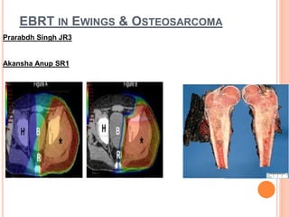
- 1. EBRT IN EWINGS & OSTEOSARCOMA Prarabdh Singh JR3 Akansha Anup SR1
- 2. FLOW OF SEMINAR Epidemiology Diagnostic staging & workup Management :Chemotherapy & local therapy RT Planning in Ewings sarcoma
- 3. EPIDEMIOLOGY 2nd most common primary bone cancer • 1 new case per 1 million population in US • Between age 10-19 yrs incidence rises to 9-10 per million • Whites affected twice as compared to Asians/Africans • Male to female: 1.3:1 • Indian Incidence: Difficult to determine as rare and highly underdiagnosed
- 4. S.J. Cotterill et al, JCO: 18;2000, 3108- 3114 Sites
- 5. • Locoregional Pain • Palpable Mass • Low grade Fever • Site Specific Symptoms • Metastatic Disease-Non Specific Symptoms(25% at diagnosis) • Median Duration Of Onset of Symptoms to definite Diagnosis:3-9 months CLINICAL FEATURES
- 6. WORKUP • Routine blood inv(CBC,Biochem,Electrolytes) • ESR • LDH • Radiology • Primary Biopsy • Bone Marrow Biopsy(BM spread in approx. 10% cases) • Molecular Pathology/Genetics Imaging • X ray • CT scan • MRI • Bone scan • PET scan
- 8. PATHOLOGY • Classic Ewing’s sarcoma appears as sheets of monotonous round cells. • Scanty cytoplasm & round nuclei with evenly distributed finely granular chromatin & inconspicuous nucleoli. • Strong,diffuse membrane staining is observed with the O13 monoclonal antibody to p30/32MIC2 (CD99).
- 9. MANAGEMENT Induction chemo Local RT/Sx Maintainence Chemo 12-24 Weeks VAC/IE AT 12/18weeks Upto 1year
- 11. CHEMOTHERAPY
- 13. LOCAL THERAPY • Surgery Radiotherapy • Types of resection: Types: I. Intralesional Pre-op II. Marginal Definitive III. Wide excison Post-op IV. Radical Safety margin -atleast Bone-1cm margin Muscle/fat-5mm margin Fascia-2mm margin
- 14. RADIOTHERAPY
- 15. Indications of Radiotherapy Definitive Tumors where Resection is Impossible For skull, face, vertebra, or pelvic primary where only an intra- lesional resection is achievable Patient with poor Surgical risk Patient refusing surgery Preoperative Narrow resection margin expected Sterilize tumor compartment Reduce risk of dissemination Postoperative For gross or microscopic positive margin For marginal Resection For wide-resection with Poor Histological response to Neo- adjuvant Chemotherapy (>10% viable tumor cells in the specimen) Based on CESS-81, CESS-86, EICESS-92 Studies : Schuck et al,IJROBP-1998 & 2003
- 16. • Gross tumor- 55-60 Gy • Microscopic Residual Disease-50 -55 Gy • No residual Disease(Post op poor response to chemo)-45-50 Gy Target: The appropriate irradiated volume is an involved field to the pretreatment tumor volume plus 2-2.5cm margin, followed by a boost to the post-induction chemotherapy tumor volume with margin https://doi.org/10.1002/pbc.10472
- 17. Laskar S ,Mallick I. et al. Pediatr Blood Cancer 2008;51:575– 580
- 18. In an analysis of patients receiving PORT in the CESS 86 and EICESS trials, Schuck et al no significant difference in the local control and survival who received RT within 60 days of surgery or later. Dunst J,et al - improved local control in CESS 86 over CESS 81 timing of RT was brought forward from the 18th week to the 10th week Dunst J, Results of CESS 81 and CESS 86. Cancer 1991;67:2818–2825 Schuck A,. Strahlenther Onkol 2002;178:25–31. Timing of Post-Operative Radiation
- 19. FIG. Changes in treatment volume. (A) Field encompassing the entire length of the medullary cavity for a tumor involving the proximal left humerus. (B) Tailored field encompassing only the proximal aspect of the leg for a limited tumor of the left tibia.
- 20. STEPS OF PLANNING Positioning & Immobilization Planning CT Scan Target Volumes Delineation Treatment Planning Process
- 21. CASE CAPSULE
- 22. 15 years female P.w.c.o pain in right buttock region radiating to right leg since july 2019 Difficulty in walking since sept 2019 Investigations: PETCECT(25.5.2020) FDG avid STM involving sacrum & coccyx with erosive lesion Low FDG avid mesentric LN anterior to right kidney Sacrum mass bx (1.6.20) Ewings sarcoma Bone marrow: Uninvolved Received 3# VIDE from 18.6.20 to L.D 25.7.20 PETCECT (2.8.20) Low grade FDG uptake in lytic changes with STM Plan:Definitive CTRT
- 24. Post Chemo Scan
- 25. POSITIONING Head first supine Arms overhead, No Knee rest. Abdo BP, 4 clamp Abdo orfit. All Outer clamps. Fiducials placed at Pubic symphysis level 5 mm NCCT cuts taken. Pushed to PACS.
- 26. PLANNING SCAN
- 27. CONTOURING
- 36. PLAN:CONVENTIONAL VS 3DCRT 95% Isodose volume 95% isodose volume
- 37. PLAN:CONVENTIONAL VS 3DCRT 50% Isodose volume 50% Isodose volume
- 38. PLAN:CONVENTIONAL VS IMRT 95% isodose volume 95% Isodose volume
- 39. PLAN: CONVENTIONAL VS IMRT 50% Isodose volume 50% Isodose volume
- 40. PLAN:3DCRT VS IMRT 95% isodose volume 95% isodose volume
- 41. PLAN:3DCRT VS IMRT 50% Isodose volume 50% Isodose volume
- 42. PLAN :3DCRT VS IMRT 70% Isodose volume 70% Isodose volume
- 43. DVH
- 45. OAR:DVH(BLADDER)
- 48. DVH(RECTUM)
- 51. Structure Conventional 3DCRT IMRT PTV V95%-96% 97% 96% Bladder 55Gy 25.91Gy 23Gy Rectum 52Gy 38.3Gy 36.1Gy Bowel Bag 22Gy 11.4Gy 9.1Gy Rt Femoral head 10.4Gy 7.9Gy 4.8Gy Left femoral head 5.1Gy 3.8Gy 3.1Gy
- 52. CASE CAPSULE-2 12 year female P.w.c.o swelling in forearm since 4 month X ray Forearm(A+P):Diaphyseal expansile lesion in left leg Bx :Ewings sarcoma Started with EFT 2001-Good response U/w I/C Fibulectomy on 5.6.20 HPR:88%necrosis,all margins free Planned for Post Op RT
- 53. POSITIONING Supine ,arms by side ,Leg externally rotated Thermoplastic mask
- 54. CONTOURING
- 58. PLANNING Dose prescription to tumor bed :45Gy/25# Lung bath:12.6Gy/7#
- 66. PROTON THERAPY
- 67. Retrospective review of 30 patients treated with proton therapy between april 2003-April 2009 at Massachusetts hospital 14Male & 16female patients Median dose:54Gy Median F/u:38.4months https://doi.org/10.1016/j.ijrobp.2011.03.038 3year actuarial rates EFS 60% LC 86% OS 89%
- 69. FOLLOWUP After completion of therapy all children shall be followed up: every 3 monthly for the first year every 6 monthly for the 2nd & 3rd year yearly thereafter This is applicable to all patients except when specified
- 70. Second malignant neoplasms Radiation Induced Sarcoma dose related much less frequent < 48 Gy Alkylator Induced Leukemia alkylator dose more important than the addition of etoposide Adriamycin cardiomyopathy Ifosfamide: renal toxicity Gonadal failure Chronic pain/musculoskeletal dysfunction related to local therapies Psychological Trauma - adolescents Late Effects in ESFT Survivors
- 71. OSTEOSARCOMA
- 72. • Malignant mesenchymal tumour • Characterized by production of osteoid matrix and woven bone by tumor • Molecularly: • It is a sporadic complex genotype sarcoma, (as distinguished from the balanced translocation- associated sarcomas (e.g., Ewing sarcoma)): Cell cycle regulators such as p53 and Rb have been implicated (Li-Fraumeni, Hereditary Retinoblastoma INTRODUCTION
- 73. EPIDEMIOLOGY Most commonly diagnosed primary malignancy of bone (Rare tumor nonetheless Bimodal peak of incidence: One around adolescence and another smaller peak in elderly (60+) Males affected slightly more frequently than females.
- 74. •Malignant spindle cells •Osteoblasts •Osteoid/immature bone matrix Pathologic Classification of tumor
- 75. •METASTASIS: •Disseminate through blood •1st site – lung 9.3% •2nd bone 3.9% •Lymph node involvement only <10% -poor prognostic sign. •SKIP METASTATIS: •Located within the same bone as the main tumour but not in continuity with it. •High grade sarcomas-develop by embolisation of tumour cells within the marrow sinusoids— represent local micromets •Clinical incidence -1%. Predict poor prognosis
- 76. •Site •Pain and soft tissue swelling •Night pain, no effusion in adjacent joint and movements are normal. •Age > 40 yrs – pre existing disease •Paget’s disease, irradiation, multiple hereditary exostosis, polyostotic fibrous dysplasia. •Associated syndromes- •Li fraumeni syndrome •Rothmund Thomson syndrome •Werner’s syndrome
- 77. •CENTRAL/INTRAMEDULLARY(90-95%) •CLASSIC(CONVENTIONAL) 75-80%-HIGH GRADE •OSTEOBLASTIC •CHONDROBLASTIC •FIBROBLASTIC •LOW GRADE •SMALL CELL •TELANGIECTATIC •SECONDARILY IN ABNORMAL BONE(PAGET’S) •SURFACE (5-10%) •PAROSTEAL-LOW GRADE •PAROSTEAL-HIGH GRADE •PERIOSTEAL •EXTRAOSSEOUS
- 78. IMAGING X-RAY CT SCAN MRI BONE SCAN ANGIOGRAPHY
- 81. Definitive Indications Unresectable tumor Preoperative Indications Adjuvant Indication • Margin+ • Head & Neck Cancer
- 85. 21yrs,male ,Hailing from kolkata p.w.c.o left anterior & lateral chest wall pain X5months Investigations: 1)MRI Thorax 11x5.2cm lobulated chest wallmass in left & posterior chest wall with encasement of 3rd & 6th rib underlying nodular extrapleural and overlying extraosseous soft tissue components at the intercostal muscles Receive 6# of chemo 2)CT Guided bx :Chondroblastic OGS Plan:Definitive RT
- 86. Pre-Chemo Scan
- 87. Post chemo response assesment
- 88. CONTOURING
- 93. CONVENTIONAL VS 3DCRT 95% Isodose volume 95% Isodose volume
- 94. CONVENTIONAL VERSUS 3DCRT 50% Isodose Volume 50% Isodose Volume
- 95. CONVENTIONAL VS IMRT 95% OF ISODOSE VOLUME 95% OF ISODOSE VOLUME
- 96. CONVENTIONAL VS IMRT 50% Isodose volume 50% Isodose volume
- 97. DVH(COMPARISON)
- 99. Structure Conv 3DCRT IMRT PTV V95-96% V95-96% V95-97% Lt Lung 43.9Gy 28.45Gy 22.1Gy Rt Lung 1.5Gy 5.5Gy 4.8Gy Heart 3.9Gy 2.1Gy 1.5Gy Spinal cord 65Gy 60Gy 44.5Gy
- 100. CASE CAPSULE-2 (POST OP)
- 101. 26 Year Female from Lucknow H/o nasal obstruction & epistaxis in march 20 MRI 15.10.20 :Lobulated lesion in right nasal cavity with intraorbital extension U/w FESS +Bx--.Osteosarcoma Received 4# Cis adria MRI H+N: Residual lesion U/w endoassisted resection of sinonasal osteosarcoma Plan:Adjuvant RT
- 102. IMAGING Post op MRI
- 103. CONTOURING
- 106. PLANNING Dose prescription: 63Gy/35# Technique:IMRT with IGRT technique
- 108. CONVENTIONAL VS 3DCRT 50% Isodose volume 50% Isodose volume
- 109. CONVENTIONAL VS 3DCRT 95% Isodose volume 95% Isodose volume
- 110. CONVENTIONAL VS IMRT 95% Isodose volume 95% Isodose volume
- 111. CONVENTIONAL VS IMRT 50% Isodose volume 50% Isodose volume
- 112. DVH(COMPARISON)
- 114. Structure CONV 3DCRT IMRT PTV V95%-93% 91% 92% Left eye 66.53Gy 26Gy 10.5Gy Right eye 23.1Gy 35Gy 22Gy Chiasma 45.8Gy 28Gy 23Gy Left optic nerve 28.5Gy 26.1Gy 10.5Gy Right optic nerve 62Gy 57Gy 22.2Gy
- 115. THANK YOU