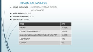
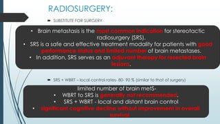
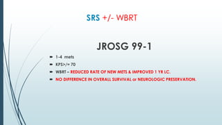
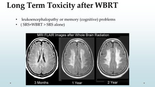
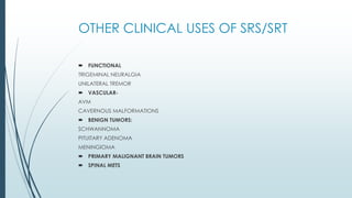
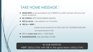
Radiosurgery
for Brain Metastases
DR SWARNITA SAHU
DNB RESIDENT
RADIATION ONCOLOGY
BATRA HOSPITAL,NEW DELHI
BRAIN METASTASIS
 RISING INCIDENCE - INCREASE IN SYSTEMIC THERAPY
MRI ADVANCES
 METS : PRIMARY – 10:1
 MEDIAN SURVIVAL < 1 YR
 MEAN AGE – 60 YRS.
LUNG 50%
BREAST 15-20%
OTHER KMOWN PRIMARY 10-15%
UNKNOWN PRIMARY (DECREASING WITH PET) 10-15%
MELANOMA 10%
COLON 5%
SYMPTOMS
 HEADACHE
 MENTAL PROBLEMS
 FOCAL WEAKNESS
 ATAXIA
 SEIZURES
 SPEECH ABNORMALITIES
IMAGING: TYPICAL PRESENTATION
 The imaging of choice is a CEMRI.
 multiple lesions.
 localization at the grey-white matter junction.
 circumscribed margins.
 vasogenic edema.
 CONFUSION – IN CASE OF SINGLE LESION.
D/D:
 BRAIN ABSCESS/ INFECTION
 MENINGIOMA
 INFARCT
CONFIRMED BY CEMRI
SINGLE LESION:
BRAIN ABSCESS- DURAL RIM SIGN DURAL TAIL SIGN
TREATMENT :
 STEROIDS – 10 mg(oral /iv) bolus f/b 6-8mg every 6-8 hrs + PPI.
(NOTE : in asymptomatic patients, steroids is reserved until the 1st neuro symptom)
 WBRT : standard of care
SURGERY:
Immediate relief (WBRT – Days to weeks)
3 studies:
 Patchell et al
 Noordijk et al
 Mintz et al
 Fallacy: all 3 trials were on pts with single brain lesion
Resection reserved for life threatening
lesions or KPS >/=70
RADIOSURGERY:
 SUBSTITUTE FOR SURGERY
 SRS + WBRT – local control rates- 80- 90 % (similar to that of surgery)
• Brain metastasis is the most common indication for stereotactic
radiosurgery (SRS).
• SRS is a safe and effective treatment modality for patients with good
performance status and limited number of brain metastases.
• In addition, SRS serves as an adjuvant therapy for resected brain
lesions.
limited number of brain metS-
• WBRT to SRS is generally not recommended.
• SRS + WBRT - local and distant brain control
• significant cognitive decline without improvement in overall
survival
Advantages of Stereotactic
Radiosurgery
Advantages of Surgery
• Treatment for larger lesions (>4cm)
• Treatment of small, deep lesions• Rapid resolution of mass effect and
edema • Minimally invasive
•
•
•
Removal of cancer • General anesthesia not required
• Outpatient procedure
• Treatment of multiple lesions at same
setting
• Short recovery (<1 week)
• Potential avoidance of whole brain XRT
• Rapid initiation of chemoRx
Histologic confirmation
Rapid tapering of steroids
Less intensive follow up•
• Lower risk of radiation necrosis
TERMINOLOGY:
 STEREOTACTIC: precise 3D mapping technique to guide a procedure.
 SRS (stereotactic radiosurgery) : stereotactically guided conformal
irradiation of a defined target volume in a SINGLE SESSION.
 FSR (fractionated stereotactic radiosurgery) : 2-5 SESSIONS.
 SRT (stereotactic radiotherapy) : MULTIPLE FRACTIONS ( >5).
KEY REQUIREMENTS FOR STEREOTACTIC IRRADIATION:
 TARGET- SMALL
SHARPLY DEFINED
 RADIATION DELIVERY - ACCURATE
HIGHLY CONFORMAL
 SENSITIVE STRUCTURES- EXCLUDED FROM TARGET.
BASIS OF HYPOFRACTIONATION
■ Fraction Size– Increased > 2 Gy/per fraction.
■ Fraction Number– Decreased
■ Total treatment Time- Decreased
■ Rationale
Treatment completed in a shorter time.
Higher dose /# gives -
better control for larger tumors.
useful for hypoxic fraction of large tumor.
■ Disadvantage-
Higher potential for late normal tissue complications.
Machines used to focus
highly targeted radiation or
radiosurgery (SRS or
stereotactic radiosurgery)
VitalBeam Linac
Gamma Knife
Cyberknife
Tomotherapy
CYBERKNIFE:
 image guided frameless stereotactic
radiosurgery system for treating cranial &
extracranial lesions.
 Two diagnostic xray tube mounted orthogonally
in the ceiling and 2 opposing Si-flat panel
detectors.
 The robotic arm has 6 degrees of freedom and is
capable of pointing the linac beam almost
anywhere in space.
 Not restricted to isocenter geometry, it can be
directed independently without a fixed
isocenter.
GAMMA KNIFE:
 Hemispherical array of multiple fixed Co 60 beams (201 in most models)
that are sharply collimated to create small, relatively spherical treatment
volumes of varied diameter with sharp dose fall off.
TOMOTHERAPY:
 IMRT delivery technique – combines
features of linear accelerator + helical
CT scanner.
 The linear accelerator is mounted on a
CT like gantry and rotates through a full
circle.
 Treatment couch is translated slowly
through a doughnut like aperture.
 Creating a helical motion of the beam
with respect to the patient.
LINAC RADIOSURGERY
PROTON RADIOSURGERY
IMMOBILISATION :
PATIENT SELECTION
 (KPS ≥70).
( KPS <70 have poor overall prognosis - (WBRT) or best supportive care )
 Indications for SRS :
 1–4 brain metastases (Sx not possible).
 POST OP pts with few brain metastases.
 considered for patients with good performance status and 4–10 brain metastases
with low tumor burden.
TREATMENT PLANNING CONSIDERATIONS :
Simulation:
 Position: Supine
 Immobilization: Customized head cast
 1 mm thick CT slices
 Fuse pre- and postoperative MR for surgical bed treatment
Margins :
 No CTV or PTV expansions for intact brain metastasis.
 Post op cases – discussed later.
Tumor coverage considerations :
 100% of GTV (or CTV for postoperative cases) receives 100% of dose (if GTV/CTV ≤20 mm)
 ≥95% of GTV (or CTV for postoperative cases) receives 100% of dose (if GTV/CTV >20 mm)
Recommendations for CTV contouring for postoperative
completely resected cavity SRS (Soliman IJROBP 2018;100:436)
• CTV should include the entire contrast-enhancing surgical cavity using the
T1-weighted gadolinium-enhanced axial MRI scan, excluding edema
determined by MRI
• CTV should include entire surgical tract seen on postoperative CT or MRI
• If the tumor was in contact with the dura preoperatively, CTV should
include a 5- to 10-mm margin along the bone flap beyond the initial region
of preoperative tumor contact.
• If the tumor was not in contact with the dura, CTV should include a
margin of 1 to 5 mm along the bone flap
• If the tumor was in contact with a venous sinus preoperatively, CTV
should include a margin of 1 to 5 mm along the sinus.
DOSES
• SRS FOR INTACT LESIONS:
RTOG 90-05 2cm = 24 Gy
2.1 – 3cm = 18 Gy
3.1 - 4cm = 15 Gy
FOR RADIORESISTANT HISTOLOGIES: RCC, MELANOMA, SARCOMA
Dose escalation with 30 Gy/3# can be considered.
• POSTOP SRS:
MAHAJAN 2016
</=10 cc = 16 Gy
10-15cc = 14 Gy
>/= 15cc = 12 Gy
NORMAL TISSUE TOLERANCE:
NORMAL STRUCTURE : QUANTEC :
BRAIN PARENCHYMA V 12 < 5- 10cc
OPTIC APPARATUS Dmax < 12 Gy
BRAIN STEM Dmax < 12.5 Gy
SPINAL CORD Dmax = 13 Gy
COCHLEA Dmax </= 14 Gy
WBRT + / - Sx
Patchell NEJM 1990
RT + Sx ARM
Local recurrence less
Median survival more
Functional independence more
Sx +/ - WBRT
Sx +/ - SRS :
SAME OS LC BETTER
MAHAJAN 2016 :
1. SRS – 17-month median OS.
72% 1-year LC.
2. Observation – 18-month median OS.
– 43% 1-year LC.
WBRT +/- SRS BOOST
RTOG 95-08
WBRT + SRS WBRT
OS- 6.5 months
better local contrl
better performance status
5.7 months
OS better in
RPA I
Non small cell lung cancer
Metastatic squamous histology
DEMONSTRATED : lower oedema & corticosteroid use
CONCLUSION : SRS indicated in single mets
Routine use in multiple mets – not indicated
SRS +/- WBRT
JROSG 99-1
 1-4 mets
 KPS>/= 70
 WBRT – REDUCED RATE OF NEW METS & IMPROVED 1 YR LC.
 NO DIFFERENCE IN OVERALL SURVIVAL or NEUROLOGIC PRESERVATION.
WBRT FRACTIONATION
 NEUROCOGNITIVE DELAY:
1) anticonvulsants 4)chemotherapy
2) benzodiazepines 5)craniotomy
3) opioids 6)brain tumor itself
 There has been efforts to reduce the neuro toxicity with hippocampal
sparing (RTOG 0933) or Memantine (RTOG 0614)
ANTICONVULSANTS –
Have a negative impact on QOL & Neurocognition
NOT TO BE STARTED UNTILL CONVULSION STARTS
MEMANTINE DURING WBRT-RTOG 0614
 DURING & AFTER WBRT IS STANDARD OF CARE.
 20mg/d within 3 days of initiating radiotherapy for 24 weeks.
 Memantine – treat moderate to severe confusion in alzeimers.
In WBRT- Better cognitive function over time,
specifically delaying time to cognitive decline and
reducing the rates of decline in memory, executive
function and processing speed.
Brain metastasis that progressed after prior
whole-brain radiotherapy (WBXRT).
 RTOG 95-08
 Phase III randomized trial
 Established that Radiosurgery immediately following standard WBXRT (37.5
Gy in 15 fractions) improves LC and QOL for patients with one to three brain
metastases
 while
 improving OS for patients with solitary metastasis,
 (all compared with patients initially managed with WBXRT only.)
For a single lesion, radiosurgery alone may be used, but there is a higher
risk of a new lesion showing up in the brain
Aug 2012 –Treated tumor is virtually gone, but there
is a new tumor on the opposite side of the brainJan 2011 - Radiosurgery
Treatment for Patients
with Multiple Brain
Metastases
Median overall survival after stereotactic radiosurgery:
13.9 m – 1 tumor.
10.8 m – 2-4 tumors.
10.8 m – 5-10 tumors.
SRS FOR PTS WITH MULTIPLE BRAIN METS
(JLGK0901): a multi-institutional prospective observational study.
Yamamoto Lancet Oncol 2014:15:387
• <4 mL were irradiated with 22 Gy
• 4-10 mL with 20 Gy.
• 1194 eligible patients
• largest tumour <10 mL .
• <3 cm
• total cumulative volume ≤15 mL.
Results with Radiosurgery
• In controlled studies in patients-
with tumors up to 3 cm in diameter,
 SRS – LC -70% at 1 yr.
 SRS + WBRT- 90% at 1 yr.
• Prospective nonrandomized data in patients-
with newly diagnosed brain metastases suggest that up to 10 tumors with a total
cumulative volume ≤15 mL may be treated in a single session with similar efficacy
and no increase in toxicity.
• When patients are treated with SRS alone, new or recurrent brain
metastases develop in approximately 25 to 50 % of patients within
the first 6 to 12 m.
Survival and Prognosis for People with Brain
Metastases
Karnofsky Score (KPS) = 70
Cares for self; unable to carry on normal activity or
do active work
Best prognosis In patients with
• KPS>70
• brain only mets
• age< 65 yrs
PROGNOSIS
 GASPER ET AL : KPS & extracranial disease
 3 recursive partitioning analysis classes:
RPA I KPS > 70 CONTROLLED PRIMARY
AGE< 65 YRS.
BRAIN METS ONLY
7.1 MONTHS
RPA II NOT I OR II 4.2 MONTHS
RPA III KPS< 70 2.3 MONTHS
Side Effects and
Toxicity
Complications of Radiosurgery
• Short term side effects are uncommon (2%) with worsening symptoms or
new seizures
• About one third- mild swelling (headaches, nausea)
• Radionecrosis in 5% to 10%
Radionecrosis
• Symptoms:
i. asymptomatic (50%)
ii. focal neurologic signs
iii. symptoms related to cerebral edema.
• Imaging:
increased enhancement at the site of prior SRS
accompanied by surrounding edema.
• Treatment:
(largely symptomatic)
i. corticosteroids.
ii. Resection may be required
iii. bevacizumab in severe cases.
Sometimes the MRI will look worse after
radiosurgery due to radionecrosis of the
cancer
but with time this should fade away.
Long Term Toxicity after WBRT
• leukoencephalopathy or memory (cognitive) problems
• ( SRS+WBRT > SRS alone)
FOLLOW UP:
 As per NCCN guidelines :
 Brain MRI q2–3 months for the first year
 Follow-up and imaging as clinically indicated after 1 year.
OTHER CLINICAL USES OF SRS/SRT
 FUNCTIONAL
TRIGEMINAL NEURALGIA
UNILATERAL TREMOR
 VASCULAR-
AVM
CAVERNOUS MALFORMATIONS
 BENIGN TUMORS:
SCHWANNOMA
PITUITARY ADENOMA
MENINGIOMA
 PRIMARY MALIGNANT BRAIN TUMORS
 SPINAL METS
TAKE HOME MESSAGE :
 BRAIN METS to be excluded in pts of BREAST & LUNG cancers with any new
neuro symptoms .
 BE CAREFUL WITH SINGLE BRAIN LESIONS.
 SRS/Sx alone = new lesions in 6-12 months
 SRS/Sx + WBRT =
better local control but AT THE COST OF COGNITIVE DECLINE
no improvement in OS
 SRS for every new lesion – COST ISSUE
 RADIONECROSIS- Imp side effect of SRS.
IN OUR INSTITUTE-
WBRT (30Gy/10#) with SIB to the gross lesion (45Gy/10#)
Brain metastasis

Brain metastasis

  • 1.
    Radiosurgery for Brain Metastases DRSWARNITA SAHU DNB RESIDENT RADIATION ONCOLOGY BATRA HOSPITAL,NEW DELHI
  • 2.
    BRAIN METASTASIS  RISINGINCIDENCE - INCREASE IN SYSTEMIC THERAPY MRI ADVANCES  METS : PRIMARY – 10:1  MEDIAN SURVIVAL < 1 YR  MEAN AGE – 60 YRS. LUNG 50% BREAST 15-20% OTHER KMOWN PRIMARY 10-15% UNKNOWN PRIMARY (DECREASING WITH PET) 10-15% MELANOMA 10% COLON 5%
  • 3.
    SYMPTOMS  HEADACHE  MENTALPROBLEMS  FOCAL WEAKNESS  ATAXIA  SEIZURES  SPEECH ABNORMALITIES
  • 4.
    IMAGING: TYPICAL PRESENTATION The imaging of choice is a CEMRI.  multiple lesions.  localization at the grey-white matter junction.  circumscribed margins.  vasogenic edema.
  • 5.
     CONFUSION –IN CASE OF SINGLE LESION.
  • 6.
    D/D:  BRAIN ABSCESS/INFECTION  MENINGIOMA  INFARCT CONFIRMED BY CEMRI
  • 7.
  • 8.
    BRAIN ABSCESS- DURALRIM SIGN DURAL TAIL SIGN
  • 9.
    TREATMENT :  STEROIDS– 10 mg(oral /iv) bolus f/b 6-8mg every 6-8 hrs + PPI. (NOTE : in asymptomatic patients, steroids is reserved until the 1st neuro symptom)  WBRT : standard of care
  • 10.
    SURGERY: Immediate relief (WBRT– Days to weeks) 3 studies:  Patchell et al  Noordijk et al  Mintz et al  Fallacy: all 3 trials were on pts with single brain lesion Resection reserved for life threatening lesions or KPS >/=70
  • 11.
    RADIOSURGERY:  SUBSTITUTE FORSURGERY  SRS + WBRT – local control rates- 80- 90 % (similar to that of surgery) • Brain metastasis is the most common indication for stereotactic radiosurgery (SRS). • SRS is a safe and effective treatment modality for patients with good performance status and limited number of brain metastases. • In addition, SRS serves as an adjuvant therapy for resected brain lesions. limited number of brain metS- • WBRT to SRS is generally not recommended. • SRS + WBRT - local and distant brain control • significant cognitive decline without improvement in overall survival
  • 12.
    Advantages of Stereotactic Radiosurgery Advantagesof Surgery • Treatment for larger lesions (>4cm) • Treatment of small, deep lesions• Rapid resolution of mass effect and edema • Minimally invasive • • • Removal of cancer • General anesthesia not required • Outpatient procedure • Treatment of multiple lesions at same setting • Short recovery (<1 week) • Potential avoidance of whole brain XRT • Rapid initiation of chemoRx Histologic confirmation Rapid tapering of steroids Less intensive follow up• • Lower risk of radiation necrosis
  • 13.
    TERMINOLOGY:  STEREOTACTIC: precise3D mapping technique to guide a procedure.  SRS (stereotactic radiosurgery) : stereotactically guided conformal irradiation of a defined target volume in a SINGLE SESSION.  FSR (fractionated stereotactic radiosurgery) : 2-5 SESSIONS.  SRT (stereotactic radiotherapy) : MULTIPLE FRACTIONS ( >5).
  • 14.
    KEY REQUIREMENTS FORSTEREOTACTIC IRRADIATION:  TARGET- SMALL SHARPLY DEFINED  RADIATION DELIVERY - ACCURATE HIGHLY CONFORMAL  SENSITIVE STRUCTURES- EXCLUDED FROM TARGET.
  • 16.
    BASIS OF HYPOFRACTIONATION ■Fraction Size– Increased > 2 Gy/per fraction. ■ Fraction Number– Decreased ■ Total treatment Time- Decreased ■ Rationale Treatment completed in a shorter time. Higher dose /# gives - better control for larger tumors. useful for hypoxic fraction of large tumor. ■ Disadvantage- Higher potential for late normal tissue complications.
  • 18.
    Machines used tofocus highly targeted radiation or radiosurgery (SRS or stereotactic radiosurgery) VitalBeam Linac Gamma Knife Cyberknife Tomotherapy
  • 19.
    CYBERKNIFE:  image guidedframeless stereotactic radiosurgery system for treating cranial & extracranial lesions.  Two diagnostic xray tube mounted orthogonally in the ceiling and 2 opposing Si-flat panel detectors.  The robotic arm has 6 degrees of freedom and is capable of pointing the linac beam almost anywhere in space.  Not restricted to isocenter geometry, it can be directed independently without a fixed isocenter.
  • 20.
    GAMMA KNIFE:  Hemisphericalarray of multiple fixed Co 60 beams (201 in most models) that are sharply collimated to create small, relatively spherical treatment volumes of varied diameter with sharp dose fall off.
  • 21.
    TOMOTHERAPY:  IMRT deliverytechnique – combines features of linear accelerator + helical CT scanner.  The linear accelerator is mounted on a CT like gantry and rotates through a full circle.  Treatment couch is translated slowly through a doughnut like aperture.  Creating a helical motion of the beam with respect to the patient.
  • 22.
  • 23.
  • 24.
    PATIENT SELECTION  (KPS≥70). ( KPS <70 have poor overall prognosis - (WBRT) or best supportive care )  Indications for SRS :  1–4 brain metastases (Sx not possible).  POST OP pts with few brain metastases.  considered for patients with good performance status and 4–10 brain metastases with low tumor burden.
  • 25.
    TREATMENT PLANNING CONSIDERATIONS: Simulation:  Position: Supine  Immobilization: Customized head cast  1 mm thick CT slices  Fuse pre- and postoperative MR for surgical bed treatment Margins :  No CTV or PTV expansions for intact brain metastasis.  Post op cases – discussed later. Tumor coverage considerations :  100% of GTV (or CTV for postoperative cases) receives 100% of dose (if GTV/CTV ≤20 mm)  ≥95% of GTV (or CTV for postoperative cases) receives 100% of dose (if GTV/CTV >20 mm)
  • 26.
    Recommendations for CTVcontouring for postoperative completely resected cavity SRS (Soliman IJROBP 2018;100:436) • CTV should include the entire contrast-enhancing surgical cavity using the T1-weighted gadolinium-enhanced axial MRI scan, excluding edema determined by MRI • CTV should include entire surgical tract seen on postoperative CT or MRI • If the tumor was in contact with the dura preoperatively, CTV should include a 5- to 10-mm margin along the bone flap beyond the initial region of preoperative tumor contact. • If the tumor was not in contact with the dura, CTV should include a margin of 1 to 5 mm along the bone flap • If the tumor was in contact with a venous sinus preoperatively, CTV should include a margin of 1 to 5 mm along the sinus.
  • 27.
    DOSES • SRS FORINTACT LESIONS: RTOG 90-05 2cm = 24 Gy 2.1 – 3cm = 18 Gy 3.1 - 4cm = 15 Gy FOR RADIORESISTANT HISTOLOGIES: RCC, MELANOMA, SARCOMA Dose escalation with 30 Gy/3# can be considered. • POSTOP SRS: MAHAJAN 2016 </=10 cc = 16 Gy 10-15cc = 14 Gy >/= 15cc = 12 Gy
  • 28.
    NORMAL TISSUE TOLERANCE: NORMALSTRUCTURE : QUANTEC : BRAIN PARENCHYMA V 12 < 5- 10cc OPTIC APPARATUS Dmax < 12 Gy BRAIN STEM Dmax < 12.5 Gy SPINAL CORD Dmax = 13 Gy COCHLEA Dmax </= 14 Gy
  • 30.
    WBRT + /- Sx Patchell NEJM 1990 RT + Sx ARM Local recurrence less Median survival more Functional independence more Sx +/ - WBRT
  • 31.
    Sx +/ -SRS : SAME OS LC BETTER MAHAJAN 2016 : 1. SRS – 17-month median OS. 72% 1-year LC. 2. Observation – 18-month median OS. – 43% 1-year LC.
  • 32.
    WBRT +/- SRSBOOST RTOG 95-08 WBRT + SRS WBRT OS- 6.5 months better local contrl better performance status 5.7 months OS better in RPA I Non small cell lung cancer Metastatic squamous histology DEMONSTRATED : lower oedema & corticosteroid use CONCLUSION : SRS indicated in single mets Routine use in multiple mets – not indicated
  • 33.
    SRS +/- WBRT JROSG99-1  1-4 mets  KPS>/= 70  WBRT – REDUCED RATE OF NEW METS & IMPROVED 1 YR LC.  NO DIFFERENCE IN OVERALL SURVIVAL or NEUROLOGIC PRESERVATION.
  • 34.
  • 35.
     NEUROCOGNITIVE DELAY: 1)anticonvulsants 4)chemotherapy 2) benzodiazepines 5)craniotomy 3) opioids 6)brain tumor itself  There has been efforts to reduce the neuro toxicity with hippocampal sparing (RTOG 0933) or Memantine (RTOG 0614) ANTICONVULSANTS – Have a negative impact on QOL & Neurocognition NOT TO BE STARTED UNTILL CONVULSION STARTS
  • 36.
    MEMANTINE DURING WBRT-RTOG0614  DURING & AFTER WBRT IS STANDARD OF CARE.  20mg/d within 3 days of initiating radiotherapy for 24 weeks.  Memantine – treat moderate to severe confusion in alzeimers. In WBRT- Better cognitive function over time, specifically delaying time to cognitive decline and reducing the rates of decline in memory, executive function and processing speed.
  • 37.
    Brain metastasis thatprogressed after prior whole-brain radiotherapy (WBXRT).  RTOG 95-08  Phase III randomized trial  Established that Radiosurgery immediately following standard WBXRT (37.5 Gy in 15 fractions) improves LC and QOL for patients with one to three brain metastases  while  improving OS for patients with solitary metastasis,  (all compared with patients initially managed with WBXRT only.)
  • 38.
    For a singlelesion, radiosurgery alone may be used, but there is a higher risk of a new lesion showing up in the brain Aug 2012 –Treated tumor is virtually gone, but there is a new tumor on the opposite side of the brainJan 2011 - Radiosurgery
  • 39.
    Treatment for Patients withMultiple Brain Metastases
  • 40.
    Median overall survivalafter stereotactic radiosurgery: 13.9 m – 1 tumor. 10.8 m – 2-4 tumors. 10.8 m – 5-10 tumors. SRS FOR PTS WITH MULTIPLE BRAIN METS (JLGK0901): a multi-institutional prospective observational study. Yamamoto Lancet Oncol 2014:15:387 • <4 mL were irradiated with 22 Gy • 4-10 mL with 20 Gy. • 1194 eligible patients • largest tumour <10 mL . • <3 cm • total cumulative volume ≤15 mL.
  • 41.
    Results with Radiosurgery •In controlled studies in patients- with tumors up to 3 cm in diameter,  SRS – LC -70% at 1 yr.  SRS + WBRT- 90% at 1 yr. • Prospective nonrandomized data in patients- with newly diagnosed brain metastases suggest that up to 10 tumors with a total cumulative volume ≤15 mL may be treated in a single session with similar efficacy and no increase in toxicity. • When patients are treated with SRS alone, new or recurrent brain metastases develop in approximately 25 to 50 % of patients within the first 6 to 12 m.
  • 42.
    Survival and Prognosisfor People with Brain Metastases Karnofsky Score (KPS) = 70 Cares for self; unable to carry on normal activity or do active work Best prognosis In patients with • KPS>70 • brain only mets • age< 65 yrs
  • 43.
    PROGNOSIS  GASPER ETAL : KPS & extracranial disease  3 recursive partitioning analysis classes: RPA I KPS > 70 CONTROLLED PRIMARY AGE< 65 YRS. BRAIN METS ONLY 7.1 MONTHS RPA II NOT I OR II 4.2 MONTHS RPA III KPS< 70 2.3 MONTHS
  • 44.
  • 45.
    Complications of Radiosurgery •Short term side effects are uncommon (2%) with worsening symptoms or new seizures • About one third- mild swelling (headaches, nausea) • Radionecrosis in 5% to 10%
  • 46.
    Radionecrosis • Symptoms: i. asymptomatic(50%) ii. focal neurologic signs iii. symptoms related to cerebral edema. • Imaging: increased enhancement at the site of prior SRS accompanied by surrounding edema. • Treatment: (largely symptomatic) i. corticosteroids. ii. Resection may be required iii. bevacizumab in severe cases. Sometimes the MRI will look worse after radiosurgery due to radionecrosis of the cancer but with time this should fade away.
  • 47.
    Long Term Toxicityafter WBRT • leukoencephalopathy or memory (cognitive) problems • ( SRS+WBRT > SRS alone)
  • 48.
    FOLLOW UP:  Asper NCCN guidelines :  Brain MRI q2–3 months for the first year  Follow-up and imaging as clinically indicated after 1 year.
  • 49.
    OTHER CLINICAL USESOF SRS/SRT  FUNCTIONAL TRIGEMINAL NEURALGIA UNILATERAL TREMOR  VASCULAR- AVM CAVERNOUS MALFORMATIONS  BENIGN TUMORS: SCHWANNOMA PITUITARY ADENOMA MENINGIOMA  PRIMARY MALIGNANT BRAIN TUMORS  SPINAL METS
  • 50.
    TAKE HOME MESSAGE:  BRAIN METS to be excluded in pts of BREAST & LUNG cancers with any new neuro symptoms .  BE CAREFUL WITH SINGLE BRAIN LESIONS.  SRS/Sx alone = new lesions in 6-12 months  SRS/Sx + WBRT = better local control but AT THE COST OF COGNITIVE DECLINE no improvement in OS  SRS for every new lesion – COST ISSUE  RADIONECROSIS- Imp side effect of SRS. IN OUR INSTITUTE- WBRT (30Gy/10#) with SIB to the gross lesion (45Gy/10#)

Editor's Notes

  • #9 NO OEDEMA IN STROKE.
  • #17 LATE EFFECTS: FRACTION SIZE……..ACUTE: FRACTION SIZE & OVERALL TREATMENT TIME.