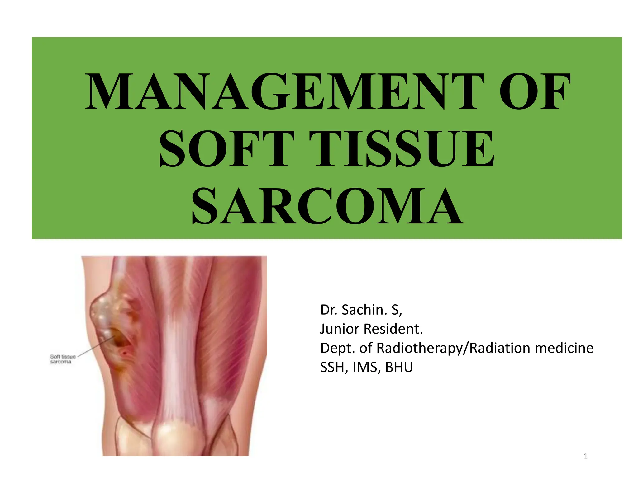
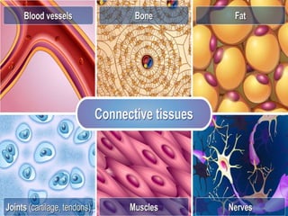
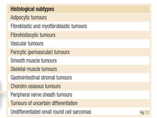
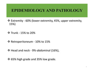
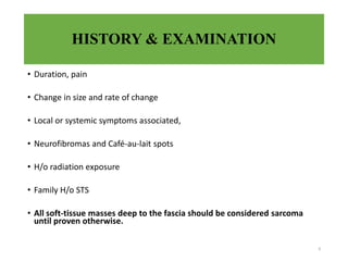
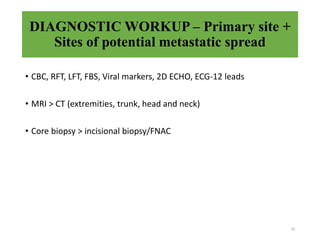
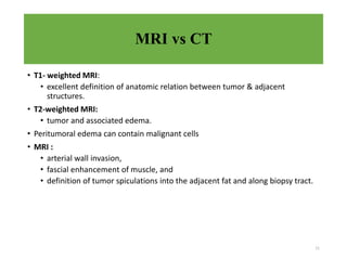
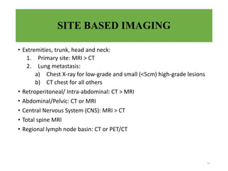
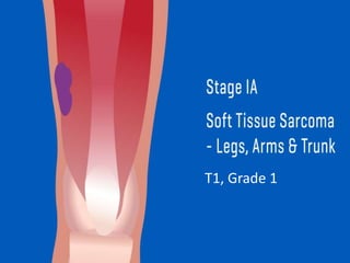
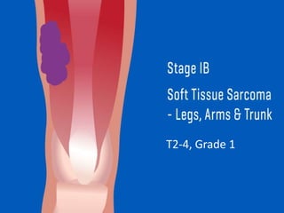
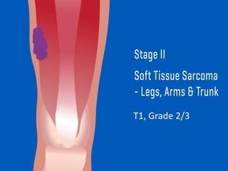
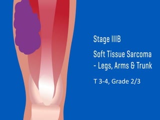
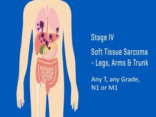
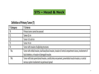
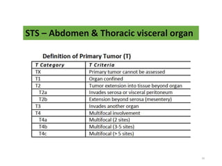
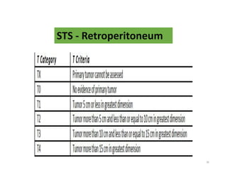
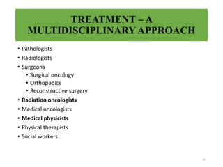
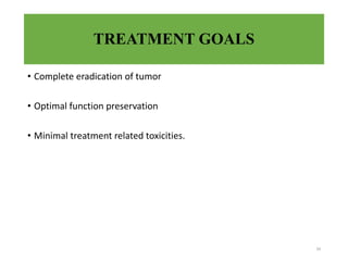
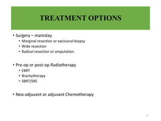
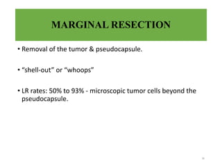
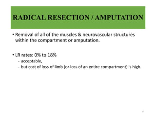
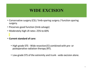
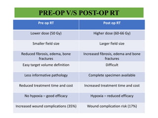
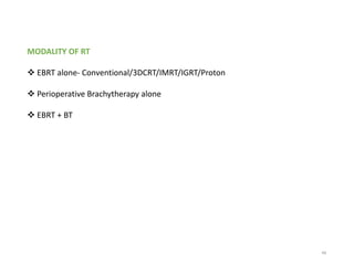
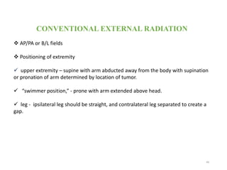
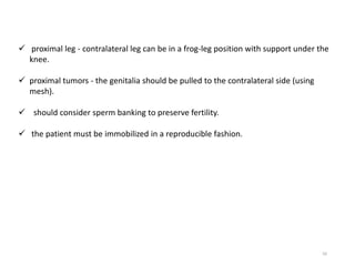
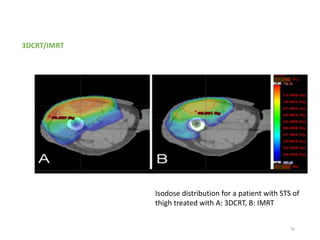
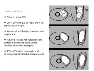
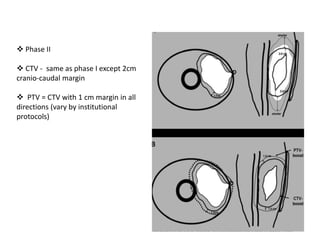
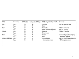
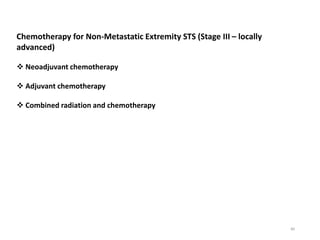
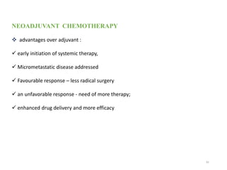
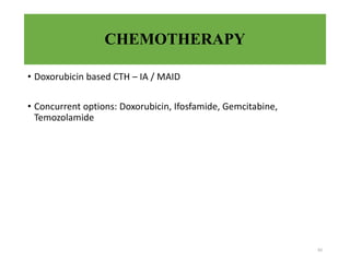
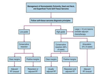
8. CLINICAL MANIFESTATIONS
most common - painless mass
pain - origin from or invasion of neurovascular structures.
Erythema and warmth.
Large slow growing, restricted joint mobility
Skin changes
Signs of neuro-vascular compromise
8
9. HISTORY & EXAMINATION
• Duration, pain
• Change in size and rate of change
• Local or systemic symptoms associated,
• Neurofibromas and Café-au-lait spots
• H/o radiation exposure
• Family H/o STS
• All soft-tissue masses deep to the fascia should be considered sarcoma
until proven otherwise.
9
10. DIAGNOSTIC WORKUP – Primary site +
Sites of potential metastatic spread
10
• CBC, RFT, LFT, FBS, Viral markers, 2D ECHO, ECG-12 leads
• MRI > CT (extremities, trunk, head and neck)
• Core biopsy > incisional biopsy/FNAC
11. MRI vs CT
• T1- weighted MRI:
• excellent definition of anatomic relation between tumor & adjacent
structures.
• T2-weighted MRI:
• tumor and associated edema.
• Peritumoral edema can contain malignant cells
• MRI :
• arterial wall invasion,
• fascial enhancement of muscle, and
• definition of tumor spiculations into the adjacent fat and along biopsy tract.
11
12. SITE BASED IMAGING
• Extremities, trunk, head and neck:
1. Primary site: MRI > CT
2. Lung metastasis:
a) Chest X-ray for low-grade and small (<5cm) high-grade lesions
b) CT chest for all others
• Retroperitoneal/ Intra-abdominal: CT > MRI
• Abdominal/Pelvic: CT or MRI
• Central Nervous System (CNS): MRI > CT
• Total spine MRI
• Regional lymph node basin: CT or PET/CT
12
13. BIOPSY
• Performed by or in presence of the operating surgeon and radiation
oncologist
• CT-guided core biopsy:
• accurate, safe, and expeditious.
• Incisional biopsy:
• accurate and acceptable, but more invasive.
• subsequent definitive resection in mind.
• Tumor cells can potentially seed an incision, thereby necessitating
removal of skin incisions at the time of surgical resection.
13
14. BIOPSY
• Biopsy should not transgress an uninvolved compartment or
joint
• much more radical resection.
• subsequent loss of function,
• LR, and death.
• FNAC for recurrent disease, no enough tissue to establish an
initial diagnosis.
14
21. 21
TNM Classification of STS of
extremities and trunk
except Kaposi’s sarcoma,
fibromatosis (desmoid
tumor), and sarcoma arising from
the dura mater, brain, and
parenchymatous organs
32. PROGNOSTIC FACTORS
• AJCC TNMG stage
• High grade
• Tumor size and site
• Histologic subtype
• Older age at presentation
• Positive resection margins
• Bone or neurovascular invasion
• Gender and race
• Lymph node involvement
32
33. TREATMENT – A
MULTIDISCIPLINARY APPROACH
• Pathologists
• Radiologists
• Surgeons
• Surgical oncology
• Orthopedics
• Reconstructive surgery
• Radiation oncologists
• Medical oncologists
• Medical physicists
• Physical therapists
• Social workers.
33
34. TREATMENT GOALS
• Complete eradication of tumor
• Optimal function preservation
• Minimal treatment related toxicities.
34
35. TREATMENT OPTIONS
• Surgery – mainstay
• Marginal resection or excisional biopsy
• Wide resection
• Radical resection or amputation.
• Pre-op or post-op Radiotherapy
• EBRT
• Brachytherapy
• SBRT/SRS
• Neo-adjuvant or adjuvant Chemotherapy
35
36. MARGINAL RESECTION
• Removal of the tumor & pseudocapsule.
• “shell-out” or “whoops”
• LR rates: 50% to 93% - microscopic tumor cells beyond the
pseudocapsule.
36
37. RADICAL RESECTION / AMPUTATION
• Removal of all of the muscles & neurovascular structures
within the compartment or amputation.
• LR rates: 0% to 18%
- acceptable,
- but cost of loss of limb (or loss of an entire compartment) is high.
37
38. WIDE EXCISION
• Conservative surgery (CS) / limb-sparing surgery / function sparing
surgery.
• Preserves good function (limb salvage)
• Moderately high LR rates: 25% to 60%
• Current standard of care:
• High-grade STS - Wide resection/CS combined with pre- or
postoperative radiation therapy (RT).
• Low-grade STS of the extremity and trunk - wide excision alone.
38
39. 39
PRIMARY THERAPY FOR EXTREMITY STS
Surgery :
Limb-sparing surgery - mainstay treatment for extremity STS.
Aim :
1. R0 resection or planned close or microscopically positive margins
2. Biopsy site should be excised en bloc with the definitive surgical specimen.
3. Surgical clips placed to mark the periphery of the surgical field - guide
potential future RT.
4. The drains should exit the skin close to the edge of the surgical incision
42. SURGERY ALONE
Low grade, negative margins
T1 and Superficial or subcutaneous
Widely excised
Margin >/=1cm or less if an intervening fascial barrier present
Surgical plane should transverse normal tissue outside the reactive tumor zone
Tumor locations amenable to limb-sparing salvage surgery for recurrence
Patient willingness to comply with appropriate follow-up
42
43. 43
Amputation vs Conservative surgery + RT
No significant difference in OS/DFS and LR rates
Amputation should be considered only –
tumor involves several compartments or invades major nerves (thigh STS
involving both sciatic and femoral nerves).
distal lower extremity lesion - below-knee amputation prosthesis may
provide better functional outcome than a limb damaged by tumor, extensive
surgery, and RT
44. 44
Benefit of adjuvant RT in terms of local control:
Trial from MSKCC - CS + BRT vs CS alone.
Trial from NCI - CS vs CS + EBRT.
Both trials showed improved local control with the addition of RT
BRT - not recommended for low-grade STS.
BRT as monotherapy - only for high-grade tumors resected with negative margins.
Positive margins – EBRT + BRT
45. POST OP RT
Start RT within 4-6 weeks of surgery.
High risk of local recurrence after surgical resection –
i. tumor size >5cm
ii. lesions deep to or invading superficial fascia
iii. high grade tumors
iv. narrow or positive surgical margins
v. local recurrence after prior surgery
45
46. POST OP RT FOR LOW GRADE TUMOURS
Positive resection margins
Locally recurrent disease following initial treatment with surgery alone
Tumor location that would not be amenable to subsequent salvage surgery.
46
47. PRE-OP V/S POST-OP RT
Pre op RT Post op RT
Lower dose (50 Gy) Higher dose (60-66 Gy)
Smaller field size Larger field size
Reduced fibrosis, edema, bone
fractures
Increased fibrosis, edema and bone
fractures
Easy target volume definition Difficult
Less informative pathology Complete specimen available
Reduced treatment time and cost Increased treatment time and cost
No hypoxia – good efficacy Hypoxia – reduced efficacy
Increased wound complications (35%) Wound complication risk (17%)
49. CONVENTIONAL EXTERNAL RADIATION
AP/PA or B/L fields
Positioning of extremity
upper extremity – supine with arm abducted away from the body with supination
or pronation of arm determined by location of tumor.
“swimmer position,” - prone with arm extended above head.
leg - ipsilateral leg should be straight, and contralateral leg separated to create a
gap.
49
50. 50
proximal leg - contralateral leg can be in a frog-leg position with support under the
knee.
proximal tumors - the genitalia should be pulled to the contralateral side (using
mesh).
should consider sperm banking to preserve fertility.
the patient must be immobilized in a reproducible fashion.
51. 51
The “swimmer position” is often an
ideal position for treating STS of the
hand, forearm, or distal upper arm
52. Tattoo placement, protocol for
extremities - 3 anterior tattoos and 3
lateral tattoos.
3 anterior tattoos: 15 to 20 cm
apart in the same sagittal plane and
at each point, an additional lateral
mark is placed in the same axial
plane
6 tattoo points are all placed in a
stable anatomical location
52
Anterior view of the lower leg
tumor showing placement of
tattoos at isocenter and about 15
to 20 cm apart in the same
53. • Dosing guidelines
• Spare 1cm thickness to receive <20 Gy
• 3D CRT – part of limb can be entirely excluded
• IMRT – some low dose is almost always received (<20Gy preferable)
• Joint dose < 40-45 gy
• Avoid full thickness bone radiation
• V40 <64%, Dmean <37Gy, Dmax <59Gy
53
57. 57
PRE OP RT CTV
Sarcoma case without peritumoral
edema.
CTV = GTV with 1.5 cm radial and
4 cm cranio-caudal margin
Radially CTV need not expand
beyond surface of bones and fascia,
unless invaded
58. 58
Sarcoma case with peritumoral
edema,
CTV = GTV with 1.5 cm radial and 4
cm cranio-caudal margin
radially CTV need not expand beyond
surface of bones and fascia, unless
invaded
CTV - manually edited (bold dashed
line) to encompass edema zone in both
transverse and coronal planes.
PTV = CTV with 1 cm margin in all
directions (vary by institutional
protocols)
59. 59
POST OP RT CTV
Phase I – preop GTV
CTV = GTV with 1.5 cm radial and 4 cm
cranio-caudal margin
includes all visible clips, drain sites and
surgical scar
radially CTV need not expand beyond
surface of bones and fascia, unless
involved with tumor at surgery
PTV = CTV with 1 cm margin in all
directions (vary by institutional protocols)
60. 60
Phase II
CTV - same as phase I except 2cm
cranio-caudal margin
PTV = CTV with 1 cm margin in all
directions (vary by institutional
protocols)
61. 61
DOSE
Pre op RT 50 Gy @ 1.8-2 Gy (no concurrent chemotherapy)
+ve margins after surgery, a post-op external beam RT boost of 16 to 20 Gy
( 1.8- to 2-Gy fractions)
total dose – post op
R0 : 60 Gy @ 1.8-2 Gy
R1 : 64-66 Gy @ 1.8-2 Gy
R2 : 66-70 Gy @ 1.8-2 Gy
64. INDEPENDENT PREDICTORS FOR
WOUND COMPLICATIONS
• Tumor size >10 cm
• Tumor proximity to skin surface
• Vascularized flap closure
• Diabetes mellitus
64
65. BRACHYTHERAPY
Localized radiation with maximum sparing of surrounding normal tissues
radio biologically better
short and convenient
small rate of complications
65
68. BRACHYTHERAPY
intra op
nylon catheters are placed in target
area of operative tumor bed
spaced at 1-1.5 cm interval
2 cm cranio-caudal and 1-2 cm radial
margin around tumor
treatment usually starts 4-5 days after
surgery
68
69. 69
(a) Operative cavity - catheters were placed perpendicular to the wound, spaced 1-1.5
cm apart in parallel.
(b) CTV was constructed with a tumor bed expansion of >/= 2 cm craniocaudally and >/=
1 cm lateral to tumor bed
70. 70
Catheter entry point - at least 1cm away from wound incision
Fix catheter to tumor bed by –
sutures (vicryl)
anchor button to skin
keep drain on top of brachy catheters and remove after completion of brachy
Avoid –
Penetrating blood vessels during catheter placement
direct catheter contact with bones and nerves
71. 71
CT Simulation preferred – on D4-5
If CT not available, Orthogonal X-Rays
check that wound has begun to heal, not infected, drains are working well, flap
has been taken up well
CTV is reconstructed using pre op imaging and clips in post op scans
72. 73
V100 >/= 90-95% (ideal 95%)
V150 </=50-40% (ideal 40%)
D90 >/=90-100% (ideal 100%)
Dose to skin should be less than 2/3rd of prescription dose
Loading dose should be >/= 0.5 cm from skin surface
OARs should get <100% of prescription dose, unless it is at high risk of tumor
involvement
73. 74
TREATMENT PRESCRIPTION
At 0.5 cm from depth of catheter, up to 1cm can be used after consideration of doses
to OARs and RT doses
>1 cm not recommended because of increased risk of late complications
BT Monotherapy :
i. LDR/PDR (45 Gy)
ii. HDR (30-50 Gy) in 2- to 4-Gy bid fractions
IORT + EBRT :
EBRT 45-50 Gy/ 1.8-2 Gy per fraction + 10-20 Gy IORT
EBRT 45-50 Gy/ 1.8-2 Gy per fraction +
i. 15 to 25 Gy LDR BT boost or
ii. 12 to 20 Gy HDR BT boost ( 2- to 4-Gy bid fraction)
for a total of 65 Gy
74. 75
TREATMENT DELIVERY
Catheter position, tumor bed geometry (seroma/hematoma) and wound healing
should be checked before starting treatment
with immediate reconstruction and BT monotherapy – start after PO D5
With BT Boost – can start on PO D2-4
If wound breakdown or infection – delay BT
76. Limitations –
Large target volume
Restriction of catheter placement because of bone or visceral organs
site where good catheter geometry is difficult to achieve – around shoulder
77
77. BRACHYTHERAPY BOOST
EBRT +BT = LCR – 77-100%, Grade 3 toxicity – 27-30%
when target volume/ volume at risk cannot adequately be covered with BT alone
(i.e. OAR restriction, atypical geometry, skin ulceration)
high risk of recurrence –
tumor > 10 cm
positive margin
recurrent disease
Advantage of adding EBRT to BT –
safely escalate dose to a large clinical volume with cumulative RT doses - >60 Gy
(45-50 Gy EBRT +15-20 Gy BT)
78
78. BRACHYTHERAPY BOOST
Advantages:
RT starts in immediate post op period controlling tumor repopulation
In perioperative period, tissues less hypovascular/fibrosed –than in the
postoperative period - RT more effective
BT catheters in contact with tumor bed giving higher RT doses to potential areas
of microscopic disease
can safely treat STS abutting neurovascular structures
79
79. 80
Chemotherapy for Non-Metastatic Extremity STS (Stage III – locally
advanced)
Neoadjuvant chemotherapy
Adjuvant chemotherapy
Combined radiation and chemotherapy
80. 81
NEOADJUVANT CHEMOTHERAPY
advantages over adjuvant :
early initiation of systemic therapy,
Micrometastatic disease addressed
Favourable response – less radical surgery
an unfavorable response - need of more therapy;
enhanced drug delivery and more efficacy
83. 84
Retroperitoneal Sarcoma
large at presentation with a median size of 17 cm.
nonspecific abdominal pain, anorexia, and weight loss.
Metastasis to the lungs and to the liver may occur
initial assessment : CT of chest, abdomen, and pelvis.
Core biopsy with image guidance - preferred
optimal primary treatment - en bloc excision of the tumor with adjacent involved
viscera and a margin of uninvolved tissue
Adjuvant RT could be recommended, but the benefits, are uncertain.
Chemotherapy has not shown benefit
85. 86
Retroperitoneal Sarcoma….
no randomized trials have compared surgery with surgery and radiation
The typical pattern of relapse is more local than distant suggesting a role for
adjuvant RT.
no prospective randomized trials supports its role, and the toxicity is not minimal.
If RT is to be used, preoperative RT of 45 Gy to 50 Gy is preferred over postoperative
RT.
dose escalation can be done through IORT, dose 10-15 Gy
target to be treated with IORT is mainly the posterior structures (paraspinal tissues
and vessels) where surgeons have difficulty obtaining wide negative margins.