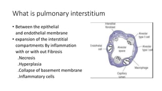
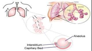
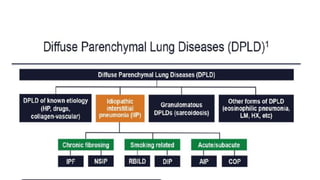
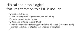
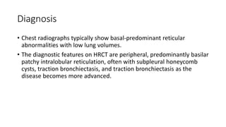
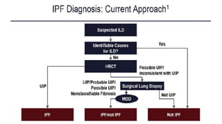
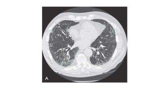
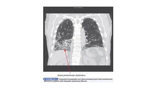
Interstitial lung disease
Dr Mustafe Hussein
Email: Mustafe.Hussein@kiu.ac.ug
Outline
• Defination
• Epidemiology
• Pathogenesis
• clinical features
• Diagnosis
• Treatment
What is pulmonary interstitium
• Between the epithelial
and endothelial membrane
• expansion of the interstitial
compartments By inflammation
with or with out Fibrosis
.Necrosis
.Hyperplasia
.Collapse of basement membrane
.Inflammatory cells
DEFINATION
• Interstitial lung disease (ILD) is an umbrella term for a large group of
disorders that cause scarring (fibrosis) of the lungs. The scarring
causes stiffness in the lungs which makes it difficult to breathe.
American lung association.
• In an apparently immunocompetent host, interstitial lung disease
(ILD) is a clinical term for a heterogenous group of acute and chronic
lower respiratory tract disorders with many potential causes. Cecil 25
edition
clinical and physiological
features common to all ILDs include
Exertional dyspnea
A restrictive pattern of pulmonary function testing
Coexisting airflow obstruction
Decreased diffusing capacity(DLCO)
Increased alveolar arterial oxygen difference (Pao2-Pao2) at rest or during
exertion and absence of pulmonary infection or neoplasm

Cont..
• The term interstitial is a misnomer because the pathologic processes
are not restricted to the interstitium, which is the microscopic space
bounded by the basement membranes of epithelial and endothelial
cells. Rather, all of the several cellular and soluble constituents that
make up the gas exchange units (alveolar wall, capillaries, alveolar
space, and acini) and the bronchiolar lumen, terminal bronchioles,
and pulmonary parenchyma beyond the gas exchange units (as well
as the pleura and lymphatics and sometimes the lymph nodes) are
involved in the pathogenesis and manifestations of ILD.
Epidemiology
• IPF accounts for 20% to half of all cases of interstitial lung
disease (ILD), and represents the most frequent and severe
of the idiopathic interstitial pneumonias (IIPs), a group of
ILDs of unknown cause . Idiopathic pulmonary fibrosis is
considered a rare disease (occurring in less than 5 per 10,000
person-years), yet its burden is high.
• In Europe alone, approximately 40,000 new cases are diagnosed each
year. Idiopathic pulmonary fibrosis is a clinically heterogeneous
disease but its
• prognosis is overall poor, with a median survival of 3–4 years [6].
Taking all of the above into account, IPF is also an expensive disease,
with direct treatment costs of around 25,000 USD/person-year, which
is a higher cost in comparison to breast cancer and many other
serious conditions
• According to the GBD’s tools, there is wide heterogeneity in the
distribution of this category of diseases by country, with prevalence
ranging 30-fold from 214.5 per 100,000 in Guam to a low of 6.8 per
100,000 in Benin; mortality ranging 150-fold from 10.5 per 100,000 in
Japan to a low of 0.064 per 100,000 in Burkina Faso; and the
combined estimate of both morbidity and mortality, the so-called
disability-adjusted life years (DALYs) ranging more than 60-fold from
173.1 per 100,000 in Guam to a low of 2.7 per 100,000 in Burkina
Faso (Figure 1).
Pathogenasis
• Interstitial lung disease are thought to result from an unknown tissue
injury and attempted repair in the lung of a genetically predisposed
person.
• Genetic variants within the hTERT or hTR components of the
telomerase gene and surfactant protein gene have been associated in
a subset of familial pulmonary fibrosis and in some sporadic cases.
• An MUC5B promotor polymorphism is associated with familial
interstitial pneumonia and idiopathic pulmonary fibrosis.
Cont.…
• Ultimately, progressive fibrosis results in honeycombing, an end-stage
finding that is often associated with increased pulmonary vascular
resistance and secondary pulmonary hypertension.
• As a reflection of these dynamic processes, histopathologic
examination of lung tissue often reveals highly heterogeneous
findings.
Clinical Manifestations
• In some patients, the initial presentation may be with peripheral cyanosis,
clubbing, or the signs and symptoms of an underlying systemic disease
• Progressive dyspnea
• Non productive cough
• Fatigue
• Pleuritic chest pain with dyspnea may represent a spontaneous
pneumothorax
• Neurofibromatosis or Langerhans cell histiocytosis
• hemoptysis suggests a diffuse alveolar hemorrhagic syndrome, SLE,
lymphangioleiomyomatosis , granulomatosis with polyangiitis or
goodpasture syndrome
Cont.….
• In patients with existing ILD, new hemoptysis should prompt
consideration of a superimposed malignancy, pulmonary embolus, or
infection such as aspergillosis.
• In some patients, the first and the only clue to the presence of an ILD
may be the finding of coarse rales (crackles) on auscultation of the
lungs.
Diagnosis
• CBC with leucocyte differential
• ESR
• Chemistry panel ( calcium, liver enzymes, electrolytes, creatinine.
• Urine analysis
• Antinuclear antibody for SLE
• Rheumatoid factor, anticitrullinated peptide antibody for RA
• Scleroderma (Scl 70
• Dermatomyositis or polymyositis ( CK, Aldolase, and anti-Jo-I antibody
• Granulomatosis or polyangiitis ( antineutrophil cytoplasmic antibodies
• Goodpasture syndrome ( anti-basement membrane antibodies )
• ABG analaysis
• Chest X-ray
• Chest Ct Scan
• Bronchoalveolar lavage
• Biobsy
•
Treatment
• Systemic corticosteroids are indicated
• Supportive oxygen
• Lung transplantation
SPECIFIC TYPES OF INTERSTITIAL LUNG DISEASE
Idiopathic Interstitial Pneumonias
• Idiopathic interstitial pneumonias, which are a subset of acute or
chronic ILDs of unknown etiology, are characterized by the presence
of varying degrees of interstitial and alveolar inflammation and
fibrosis.
• Distinct clinicopathologic forms of idiopathic interstitial pneumonia
include chronic fibrosing interstitial pneumonias (idiopathic
pulmonary fibrosis and nonspecific interstitial pneumonia), smoking-
related interstitial pneumonias (respiratory bronchiolitis–ILD and
desquamative interstitial pneumonia), and acute or subacute
idiopathic interstitial pneumonias (cryptogenic organizing pneumonia
and acute interstitial pneumonia).
CHRONIC FIBROSING INTERSTITIAL
PNEUMONIA
• Idiopathic Pulmonary Fibrosis
Idiopathic pulmonary fibrosis accounts for 50% to 60% of all idiopathic
interstitial pneumonias. Idiopathic pulmonary fibrosis occurs in adult
men and women with a mean age at onset of 62 years.
The best validated genetic risk factor is a polymorphism in the
promoter of the gene encoding mucin-5B (MUC5B), which is associated
with both familial and sporadic forms
• Idiopathic pulmonary fibrosis is limited to the lungs in adults, usually
older than 60 years, and it generally occurs in men with a history of
cigarette smoking.
• Most often patients have otherwise been in good health and have no
known connective tissue disease or exposure to drugs or
environmental factors known to cause pulmonary fibrosis, although
patients with significant cigarette smoking history may have
coexisting emphysema.
• After alveolar epithelium injury there is increased activity of type II
alveolar epithelial cells, which proliferate in order to repair the
damage. However, the repair process fails and leads to fibrosis.
• CARLONI et al. To further support the concept that IPF is a disease of
premature ageing, dehydroepiandrosterone (DHEA) and its sulfated
form (DHEA-S), which have been previously linked to the impaired
function of the immune system occurring with ageing, have been
evaluated in the serum of IPF patients.
• Previous findings that peripheral blood proteins, such as mucin-1
(MUC1-KL6), CC chemokine ligand-18 (CCL-18) and surfactant protein-
A , are related to increased mortality.
• Deregulation of humoral immunity is associated with IPF. B-cell
aggregates, activated T-cells and mature dendritic cells have been
reported in the IPF lung, increasing the likelihood of antigen
presentation activity.
• Innate immunity has been proposed to have a role in the
development and progression of IPF, mainly through the activation of
Toll-like receptor (TLR)-9 [59, 60]. TLR-3, amongst other properties,
regulates the recognition of viral pathogen-associated molecular
patterns.
Clinical features
• manifestations include a gradual onset and progression of exertional
dyspnea, restrictive abnormalities on PFTs, and a distinct pattern of
bilateral pulmonary fibrosis on HRCT.
Diagnosis
• Chest radiographs typically show basal-predominant reticular
abnormalities with low lung volumes.
• The diagnostic features on HRCT are peripheral, predominantly basilar
patchy intralobular reticulation, often with subpleural honeycomb
cysts, traction bronchiectasis, and traction bronchiectasis as the
disease becomes more advanced.
• Pulmonary function tests usually show a progressive restrictive
pattern. However, patients with milder disease may have normal lung
volumes and a small decrease in Dlco; rarely, PFT results may be
normal.
• The cellular pattern in BAL fluid, which is nonspecific, is marked by an
excess of neutrophils in proportion to the extent of reticular change
on HRCT; the percentage of eosinophils may be mildly increased. The
histopathologic pattern of usual interstitial pneumonia consists of
patchy interstitial changes alternating with zones of honeycombing,
fibrosis, minimal inflammatory cells, collagen deposition, and normal
lung.
• ALLAIX et al. [69] compared the clinical presentation, the oesophageal
function and the reflux profile in 80 patients with GORD to 22
patients with GORD and IPF. Results showed that GORD is often
asymptomatic in IPF patients, with heartburn present in ,60% of
patients with GORD and IPF.
• IPF patients had significantly higher oesophageal acid exposure
(9.25% versus 3.3% versus 0.7%) and proximal reflux events (median
51 versus 20 versus nine) compared to non-IPF patients and healthy
volunteers, respectively.
• RYERSON et al. revised this entity and attempted to better define its
clinical profile and prognosis. Clinical characteristics and outcomes of
CPFE and non-CPFE IPF patients were compared in two large cohorts
of 365 IPF patients. 29% of patients had some emphysema detectable
on HRCT, 13.4% had at least 5% emphysema.
• Patients with IPF and CPFE have less physiological restriction and
worse gas exchange compared to IPF patients without emphysema.
• It has been shown that IPF patients are more likely than controls to
have venous thromboembolism [74], and both epidemiological and
laboratory studies suggest that activation of the coagulation cascade
within the lung may be involved in the pathogenesis of IPF.
• BARGAGLI et al. [75] showed procoagulant status to correlate with IPF
and acute exacerbation of IPF compared to both NSIP and healthy
controls.
• NAVARATNAM et al. [76] investigated the presence of a pro-
thrombotic state in a large cohort of patients. They recruited 211
incident cases of IPF and 256 age- and sex-matched general
population controls and reported that IPF cases were more than four
times more likely than controls to have a pro-thrombotic state (OR
4.78 (95% CI 2.93–7.80); p,0.0001).
Treatment
• Pirfenidone(1800 mg/day for I year ) decreases the rate of decline in
forced vital capacity in clinical trails of patients who have idiopathic
pulmonary fibrosis and mild to moderate impairment in pulmonary
function and pooled data suggest an improvement and survival.
• Antoniou et all. In the past year, safety reports of pirfenidone in real-
life practice have been published both from Japan and Europe,
confirming that pirfenidone is well tolerated and can improve
progression-free survival in IPF
• The INBUILD trial.Nintedanib 150mg oraly twice daily also decreases
the rate of disease progression as measured by FVC over 52 weeks in
patients with idiopathic pulmonary fibrosis and mild to moderate
impairment in pulmonary function.
• Sildenafil 20mg orally three times a day has shown small benefits in
terms of dyspnea, oxygenation and quality of life but not exercise
capacity in pateints with idiopathic pulmonary fibrosis and severe
impairment in pulmonary function.
• Proton pump inhibitors to avoid Gastroesophageal reflux disease is
common is very common in patients with idiopathic pulmonary
fibrosis and treatment
• By comparison, N-acetylcysteine alone or as part of triple-therapy
combined with prednisone plus azathioprine is not beneficial.
• Warfarin increases respiratory hospitalizations and death in patients
with idiopathic pulmonary fibrosis.
• corticosteroids (e.g., methylprednisolone 1g intravenously as a pulse
dose once a day for 3 days and followed by hydrocortisone, 125 mg
every 6 hours for another 3 to 5 days), with further dosing dependent
on the clinical response.
• Ancillary treatment measures, including supplemental oxygen (based
on clinical and physiologic needs); prompt detection and treatment of
respiratory tract infections and pulmonary embolism, pulmonary
rehabilitation; and immunization for influenza, herpes zoster, and
pneumococcus, are all appropriate.
• Lung Transplantation is also recommended
• Cui and colleagues. Preventing Glutaminolysis A Potential Therapy for
Pulmonary Fibrosis
• Nonspecific Interstitial Pneumonia
Nonspecific interstitial pneumonia is often associated with connective
tissue diseases, but idiopathic nonspecific interstitial pneumonia is also
recognized as a distinct clinical entity. It typically occurs in middle-aged,
nonsmoking women with an average age at diagnosis of about 50
years. The prevalence of nonspecific interstitial pneumonia has been
estimated at one to nine per 100,000.
Two subgroups have been described, cellular and fibrotic.
Diagnosis
• Chest radiographs show bilateral patchy pulmonary infiltrates with a
lower lung zone predominance in all forms of nonspecific interstitial
pneumonia.
• HRCT reveals a predominant ground-glass pattern of attenuation,
usually bilateral and often associated with subpleural reticulation,
and loss of volume in the lower lobe.
• In cellular nonspecific interstitial pneumonia, HRCT shows ground-
glass opacification, consolidation, or both, but the biopsy shows mild
to moderate lymphoplasmacytic interstitial chronic inflammation.
• In contrast, fibrotic nonspecific interstitial pneumonia has a bilateral
lower lobe distribution with architectural derangement on HRCT;
histopathologically, it has uniformly dense interstitial fibrosis and may
sometimes be difficult to distinguish from idiopathic pulmonary
fibrosis and usual interstitial pneumonia in the early clinical stages.
Treatment
• Patients with cellular nonspecific interstitial pneumonia usually
respond to treatment with corticosteroids, and their prognosis is
generally better than that of patients with idiopathic pulmonary
fibrosis. Nonetheless, some patients progress over several years, and
some manifest acute exacerbations similar to patients with idiopathic
pulmonary fibrosis. Immunomodulating drugs, including prednisone,
azathioprine, and mycophenolate, have been used empirically, with
their doses based on clinical response as assessed by clinicians and
not on evidence with randomized clinical trials.
SMOKING-RELATED INTERSTITIAL
PNEUMONIAS
• Respiratory Bronchiolitis–Associated Interstitial Lung Disease
This ILD is almost invariably associated with chronic and current
cigarette smoking, and it usually manifests clinically during the fourth
or fifth decade of life.
However, it may also be detected incidentally on radiographs in
relatively younger and asymptomatic persons with a previous history of
cigarette smoking or in people passively exposed to chronic cigarette
smoke Respiratory bronchiolitis–associated ILD is always associated
with chronic exposure to cigarette smoke.
Diangosis
• Pulmonary function tests show varying degrees of airway obstruction,
mildly decreased or preserved TLC, and decreased Dlco.
• The chest radiograph typically reveals bronchial wall thickening and
areas of ground glass attenuation.
• HRCT reveals centrilobular nodules with and upper lobe
predominance, patchy ground glass attenuation and peribranchial
alveolar septal thickening.
• The characteristic finding on BAL is numerous brown-pigmented
alveolar macrophages, often with modest increase in neutrophils.
Treatment
• Progression to honeycomb lung and end-stage fibrosis seldom occurs,
and the prognosis is good with cessation of smoking. Discontinuation
of cigarette smoking is essential, and patients may benefit from low-
dose corticosteroids (e.g., prednisone, 10 to 20 mg/day) for a few
months.
• Desquamative Interstitial Pneumonia
Desquamative interstitial pneumonia is a rare entity (<3% of all ILDs)
that may represent a form of respiratory bronchiolitis–associated ILD
extending into the alveolar spaces and alveolar walls. Although most
affected individuals are cigarette smokers, the histologic pattern of
desquamative interstitial pneumonia may also occur in
pneumoconiosis, rheumatologic disease, and drugassociated ILD.
Patients are often initially seen with advanced disease and striking
hypoxemia. The histopathology features of desquamative interstitial
pneumonia are characterized by accumulation of pigmented alveolar
macrophages within the alveoli. Histologic changes within the
respiratory bronchioles and within the alveolar spaces can coexist and
represent a histopathologic spectrum of alveolar macrophage
accumulation.
Diagnosis
• Pulmonary function tests reveal a restrictive lung defect and
decreased Dlco with or without coexisting airway obstruction.
• The chest radiograph shows patchy basal consolidation with a lower
lobe and peripheral predominance
• HRCT shows bilateral symmetrical ground-glass opacities with a
predominantly basal and peripheral distribution as well as diffuse
alveolar septal thickening.
• Irregular linear opacities, typically associated with traction
bronchiectasis, may be noted.
• Fluid recovered from BAL quite often shows increased numbers of
pigmented alveolar macrophages, frequently with increased
neutrophils. Histopathologic findings on biopsy include diffuse
alveolar septal thickening, hyperplasia of type II pneumocytes, and
intense accumulation of intra-alveolar granular pigmented
macrophages in a uniform manner fibrosis is minimal.
Treatment
• With cessation of smoking and administration of oral corticosteroid
therapy, outcomes are generally good, with an estimated overall
survival rate of 70% at 10 years. However, a subset of patients may
progress despite cessation of cigarette smoking, and a trial of
corticosteroid therapy and lung transplantation is an appropriate
consideration for selected patients.
ACUTE OR SUBACUTE IDIOPATHIC
INTERSTITIAL PNEUMONIA
• Acute Interstitial Pneumonia
an apparent acute viral upper respiratory infection. The syndrome,
historically known as Hamman-Rich syndrome, mimics acute
respiratory distress syndrome. Acute interstitial pneumonia is a rare
and fulminant idiopathic interstitial pneumonia that presents with
acute symptoms and leads to respiratory distress or failure.
Cryptogenic Organizing Pneumonia
Cryptogenic organizing pneumonia, previously referred to as
bronchiolitis obliterans organizing pneumonia of unknown cause
(BOOP), is an idiopathic form of organizing pneumonia. Organizing
pneumonia affects the small airways, including the distal bronchioles,
respiratory bronchioles, alveolar ducts, and alveolar walls. Although the
incidence and prevalence of cryptogenic organizing pneumonia are
unknown, the estimated annual incidence in the United States is six to
seven cases per 100,000. The mean age at presentation is about 60
years, and there is no sex predominance.
Diagnosis
• Cryptogenic organizing pneumonia most commonly manifests as a
flulike illness with a nonproductive cough followed by exertional
dyspnea.
• PFTs show a restrictive defect, but 20% of patients, most of whom are
current or past smokers, also have an obstructive defect.
• Chest radiography reveals patchy unilateral or bilateral alveolar
opacities that may be peripheral or migratory; small nodular opacities
are seen in 10% to 50% of cases
• In about 90% of patients, HRCT shows areas of air space consolidation
with lower lung zone predominance, frequently in a subpleural or
peribronchial distribution other features include small nodules along
bronchovascular bundles and ground-glass attenuation. BAL is
nonspecific; increased lymphocytes, neutrophils, and eosinophils may
be seen. On biopsy, key histologic features are excessive proliferation
of granulation tissue within the small airways and alveolar ducts as
well as chronic inflammation in the surrounding alveoli.
Treatment
• Most patients recover rapidly and completely when treated with oral
corticosteroids for 6 months but may relapse after discontinuation
and require oral corticosteroids for longer periods and sometimes
indefinitely, often with adjunct immunosuppressive agents such as
azathioprine. A small subset of patients in whom pulmonary fibrosis
develops despite corticosteroids and azathioprine behave similarly to
patients with idiopathic pulmonary fibrosis. Spontaneous remissions
are known to occur.
• Lymphoid and Lymphocytic Interstitial Pneumonia
This condition is more common in women, especially in the fifth
decade of life, but it may occur at any age. Patients should be evaluated
for concurrent connective tissue disease, an autoimmune disorder
(especially Sjögren syndrome), or common variable immunoglobulin
deficiency because idiopathic LIP is very rare. Symptoms are
nonspecific and include a gradual onset of cough and exertional
dyspnea. Lymphoid and LIP is within the spectrum of benign
lymphoproliferative disorders, and some patients manifest pseudo
lymphoma or lymphoma as a complication.
Diagnosis
• Chest radiographs show a reticular or reticulonodular pattern
predominantly involving the lower lung zones. HRCT reveals bilateral
ground-glass attenuation, small or large nodules, and scattered cysts;
perivascular honeycombing and reticular abnormalities may also be
seen (E-Fig. 92-E5). Increased numbers of lymphocytes are found on
BAL, and biopsy reveals a dense interstitial lymphocytic infiltrate.
Treatment
• Some patients respond to or stabilize with oral corticosteroids. The
prognosis is variable, with more than one third of patients
progressing to diffuse pulmonary fibrosis.
Interstitial Lung Disease Associated with
Connective
Tissue Disease
• Many of the connective tissue diseases, including progressive systemic
sclerosis, rheumatoid arthritis, SLE, dermatomyositis and polymyositis,
Sjögren syndrome and mixed connective tissue disorder, may have ILD
as one of their manifestations. In fact, up to 20% of patients with
connective tissue diseases may initially be thought to have an ILD alone.
Therefore, these diagnoses must be considered in patients with ILD,
even in the absence of extrathoracic findings.
• PROGRESSIVE SYSTEMIC SCLEROSIS Of the connective tissue diseases,
progressive systemic sclerosis is most frequently associated with ILD.
Pulmonary symptoms may antedate cutaneous or digital manifestations of
the disease by several years. Most patients affected have nonspecific
interstitial pneumonia, with a minority having a usual interstitial
pneumonia pattern, and Dlco levels correlate with mortality. Pulmonary
hypertension, which can occur in the absence of pulmonary fibrosis, may
result in cor pulmonale. Patients with chronic pulmonary fibrosis also have
an increased risk for bronchogenic carcinoma, usually either
bronchoalveolar cell carcinoma or adenocarcinoma. In a controlled trial,
treatment with cyclophosphamide (50-100 mg/day orally) for 1 year
stabilized the PFT findings and improved health-related quality of life in
patients with scleroderma-related ILD.
• RHEUMATOID ARTHRITIS
Although rheumatoid arthritis is more common in women (2 : 1 to 4 : 1
ratio), ILD associated with rheumatoid arthritis is more common in men
(3 : 1 ratio). Most cases occur at 50 to 60 years of age, and pulmonary
symptoms follow the onset of arthritis in about 75% of cases. Lung
involvement in rheumatoid arthritis may take many forms, but
bronchiectasis, bronchiolitis, idiopathic interstitial pneumonias, and
pleural effusions or pleural thickenin are some of the most common.
• Early in the course, the histologic changes are similar to those of idiopathic
interstitial pneumonias, including pulmonary fibrosis, but are distinguished by a
prominent lymphocytic infiltrate that may contain germinal follicles adjacent to
vessels and airways.
• As the disease progresses, the infiltration becomes less pronounced and is
replaced by fibrous tissue, honeycomb changes, or both.
• Other pulmonary manifestations include pulmonary nodules, vasculitis,
pulmonary hypertension, and Caplan syndrome (progressive upper lobe nodular
pulmonary fibrosis in a coal miner with rheumatoid arthritis) but are relatively
rare. Treatment is directed at the underlying rheumatoid arthritis
• SYSTEMIC LUPUS ERYTHEMATOSUS
Pulmonary abnormalities complicating SLE may vary greatly. Pleural
disease or pleural effusions (or both) are commonly present in lung
disease that complicates SLE. Acute lupus pneumonitis may mimic
acute interstitial pneumonia, with widespread ground-glass attenuation
admixed with consolidation, or it may manifest as diffuse alveolar
hemorrhage.
• Chronic ILD may also occur. Infection must always be considered in
acutely ill patients who have received steroids or other
immunosuppressive therapy. Rarely, the restrictive lung defect, which
may be predominantly a result of diaphragmatic weakness, leads to a
chest radiographic pattern of small appearing lungs that may look
progressively smaller over time. This so-called “shrinking lung” is
generally resistant to corticosteroids or other immunosuppressive
agents used to treat SLE. Otherwise, treatment of the ILD is similar to
treatment of the underlying SLE.
• DERMATOMYOSITIS AND POLYMYOSITIS
the pattern of lung involvement in dermatomyositis and polymyositis is
more heterogeneous. Usual interstitial pneumonia, nonspecific
interstitial pneumonia, and organizing pneumonia have all been
reported.
Most patients have anti–Jo-1 antibody, and the disease is typically
progressive over time.
ILD may precede the muscular manifestations by months to years or be
superimposed on established muscle disease. Treatment isdirected at
the underlying disease
• SJÖGREN SYNDROME
Particularly those with the primary form of the disease. LIP is the most
frequent subtype, but cryptogenic organizing pneumonia may also be
present. Respiratory infections and bronchiectasis are common in
advanced stages, perhaps because of inspissated mucus. Response to
corticosteroid or immunosuppressive therapy is usually good
• MIXED CONNECTIVE TISSUE DISEASE
This overlap syndrome combines features of progressive systemic
sclerosis, SLE, rheumatoid arthritis, and polymyositis or
dermatomyositis. Pulmonary disease is common, but it is most often
subclinical and identified only radiographically. Treatment includes
corticosteroids and other immune-modulating agents for the
underlying disease.
• ANKYLOSING SPONDYLITIS
The most common pulmonary manifestation of ankylosing spondylitis
is upper lobe, bilateral reticulonodular infiltrates with cyst formation as
a result of parenchymal destruction. There is no known effective
therapy for this apical fibrobullous disease.
HYPERSENSITIVITY PNEUMONITIS
• PATHOBIOLOGY
Hypersensitivity pneumonitis, also known as extrinsic allergic alveolitis,
is a syndrome caused by repeated inhalation of specific antigens from
occupational or environmental exposure in sensitized individuals.
Within a short period after inhalation of an inciting agent, patients
develop a nonspecific diffuse pneumonitis with inflammatory cell
infiltration of the bronchioles, alveoli, and interstitium, sometimes
associated with a pleural effusion.
• In the subacute and chronic stages, loosely formed, noncaseating,
epithelioid cell granulomas and a mononuclear infiltrate may be
dispersed in the interstitium.
• Some of the more common exposures are farmer’s lung, bird fancier’s
lung, parakeet keeper’s lung, and pigeon breeder’s lung. The
exposure to an avian antigen may be occult and related to bird
droppings or bird nests. Hobbies (woodworker’s lung) and
recreational activities (sauna taker’s lung; hot tub) may be implicated
as well as occupations.
CLINICAL MANIFESTATIONS
• A history of exposure to potential agents or changes in the domestic
and other environments (or both) is essential to diagnosis and
treatment.
• symptoms canoften temporarily related to the workplace or to
hobbies, and may actually disappear on vacations or during absence
from the site of exposure only to recur when exposure is resumed
• In more chronic and low-level exposures, however the onset is
insidious.
• occur as soon as 4 to 12 hours after exposure. Fever and chills are
common symptoms, are
Diagnosis
• Findings on chest radiography are diverse, with focal patchy
consolidation or a diffuse ground-glass appearance in acute
hypersensitivity pneumonitis micronodular and reticular shadowing in
subacute forms; and diffuse, predominantly upper lung zone
reticulation with honeycombing in the chronic form. Chest radiograph
results may be normal in up to 30% of patients with significant
physiologic abnormalities.
• On HRCT, small centrilobular ill-defined nodules of ground-glass
densities are seen, along with evidence of mosaic attenuation
(trapped air) as a result of concomitant bronchiolitis and upper lobe
predominance of the parenchymal abnormalities. Chronically, findings
of lung fibrosis may be indistinguishable from the patterns seen in
usual interstitial pneumonia and idiopathic pulmonary fibrosis
• Precipitating serum antibodies for potential causes of hypersensitivity
pneumonitis confirm exposure but not cause and effect, and the
absence of antibodies does not exclude hypersensitivity pneumonitis.
• In some cases, a thorough investigation of the patient’s home and
workplace by an industrial hygienist may reveal occult molds, spores,
Thermoactinomycetes spp., Aureobasidium pullulans, and other
precipitating causes.
• BAL may be quite helpful by showing the most marked increase in T
lymphocytes and an increased number of plasma cells.
• The characteristic histologic triad in hypersensitivity pneumonitis is
cellular nonspecific interstitial pneumonia, cellular bronchiolitis, and
granulomatous inflammation; however, this triad is seen in no more
than 75% of affected patients.
Treatment
• A thorough investigation must be undertaken to identify the antigen
in the patient’s environment. Sometimes an industrial hygienist is
needed to obtain samples for culture from potential sources in the
patient’s domestic or workplace environment.
• The identified antigen must be eradicated from the patient’s
environment. Avoidance of exposure to the identified antigen or
antigens and treatment with corticosteroids are important if
improvement is to be obtained.
• Continued exposure to the unidentifiable antigens, prolonged
exposure to antigens, or both can lead to chronic hypersensitivity
pneumonitis and irreversible fibrosis that may not respond to any
treatment regimen. In the fibrotic stages, the prognosis and clinical
course may be similar to those of idiopathic pulmonary fibrosis.
DRUG-INDUCED INTERSTITIAL LUNG DISEASE
• More than 300 drugs, biomolecules, or homeopathic remedies can
cause acute, subacute, or chronic ILD, and the list continues to
increase as new medications are introduced.
• The clinical and radiographic manifestations are quite varied.
Examples of known syndromes include chronic nitrofurantoin-induced
ILD that mimics idiopathic pulmonary fibrosis (and is fatal in ≈8% of
cases), granulomatous pneumonitis secondary to methotrexate (<5%)
• sarcoid-like granulomatous ILD induced by interferon-α and tumor necrosis factor
modulating agents, nonspecific bilateral alveolar and interstitial inflammatory
and fibrotic abnormalities caused by bleomycin and other chemotherapeutic
agents, and alveolar and interstitial abnormalities and nodular densities in acute
and chronic amiodarone pulmonary toxicity.
• Most drug-induced ILD is reversible if recognized early and if use of the
responsible drug is discontinued. In addition to discontinuing the implicated drug,
treatment with corticosteroids is indicated in patients with moderate to severe
functional impairment.
Alveolar Filling Disorders
• In alveolar filling disorders, air spaces distal to the terminal
bronchioles are filled with blood, lipid, protein, water, or
inflammatory cells. The radiographic appearance is that of an alveolar
infiltrate with small nodular densities and ill-defined margins; hence,
the radiographic picture is similar to an ILD, and virtually all the
alveolar filling disorders may result in ILD, including Goodpasture
syndrome, pulmonary alveolar proteinosis (primary and secondary),
alveolar hemorrhage syndromes acute interstitial pneumonia, and
bronchoalveolar cell carcinoma.
IDIOPATHIC PULMONARY HEMOSIDEROSIS
• This rare disorder of children and young adults is characterized by
intermittent, diffuse alveolar hemorrhage without evidence of
vasculitis, inflammation, granulomas, or necrosis. The etiology is
poorly understood. Anemia and hepatosplenomegaly may be present.
Hemosiderin-laden macrophages in BAL fluid and lung tissue are part
of the diagnostic picture. The chest radiograph reveals diffuse,
bilateral alveolar infiltrates. A chronic interstitial infiltrate may
develop after repeated episodes, infrequently with hilar and
mediastinal adenopathy. Systemic corticosteroids may be beneficial in
treating acute disease.
CHRONIC EOSINOPHILIC PNEUMONIA
• The clinical manifestation of chronic eosinophilic pneumonia varies
over a wide spectrum, from asymptomatic to respiratory failure. The
disease often occurs in women in the second to fourth decades of life;
such women often manifest constitutional symptoms of fevers,
sweats, weight loss, fatigue, dyspnea, and cough. Peripheral blood
eosinophilia, usually at levels of 10% to 40%, is common but may be
absent in up to one third of affected patients at initial evaluation.
• On chest radiography and HRCT, the cardinal feature is peripheral
multifocal consolidation, predominantly in the upper and mid lung
zones. These dense peripheral infiltrates, which have sometimes been
called the “photographic negative of pulmonary edema,” often
resolve dramatically after treatment with corticosteroids. Ground-
glass attenuation commonly accompanies the consolidation. BAL fluid
may show greater than 40% eosinophils during exacerbations.
• Treatment with corticosteroids results in a rapid response, frequently
within hours; in fact, such a dramatic resolution of symptoms with
radiographic clearance of infiltrates shortly after initiation of
corticosteroid therapy is considered “diagnostic.” However, the rate of
relapse is high, so most patients require prolonged treatment with
low-dose corticosteroids (prednisone, 5-10 mg/day) to stay in
remission.
Interstitial Lung Disease Associated with
Pulmonary Vasculitides
• GRANULOMATOSIS WITH POLYANGIITIS (FORMERLY KNOWN AS
WEGENER GRANULOMATOSIS)
Granulomatosis with polyangiitis is the most common form of vasculitis
that involves the lung.
The systemic necrotizing granulomatous inflammation and small-vessel
vasculitis are often manifested first in the upper respiratory tract as
chronic rhinitis or sinusitis (or both), epistaxis, oropharyngeal
ulcerations, gingival hyperplasia with clefting, or serous otitis media.
• Destruction of the nasal cartilage may lead to septal perforation or a
saddle nose deformity. Ulcerative lesions of the tracheobronchial
tree, cavitating nodules within the lung parenchyma, and diffuse
alveolar hemorrhage caused by pulmonary capillaritis are lower
respiratory tract manifestations.
• Focal segmental necrotizing glomerulonephritis is the most common
extrathoracic manifestation, although pulmonary involvement may
occur without renal disease.
• Chest radiography usually reveals multiple nodular or cavitating
infiltrates, but single nodules may be found as well. The diagnosis is
most commonly made serologically, with demonstration of
antineutrophil cytoplasmic antibodies, although a negative test result
does not exclude the disease.
• Treatment is usually with cyclophosphamide (50-100 mg/day or 2
mg/kg of ideal body weight per day but not more than 150 mg/day)
in conjunction with oral corticosteroids (prednisone, 10-40 mg/day).
Initial remission occurs in more than 90% of patients, but most
patients require treatment for several years.
• Rituximab is an alternative if cyclophosphamide is unsuccessful or not
tolerated. Prophylaxis for Pneumocystis jiroveci infection is indicated
in patients receiving chronic treatment.
• CHURG-STRAUSS SYNDROME (ALLERGIC ANGIITIS)
This systemic necrotizing vasculitis affects both the upper and lower
respiratory tracts and is almost invariably preceded by allergic disorders
such as asthma, allergic rhinitis, sinusitis, or a drug reaction. Peripheral
and lung eosinophilia, bronchospasm, increased immunoglobulin E
levels, and rashes are common manifestations.
• The pulmonary radiographic findings are bilateral patchy, fleeting
infiltrates; diffuse nodular infiltrates; or diffuse reticulonodular
disease. Histopathologic examination of lung tissue is generally
diagnostic with features of granulomatous angiitis or vasculitis.
Although treatment with corticosteroids is indicated, the dosage and
duration are unclear
• IDIOPATHIC PULMONARY CAPILLARITIS
Idiopathic pulmonary capillaritis may involve the pulmonary
vasculature within the alveolar walls and be manifested as ILD. Patients
may also have subclinical alveolar hemorrhage, often associated with
the presence of perinuclear antineutrophilic cytoplasmic antibodies.
Corticosteroids are the mainstay of treatment, but the doses and
duration of treatment are unclear. Frequently, patients need adjunctive
treatment with cyclophosphamide or rituximab, similar to patients with
vasculitis and granulomatosis with polyangiitis
PULMONARY LANGERHANS CELL
HISTIOCYTOSIS
• This condition, previously known as pulmonary histiocytosis X or
eosinophilic granuloma of the lung, is an idiopathic, granulomatous
ILD that typically occurs in the second or third decade of life; there is
a male preponderance.
• The large majority (~90%) of affected individuals are male smokers,
and current evidence suggests that the disorder results from an
abnormal immune response to a component or derivative of cigarette
smoke.
Clinical manifestation
• Abnormal chest radiography in an asymptomatic patient to
Progressive dyspnea with non productive cough
• Systemic symptoms of malaise, fever, and weight loss may be present
• Rare Hemoptysis
• Spontaneous pneumothorax approximately 25% of patients due to
rupture of subpleural cysts
Diagnosis
• The chest radiograph shows diffuse symmetrical reticulonodular
opacities superimposed on multiple small cysts in the upper and mid
lung zones.
• HRCT reveals subpleural nodules; scattered ground-glass densities;
and irregular cysts of varying number, size, and configuration in both
lungs, with sparing of the lung bases.
• As the disease progresses, the increase in fibrosis and cysts may lead
to honeycombing of the lung.
• PFTs are characterized by a mixed restrictive and obstructive pattern,
including a reduction in diffusing capacity.
• Vital capacity is disproportionately reduced compared with TLC
because of air trapping within the cysts; the result is an increased
residual volume.
• BAL reveals Langerhans cells (atypical histiocytes) that have the
characteristic “x body” (i.e., Birbeck granule) on electron microscopy;
immunostaining shows CD1 antigen on the cell surface and S-100
protein in the cytoplasm.
• However, the absence of these findings does not exclude the
diagnosis. The diagnosis is usually made by transbronchial biopsy or
open lung biopsy, which reveals interstitial and peribronchiolar
collections of histiocytes, eosinophils, and lymphocytes;
peribronchiolar nodules; and cysts with areas of central stellate
fibrosis.
Treatment
• Although definitive regression after discontinuation of smoking has
not been proved, small series report improvement, so patients should
be encouraged to discontinue smoking. The prognosis in pulmonary
Langerhans cell histiocytosis is usually favorable, with approximately
75% of patients improving or stabilizing, especially with cessation of
smoking; some patients, however, may progress to end-stage lung
disease.
• In patients with progressive disease, corticosteroids with or without
vincristine, arabinoside, cyclosporine, cyclophosphamide, and
azathioprine have been used with some anecdotal reports of success.
Lung transplantation has been performed, but recurrent disease has
been reported in the allograft.
LYMPHANGIOLEIOMYOMATOSIS
• This rare ILD is limited to women, primarily of childbearing age.
Proliferation of abnormal smooth muscle around bronchioles leads to
bilateral small cysts, which give an appearance of ILD on chest
radiographs, and progressive impairment of lung function.
• Hemoptysis, pneumothorax (from rupture of subpleural cysts), and
chylothorax (from lymphatic obstruction) may be initial symptoms
that distinguish this disorder from other diffuse lung diseases.
• Although lymphangioleiomyomatosis is usually limited to the lungs,
an association with angiomyolipomas of the mediastinal and
retroperitoneal lymph nodes and kidney has been described, so the
disease may mimic the manifestations of tuberous sclerosis
Diagnosis
• Coarse reticulonodular infiltrates, often with cysts or bullae, are
typically seen on chest radiographs.
• In contrast to most other ILDs, increased lung volumes may be
present and should prompt consideration of this diagnosis in a
nonsmoking woman of reproductive age.
• HRCT shows characteristic diffuse thin-walled cysts, generally less
than 2 cm in diameter. BAL may show occult alveolar hemorrhage.
• Lung biopsy reveals abnormal smooth muscle cells lining the airways,
lymphatics, and blood vessels, with concurrent airflow obstruction
and replacement of the lung parenchyma with cysts.
Treatment
• In a randomized trial, sirolimus, an inhibitor of rapamycin signaling,
initially at 2 mg/day and titrated to maintain trough levels between 5
and 15 ng/mL, was safe and stabilized lung function. Treatment with
progesterone or tamoxifen has been tried, but no randomized trials
support the use of interventions to alter the estrogen–progesterone
balance.
• Although lung trans-plantation is indicated as the patient reaches
severe functional impairment, the disease may recur in the
transplanted lung. Most patients currently die of respiratory failure
about 10 years after the onset of symptoms.
REVIEW QUESTIONS
• 1. The diagnosis of idiopathic pulmonary fibrosis is ascertained by
• A. bronchoalveolar lavage (BAL) cellular analyses.
• B. the mere pattern of usual interstitial pneumonia (UIP) in the
surgical lung biopsy.
• C. the mere pattern of usual interstitial pneumonia (UIP) pattern in
HRCT image of the chest.
• D. exclusion of environmental and other clinical conditions of
interstitial lung disease (ILD).
• E. all of the above.
• Answer: D It is imperative to exclude all possible factors known to
cause or be associated with ILD and pulmonary fibrosis, including the
patient’s environment (work and domestic), where the patient spends
time, connective tissue diseases, and the use of licit and illicit drugs.
The clinical features of idiopathic pulmonary fibrosis, including HRCT
findings and histopathologic patterns of usual interstitial pneumonia,
are nonspecific. A recent prospective study in one center with
experienced ILD experts documented that 43% of patients who were
diagnosed with idiopathic pulmonary fibrosis based on current 2011
guidelines turned out to have chronic hypersensitivity pneumonitis.
• 2. Cigarette smoking is not known to be a risk factor for
• A. idiopathic pulmonary fibrosis.
• B. respiratory bronchiolitis.
• C. pulmonary Langerhans cell histiocytosis.
• D. desquamative interstitial pneumonia.
• E. acute interstitial pneumonia.
• Answer: E Acute interstitial pneumonia, which is a rare interstitial
pneumonia of unknown etiology, occurs acutely in otherwise
healthy adults and rapidly progresses to respiratory failure. The
radiologic and histopathologic features are of diffuse alveolar
damage, similar to patients with acute respiratory distress
syndrome. There are no known risk factors for acute interstitial
pneumonia, not even cigarette smoking.

Interstitial lung disease

  • 1.
    Interstitial lung disease DrMustafe Hussein Email: Mustafe.Hussein@kiu.ac.ug
  • 2.
    Outline • Defination • Epidemiology •Pathogenesis • clinical features • Diagnosis • Treatment
  • 3.
    What is pulmonaryinterstitium • Between the epithelial and endothelial membrane • expansion of the interstitial compartments By inflammation with or with out Fibrosis .Necrosis .Hyperplasia .Collapse of basement membrane .Inflammatory cells
  • 5.
    DEFINATION • Interstitial lungdisease (ILD) is an umbrella term for a large group of disorders that cause scarring (fibrosis) of the lungs. The scarring causes stiffness in the lungs which makes it difficult to breathe. American lung association. • In an apparently immunocompetent host, interstitial lung disease (ILD) is a clinical term for a heterogenous group of acute and chronic lower respiratory tract disorders with many potential causes. Cecil 25 edition
  • 8.
    clinical and physiological featurescommon to all ILDs include Exertional dyspnea A restrictive pattern of pulmonary function testing Coexisting airflow obstruction Decreased diffusing capacity(DLCO) Increased alveolar arterial oxygen difference (Pao2-Pao2) at rest or during exertion and absence of pulmonary infection or neoplasm 
  • 9.
    Cont.. • The terminterstitial is a misnomer because the pathologic processes are not restricted to the interstitium, which is the microscopic space bounded by the basement membranes of epithelial and endothelial cells. Rather, all of the several cellular and soluble constituents that make up the gas exchange units (alveolar wall, capillaries, alveolar space, and acini) and the bronchiolar lumen, terminal bronchioles, and pulmonary parenchyma beyond the gas exchange units (as well as the pleura and lymphatics and sometimes the lymph nodes) are involved in the pathogenesis and manifestations of ILD.
  • 10.
    Epidemiology • IPF accountsfor 20% to half of all cases of interstitial lung disease (ILD), and represents the most frequent and severe of the idiopathic interstitial pneumonias (IIPs), a group of ILDs of unknown cause . Idiopathic pulmonary fibrosis is considered a rare disease (occurring in less than 5 per 10,000 person-years), yet its burden is high.
  • 11.
    • In Europealone, approximately 40,000 new cases are diagnosed each year. Idiopathic pulmonary fibrosis is a clinically heterogeneous disease but its • prognosis is overall poor, with a median survival of 3–4 years [6]. Taking all of the above into account, IPF is also an expensive disease, with direct treatment costs of around 25,000 USD/person-year, which is a higher cost in comparison to breast cancer and many other serious conditions
  • 12.
    • According tothe GBD’s tools, there is wide heterogeneity in the distribution of this category of diseases by country, with prevalence ranging 30-fold from 214.5 per 100,000 in Guam to a low of 6.8 per 100,000 in Benin; mortality ranging 150-fold from 10.5 per 100,000 in Japan to a low of 0.064 per 100,000 in Burkina Faso; and the combined estimate of both morbidity and mortality, the so-called disability-adjusted life years (DALYs) ranging more than 60-fold from 173.1 per 100,000 in Guam to a low of 2.7 per 100,000 in Burkina Faso (Figure 1).
  • 14.
    Pathogenasis • Interstitial lungdisease are thought to result from an unknown tissue injury and attempted repair in the lung of a genetically predisposed person. • Genetic variants within the hTERT or hTR components of the telomerase gene and surfactant protein gene have been associated in a subset of familial pulmonary fibrosis and in some sporadic cases. • An MUC5B promotor polymorphism is associated with familial interstitial pneumonia and idiopathic pulmonary fibrosis.
  • 15.
    Cont.… • Ultimately, progressivefibrosis results in honeycombing, an end-stage finding that is often associated with increased pulmonary vascular resistance and secondary pulmonary hypertension. • As a reflection of these dynamic processes, histopathologic examination of lung tissue often reveals highly heterogeneous findings.
  • 16.
    Clinical Manifestations • Insome patients, the initial presentation may be with peripheral cyanosis, clubbing, or the signs and symptoms of an underlying systemic disease • Progressive dyspnea • Non productive cough • Fatigue • Pleuritic chest pain with dyspnea may represent a spontaneous pneumothorax • Neurofibromatosis or Langerhans cell histiocytosis • hemoptysis suggests a diffuse alveolar hemorrhagic syndrome, SLE, lymphangioleiomyomatosis , granulomatosis with polyangiitis or goodpasture syndrome
  • 17.
    Cont.…. • In patientswith existing ILD, new hemoptysis should prompt consideration of a superimposed malignancy, pulmonary embolus, or infection such as aspergillosis. • In some patients, the first and the only clue to the presence of an ILD may be the finding of coarse rales (crackles) on auscultation of the lungs.
  • 19.
    Diagnosis • CBC withleucocyte differential • ESR • Chemistry panel ( calcium, liver enzymes, electrolytes, creatinine. • Urine analysis • Antinuclear antibody for SLE • Rheumatoid factor, anticitrullinated peptide antibody for RA • Scleroderma (Scl 70 • Dermatomyositis or polymyositis ( CK, Aldolase, and anti-Jo-I antibody
  • 21.
    • Granulomatosis orpolyangiitis ( antineutrophil cytoplasmic antibodies • Goodpasture syndrome ( anti-basement membrane antibodies ) • ABG analaysis
  • 22.
    • Chest X-ray •Chest Ct Scan • Bronchoalveolar lavage • Biobsy •
  • 24.
    Treatment • Systemic corticosteroidsare indicated • Supportive oxygen • Lung transplantation
  • 25.
    SPECIFIC TYPES OFINTERSTITIAL LUNG DISEASE
  • 26.
    Idiopathic Interstitial Pneumonias •Idiopathic interstitial pneumonias, which are a subset of acute or chronic ILDs of unknown etiology, are characterized by the presence of varying degrees of interstitial and alveolar inflammation and fibrosis.
  • 27.
    • Distinct clinicopathologicforms of idiopathic interstitial pneumonia include chronic fibrosing interstitial pneumonias (idiopathic pulmonary fibrosis and nonspecific interstitial pneumonia), smoking- related interstitial pneumonias (respiratory bronchiolitis–ILD and desquamative interstitial pneumonia), and acute or subacute idiopathic interstitial pneumonias (cryptogenic organizing pneumonia and acute interstitial pneumonia).
  • 28.
    CHRONIC FIBROSING INTERSTITIAL PNEUMONIA •Idiopathic Pulmonary Fibrosis Idiopathic pulmonary fibrosis accounts for 50% to 60% of all idiopathic interstitial pneumonias. Idiopathic pulmonary fibrosis occurs in adult men and women with a mean age at onset of 62 years. The best validated genetic risk factor is a polymorphism in the promoter of the gene encoding mucin-5B (MUC5B), which is associated with both familial and sporadic forms
  • 30.
    • Idiopathic pulmonaryfibrosis is limited to the lungs in adults, usually older than 60 years, and it generally occurs in men with a history of cigarette smoking. • Most often patients have otherwise been in good health and have no known connective tissue disease or exposure to drugs or environmental factors known to cause pulmonary fibrosis, although patients with significant cigarette smoking history may have coexisting emphysema.
  • 31.
    • After alveolarepithelium injury there is increased activity of type II alveolar epithelial cells, which proliferate in order to repair the damage. However, the repair process fails and leads to fibrosis.
  • 32.
    • CARLONI etal. To further support the concept that IPF is a disease of premature ageing, dehydroepiandrosterone (DHEA) and its sulfated form (DHEA-S), which have been previously linked to the impaired function of the immune system occurring with ageing, have been evaluated in the serum of IPF patients.
  • 33.
    • Previous findingsthat peripheral blood proteins, such as mucin-1 (MUC1-KL6), CC chemokine ligand-18 (CCL-18) and surfactant protein- A , are related to increased mortality. • Deregulation of humoral immunity is associated with IPF. B-cell aggregates, activated T-cells and mature dendritic cells have been reported in the IPF lung, increasing the likelihood of antigen presentation activity.
  • 34.
    • Innate immunityhas been proposed to have a role in the development and progression of IPF, mainly through the activation of Toll-like receptor (TLR)-9 [59, 60]. TLR-3, amongst other properties, regulates the recognition of viral pathogen-associated molecular patterns.
  • 35.
    Clinical features • manifestationsinclude a gradual onset and progression of exertional dyspnea, restrictive abnormalities on PFTs, and a distinct pattern of bilateral pulmonary fibrosis on HRCT.
  • 36.
    Diagnosis • Chest radiographstypically show basal-predominant reticular abnormalities with low lung volumes. • The diagnostic features on HRCT are peripheral, predominantly basilar patchy intralobular reticulation, often with subpleural honeycomb cysts, traction bronchiectasis, and traction bronchiectasis as the disease becomes more advanced.
  • 42.
    • Pulmonary functiontests usually show a progressive restrictive pattern. However, patients with milder disease may have normal lung volumes and a small decrease in Dlco; rarely, PFT results may be normal. • The cellular pattern in BAL fluid, which is nonspecific, is marked by an excess of neutrophils in proportion to the extent of reticular change on HRCT; the percentage of eosinophils may be mildly increased. The histopathologic pattern of usual interstitial pneumonia consists of patchy interstitial changes alternating with zones of honeycombing, fibrosis, minimal inflammatory cells, collagen deposition, and normal lung.
  • 45.
    • ALLAIX etal. [69] compared the clinical presentation, the oesophageal function and the reflux profile in 80 patients with GORD to 22 patients with GORD and IPF. Results showed that GORD is often asymptomatic in IPF patients, with heartburn present in ,60% of patients with GORD and IPF. • IPF patients had significantly higher oesophageal acid exposure (9.25% versus 3.3% versus 0.7%) and proximal reflux events (median 51 versus 20 versus nine) compared to non-IPF patients and healthy volunteers, respectively.
  • 46.
    • RYERSON etal. revised this entity and attempted to better define its clinical profile and prognosis. Clinical characteristics and outcomes of CPFE and non-CPFE IPF patients were compared in two large cohorts of 365 IPF patients. 29% of patients had some emphysema detectable on HRCT, 13.4% had at least 5% emphysema. • Patients with IPF and CPFE have less physiological restriction and worse gas exchange compared to IPF patients without emphysema.
  • 47.
    • It hasbeen shown that IPF patients are more likely than controls to have venous thromboembolism [74], and both epidemiological and laboratory studies suggest that activation of the coagulation cascade within the lung may be involved in the pathogenesis of IPF. • BARGAGLI et al. [75] showed procoagulant status to correlate with IPF and acute exacerbation of IPF compared to both NSIP and healthy controls.
  • 48.
    • NAVARATNAM etal. [76] investigated the presence of a pro- thrombotic state in a large cohort of patients. They recruited 211 incident cases of IPF and 256 age- and sex-matched general population controls and reported that IPF cases were more than four times more likely than controls to have a pro-thrombotic state (OR 4.78 (95% CI 2.93–7.80); p,0.0001).
  • 50.
    Treatment • Pirfenidone(1800 mg/dayfor I year ) decreases the rate of decline in forced vital capacity in clinical trails of patients who have idiopathic pulmonary fibrosis and mild to moderate impairment in pulmonary function and pooled data suggest an improvement and survival. • Antoniou et all. In the past year, safety reports of pirfenidone in real- life practice have been published both from Japan and Europe, confirming that pirfenidone is well tolerated and can improve progression-free survival in IPF
  • 51.
    • The INBUILDtrial.Nintedanib 150mg oraly twice daily also decreases the rate of disease progression as measured by FVC over 52 weeks in patients with idiopathic pulmonary fibrosis and mild to moderate impairment in pulmonary function.
  • 54.
    • Sildenafil 20mgorally three times a day has shown small benefits in terms of dyspnea, oxygenation and quality of life but not exercise capacity in pateints with idiopathic pulmonary fibrosis and severe impairment in pulmonary function. • Proton pump inhibitors to avoid Gastroesophageal reflux disease is common is very common in patients with idiopathic pulmonary fibrosis and treatment
  • 55.
    • By comparison,N-acetylcysteine alone or as part of triple-therapy combined with prednisone plus azathioprine is not beneficial. • Warfarin increases respiratory hospitalizations and death in patients with idiopathic pulmonary fibrosis. • corticosteroids (e.g., methylprednisolone 1g intravenously as a pulse dose once a day for 3 days and followed by hydrocortisone, 125 mg every 6 hours for another 3 to 5 days), with further dosing dependent on the clinical response.
  • 56.
    • Ancillary treatmentmeasures, including supplemental oxygen (based on clinical and physiologic needs); prompt detection and treatment of respiratory tract infections and pulmonary embolism, pulmonary rehabilitation; and immunization for influenza, herpes zoster, and pneumococcus, are all appropriate. • Lung Transplantation is also recommended
  • 57.
    • Cui andcolleagues. Preventing Glutaminolysis A Potential Therapy for Pulmonary Fibrosis
  • 58.
    • Nonspecific InterstitialPneumonia Nonspecific interstitial pneumonia is often associated with connective tissue diseases, but idiopathic nonspecific interstitial pneumonia is also recognized as a distinct clinical entity. It typically occurs in middle-aged, nonsmoking women with an average age at diagnosis of about 50 years. The prevalence of nonspecific interstitial pneumonia has been estimated at one to nine per 100,000. Two subgroups have been described, cellular and fibrotic.
  • 59.
    Diagnosis • Chest radiographsshow bilateral patchy pulmonary infiltrates with a lower lung zone predominance in all forms of nonspecific interstitial pneumonia. • HRCT reveals a predominant ground-glass pattern of attenuation, usually bilateral and often associated with subpleural reticulation, and loss of volume in the lower lobe.
  • 60.
    • In cellularnonspecific interstitial pneumonia, HRCT shows ground- glass opacification, consolidation, or both, but the biopsy shows mild to moderate lymphoplasmacytic interstitial chronic inflammation. • In contrast, fibrotic nonspecific interstitial pneumonia has a bilateral lower lobe distribution with architectural derangement on HRCT; histopathologically, it has uniformly dense interstitial fibrosis and may sometimes be difficult to distinguish from idiopathic pulmonary fibrosis and usual interstitial pneumonia in the early clinical stages.
  • 62.
    Treatment • Patients withcellular nonspecific interstitial pneumonia usually respond to treatment with corticosteroids, and their prognosis is generally better than that of patients with idiopathic pulmonary fibrosis. Nonetheless, some patients progress over several years, and some manifest acute exacerbations similar to patients with idiopathic pulmonary fibrosis. Immunomodulating drugs, including prednisone, azathioprine, and mycophenolate, have been used empirically, with their doses based on clinical response as assessed by clinicians and not on evidence with randomized clinical trials.
  • 63.
    SMOKING-RELATED INTERSTITIAL PNEUMONIAS • RespiratoryBronchiolitis–Associated Interstitial Lung Disease This ILD is almost invariably associated with chronic and current cigarette smoking, and it usually manifests clinically during the fourth or fifth decade of life. However, it may also be detected incidentally on radiographs in relatively younger and asymptomatic persons with a previous history of cigarette smoking or in people passively exposed to chronic cigarette smoke Respiratory bronchiolitis–associated ILD is always associated with chronic exposure to cigarette smoke.
  • 64.
    Diangosis • Pulmonary functiontests show varying degrees of airway obstruction, mildly decreased or preserved TLC, and decreased Dlco. • The chest radiograph typically reveals bronchial wall thickening and areas of ground glass attenuation. • HRCT reveals centrilobular nodules with and upper lobe predominance, patchy ground glass attenuation and peribranchial alveolar septal thickening. • The characteristic finding on BAL is numerous brown-pigmented alveolar macrophages, often with modest increase in neutrophils.
  • 65.
    Treatment • Progression tohoneycomb lung and end-stage fibrosis seldom occurs, and the prognosis is good with cessation of smoking. Discontinuation of cigarette smoking is essential, and patients may benefit from low- dose corticosteroids (e.g., prednisone, 10 to 20 mg/day) for a few months.
  • 66.
    • Desquamative InterstitialPneumonia Desquamative interstitial pneumonia is a rare entity (<3% of all ILDs) that may represent a form of respiratory bronchiolitis–associated ILD extending into the alveolar spaces and alveolar walls. Although most affected individuals are cigarette smokers, the histologic pattern of desquamative interstitial pneumonia may also occur in pneumoconiosis, rheumatologic disease, and drugassociated ILD.
  • 67.
    Patients are ofteninitially seen with advanced disease and striking hypoxemia. The histopathology features of desquamative interstitial pneumonia are characterized by accumulation of pigmented alveolar macrophages within the alveoli. Histologic changes within the respiratory bronchioles and within the alveolar spaces can coexist and represent a histopathologic spectrum of alveolar macrophage accumulation.
  • 68.
    Diagnosis • Pulmonary functiontests reveal a restrictive lung defect and decreased Dlco with or without coexisting airway obstruction. • The chest radiograph shows patchy basal consolidation with a lower lobe and peripheral predominance • HRCT shows bilateral symmetrical ground-glass opacities with a predominantly basal and peripheral distribution as well as diffuse alveolar septal thickening. • Irregular linear opacities, typically associated with traction bronchiectasis, may be noted.
  • 69.
    • Fluid recoveredfrom BAL quite often shows increased numbers of pigmented alveolar macrophages, frequently with increased neutrophils. Histopathologic findings on biopsy include diffuse alveolar septal thickening, hyperplasia of type II pneumocytes, and intense accumulation of intra-alveolar granular pigmented macrophages in a uniform manner fibrosis is minimal.
  • 72.
    Treatment • With cessationof smoking and administration of oral corticosteroid therapy, outcomes are generally good, with an estimated overall survival rate of 70% at 10 years. However, a subset of patients may progress despite cessation of cigarette smoking, and a trial of corticosteroid therapy and lung transplantation is an appropriate consideration for selected patients.
  • 73.
    ACUTE OR SUBACUTEIDIOPATHIC INTERSTITIAL PNEUMONIA • Acute Interstitial Pneumonia an apparent acute viral upper respiratory infection. The syndrome, historically known as Hamman-Rich syndrome, mimics acute respiratory distress syndrome. Acute interstitial pneumonia is a rare and fulminant idiopathic interstitial pneumonia that presents with acute symptoms and leads to respiratory distress or failure.
  • 74.
    Cryptogenic Organizing Pneumonia Cryptogenicorganizing pneumonia, previously referred to as bronchiolitis obliterans organizing pneumonia of unknown cause (BOOP), is an idiopathic form of organizing pneumonia. Organizing pneumonia affects the small airways, including the distal bronchioles, respiratory bronchioles, alveolar ducts, and alveolar walls. Although the incidence and prevalence of cryptogenic organizing pneumonia are unknown, the estimated annual incidence in the United States is six to seven cases per 100,000. The mean age at presentation is about 60 years, and there is no sex predominance.
  • 75.
    Diagnosis • Cryptogenic organizingpneumonia most commonly manifests as a flulike illness with a nonproductive cough followed by exertional dyspnea. • PFTs show a restrictive defect, but 20% of patients, most of whom are current or past smokers, also have an obstructive defect. • Chest radiography reveals patchy unilateral or bilateral alveolar opacities that may be peripheral or migratory; small nodular opacities are seen in 10% to 50% of cases
  • 76.
    • In about90% of patients, HRCT shows areas of air space consolidation with lower lung zone predominance, frequently in a subpleural or peribronchial distribution other features include small nodules along bronchovascular bundles and ground-glass attenuation. BAL is nonspecific; increased lymphocytes, neutrophils, and eosinophils may be seen. On biopsy, key histologic features are excessive proliferation of granulation tissue within the small airways and alveolar ducts as well as chronic inflammation in the surrounding alveoli.
  • 78.
    Treatment • Most patientsrecover rapidly and completely when treated with oral corticosteroids for 6 months but may relapse after discontinuation and require oral corticosteroids for longer periods and sometimes indefinitely, often with adjunct immunosuppressive agents such as azathioprine. A small subset of patients in whom pulmonary fibrosis develops despite corticosteroids and azathioprine behave similarly to patients with idiopathic pulmonary fibrosis. Spontaneous remissions are known to occur.
  • 79.
    • Lymphoid andLymphocytic Interstitial Pneumonia This condition is more common in women, especially in the fifth decade of life, but it may occur at any age. Patients should be evaluated for concurrent connective tissue disease, an autoimmune disorder (especially Sjögren syndrome), or common variable immunoglobulin deficiency because idiopathic LIP is very rare. Symptoms are nonspecific and include a gradual onset of cough and exertional dyspnea. Lymphoid and LIP is within the spectrum of benign lymphoproliferative disorders, and some patients manifest pseudo lymphoma or lymphoma as a complication.
  • 80.
    Diagnosis • Chest radiographsshow a reticular or reticulonodular pattern predominantly involving the lower lung zones. HRCT reveals bilateral ground-glass attenuation, small or large nodules, and scattered cysts; perivascular honeycombing and reticular abnormalities may also be seen (E-Fig. 92-E5). Increased numbers of lymphocytes are found on BAL, and biopsy reveals a dense interstitial lymphocytic infiltrate.
  • 82.
    Treatment • Some patientsrespond to or stabilize with oral corticosteroids. The prognosis is variable, with more than one third of patients progressing to diffuse pulmonary fibrosis.
  • 83.
    Interstitial Lung DiseaseAssociated with Connective Tissue Disease • Many of the connective tissue diseases, including progressive systemic sclerosis, rheumatoid arthritis, SLE, dermatomyositis and polymyositis, Sjögren syndrome and mixed connective tissue disorder, may have ILD as one of their manifestations. In fact, up to 20% of patients with connective tissue diseases may initially be thought to have an ILD alone. Therefore, these diagnoses must be considered in patients with ILD, even in the absence of extrathoracic findings.
  • 84.
    • PROGRESSIVE SYSTEMICSCLEROSIS Of the connective tissue diseases, progressive systemic sclerosis is most frequently associated with ILD. Pulmonary symptoms may antedate cutaneous or digital manifestations of the disease by several years. Most patients affected have nonspecific interstitial pneumonia, with a minority having a usual interstitial pneumonia pattern, and Dlco levels correlate with mortality. Pulmonary hypertension, which can occur in the absence of pulmonary fibrosis, may result in cor pulmonale. Patients with chronic pulmonary fibrosis also have an increased risk for bronchogenic carcinoma, usually either bronchoalveolar cell carcinoma or adenocarcinoma. In a controlled trial, treatment with cyclophosphamide (50-100 mg/day orally) for 1 year stabilized the PFT findings and improved health-related quality of life in patients with scleroderma-related ILD.
  • 85.
    • RHEUMATOID ARTHRITIS Althoughrheumatoid arthritis is more common in women (2 : 1 to 4 : 1 ratio), ILD associated with rheumatoid arthritis is more common in men (3 : 1 ratio). Most cases occur at 50 to 60 years of age, and pulmonary symptoms follow the onset of arthritis in about 75% of cases. Lung involvement in rheumatoid arthritis may take many forms, but bronchiectasis, bronchiolitis, idiopathic interstitial pneumonias, and pleural effusions or pleural thickenin are some of the most common.
  • 86.
    • Early inthe course, the histologic changes are similar to those of idiopathic interstitial pneumonias, including pulmonary fibrosis, but are distinguished by a prominent lymphocytic infiltrate that may contain germinal follicles adjacent to vessels and airways. • As the disease progresses, the infiltration becomes less pronounced and is replaced by fibrous tissue, honeycomb changes, or both. • Other pulmonary manifestations include pulmonary nodules, vasculitis, pulmonary hypertension, and Caplan syndrome (progressive upper lobe nodular pulmonary fibrosis in a coal miner with rheumatoid arthritis) but are relatively rare. Treatment is directed at the underlying rheumatoid arthritis
  • 87.
    • SYSTEMIC LUPUSERYTHEMATOSUS Pulmonary abnormalities complicating SLE may vary greatly. Pleural disease or pleural effusions (or both) are commonly present in lung disease that complicates SLE. Acute lupus pneumonitis may mimic acute interstitial pneumonia, with widespread ground-glass attenuation admixed with consolidation, or it may manifest as diffuse alveolar hemorrhage.
  • 88.
    • Chronic ILDmay also occur. Infection must always be considered in acutely ill patients who have received steroids or other immunosuppressive therapy. Rarely, the restrictive lung defect, which may be predominantly a result of diaphragmatic weakness, leads to a chest radiographic pattern of small appearing lungs that may look progressively smaller over time. This so-called “shrinking lung” is generally resistant to corticosteroids or other immunosuppressive agents used to treat SLE. Otherwise, treatment of the ILD is similar to treatment of the underlying SLE.
  • 89.
    • DERMATOMYOSITIS ANDPOLYMYOSITIS the pattern of lung involvement in dermatomyositis and polymyositis is more heterogeneous. Usual interstitial pneumonia, nonspecific interstitial pneumonia, and organizing pneumonia have all been reported. Most patients have anti–Jo-1 antibody, and the disease is typically progressive over time. ILD may precede the muscular manifestations by months to years or be superimposed on established muscle disease. Treatment isdirected at the underlying disease
  • 90.
    • SJÖGREN SYNDROME Particularlythose with the primary form of the disease. LIP is the most frequent subtype, but cryptogenic organizing pneumonia may also be present. Respiratory infections and bronchiectasis are common in advanced stages, perhaps because of inspissated mucus. Response to corticosteroid or immunosuppressive therapy is usually good
  • 91.
    • MIXED CONNECTIVETISSUE DISEASE This overlap syndrome combines features of progressive systemic sclerosis, SLE, rheumatoid arthritis, and polymyositis or dermatomyositis. Pulmonary disease is common, but it is most often subclinical and identified only radiographically. Treatment includes corticosteroids and other immune-modulating agents for the underlying disease.
  • 92.
    • ANKYLOSING SPONDYLITIS Themost common pulmonary manifestation of ankylosing spondylitis is upper lobe, bilateral reticulonodular infiltrates with cyst formation as a result of parenchymal destruction. There is no known effective therapy for this apical fibrobullous disease.
  • 93.
    HYPERSENSITIVITY PNEUMONITIS • PATHOBIOLOGY Hypersensitivitypneumonitis, also known as extrinsic allergic alveolitis, is a syndrome caused by repeated inhalation of specific antigens from occupational or environmental exposure in sensitized individuals. Within a short period after inhalation of an inciting agent, patients develop a nonspecific diffuse pneumonitis with inflammatory cell infiltration of the bronchioles, alveoli, and interstitium, sometimes associated with a pleural effusion.
  • 94.
    • In thesubacute and chronic stages, loosely formed, noncaseating, epithelioid cell granulomas and a mononuclear infiltrate may be dispersed in the interstitium. • Some of the more common exposures are farmer’s lung, bird fancier’s lung, parakeet keeper’s lung, and pigeon breeder’s lung. The exposure to an avian antigen may be occult and related to bird droppings or bird nests. Hobbies (woodworker’s lung) and recreational activities (sauna taker’s lung; hot tub) may be implicated as well as occupations.
  • 95.
    CLINICAL MANIFESTATIONS • Ahistory of exposure to potential agents or changes in the domestic and other environments (or both) is essential to diagnosis and treatment. • symptoms canoften temporarily related to the workplace or to hobbies, and may actually disappear on vacations or during absence from the site of exposure only to recur when exposure is resumed • In more chronic and low-level exposures, however the onset is insidious. • occur as soon as 4 to 12 hours after exposure. Fever and chills are common symptoms, are
  • 96.
    Diagnosis • Findings onchest radiography are diverse, with focal patchy consolidation or a diffuse ground-glass appearance in acute hypersensitivity pneumonitis micronodular and reticular shadowing in subacute forms; and diffuse, predominantly upper lung zone reticulation with honeycombing in the chronic form. Chest radiograph results may be normal in up to 30% of patients with significant physiologic abnormalities.
  • 97.
    • On HRCT,small centrilobular ill-defined nodules of ground-glass densities are seen, along with evidence of mosaic attenuation (trapped air) as a result of concomitant bronchiolitis and upper lobe predominance of the parenchymal abnormalities. Chronically, findings of lung fibrosis may be indistinguishable from the patterns seen in usual interstitial pneumonia and idiopathic pulmonary fibrosis
  • 98.
    • Precipitating serumantibodies for potential causes of hypersensitivity pneumonitis confirm exposure but not cause and effect, and the absence of antibodies does not exclude hypersensitivity pneumonitis. • In some cases, a thorough investigation of the patient’s home and workplace by an industrial hygienist may reveal occult molds, spores, Thermoactinomycetes spp., Aureobasidium pullulans, and other precipitating causes.
  • 99.
    • BAL maybe quite helpful by showing the most marked increase in T lymphocytes and an increased number of plasma cells. • The characteristic histologic triad in hypersensitivity pneumonitis is cellular nonspecific interstitial pneumonia, cellular bronchiolitis, and granulomatous inflammation; however, this triad is seen in no more than 75% of affected patients.
  • 102.
    Treatment • A thoroughinvestigation must be undertaken to identify the antigen in the patient’s environment. Sometimes an industrial hygienist is needed to obtain samples for culture from potential sources in the patient’s domestic or workplace environment. • The identified antigen must be eradicated from the patient’s environment. Avoidance of exposure to the identified antigen or antigens and treatment with corticosteroids are important if improvement is to be obtained.
  • 103.
    • Continued exposureto the unidentifiable antigens, prolonged exposure to antigens, or both can lead to chronic hypersensitivity pneumonitis and irreversible fibrosis that may not respond to any treatment regimen. In the fibrotic stages, the prognosis and clinical course may be similar to those of idiopathic pulmonary fibrosis.
  • 104.
    DRUG-INDUCED INTERSTITIAL LUNGDISEASE • More than 300 drugs, biomolecules, or homeopathic remedies can cause acute, subacute, or chronic ILD, and the list continues to increase as new medications are introduced. • The clinical and radiographic manifestations are quite varied. Examples of known syndromes include chronic nitrofurantoin-induced ILD that mimics idiopathic pulmonary fibrosis (and is fatal in ≈8% of cases), granulomatous pneumonitis secondary to methotrexate (<5%)
  • 105.
    • sarcoid-like granulomatousILD induced by interferon-α and tumor necrosis factor modulating agents, nonspecific bilateral alveolar and interstitial inflammatory and fibrotic abnormalities caused by bleomycin and other chemotherapeutic agents, and alveolar and interstitial abnormalities and nodular densities in acute and chronic amiodarone pulmonary toxicity. • Most drug-induced ILD is reversible if recognized early and if use of the responsible drug is discontinued. In addition to discontinuing the implicated drug, treatment with corticosteroids is indicated in patients with moderate to severe functional impairment.
  • 106.
    Alveolar Filling Disorders •In alveolar filling disorders, air spaces distal to the terminal bronchioles are filled with blood, lipid, protein, water, or inflammatory cells. The radiographic appearance is that of an alveolar infiltrate with small nodular densities and ill-defined margins; hence, the radiographic picture is similar to an ILD, and virtually all the alveolar filling disorders may result in ILD, including Goodpasture syndrome, pulmonary alveolar proteinosis (primary and secondary), alveolar hemorrhage syndromes acute interstitial pneumonia, and bronchoalveolar cell carcinoma.
  • 107.
    IDIOPATHIC PULMONARY HEMOSIDEROSIS •This rare disorder of children and young adults is characterized by intermittent, diffuse alveolar hemorrhage without evidence of vasculitis, inflammation, granulomas, or necrosis. The etiology is poorly understood. Anemia and hepatosplenomegaly may be present. Hemosiderin-laden macrophages in BAL fluid and lung tissue are part of the diagnostic picture. The chest radiograph reveals diffuse, bilateral alveolar infiltrates. A chronic interstitial infiltrate may develop after repeated episodes, infrequently with hilar and mediastinal adenopathy. Systemic corticosteroids may be beneficial in treating acute disease.
  • 108.
    CHRONIC EOSINOPHILIC PNEUMONIA •The clinical manifestation of chronic eosinophilic pneumonia varies over a wide spectrum, from asymptomatic to respiratory failure. The disease often occurs in women in the second to fourth decades of life; such women often manifest constitutional symptoms of fevers, sweats, weight loss, fatigue, dyspnea, and cough. Peripheral blood eosinophilia, usually at levels of 10% to 40%, is common but may be absent in up to one third of affected patients at initial evaluation.
  • 109.
    • On chestradiography and HRCT, the cardinal feature is peripheral multifocal consolidation, predominantly in the upper and mid lung zones. These dense peripheral infiltrates, which have sometimes been called the “photographic negative of pulmonary edema,” often resolve dramatically after treatment with corticosteroids. Ground- glass attenuation commonly accompanies the consolidation. BAL fluid may show greater than 40% eosinophils during exacerbations.
  • 110.
    • Treatment withcorticosteroids results in a rapid response, frequently within hours; in fact, such a dramatic resolution of symptoms with radiographic clearance of infiltrates shortly after initiation of corticosteroid therapy is considered “diagnostic.” However, the rate of relapse is high, so most patients require prolonged treatment with low-dose corticosteroids (prednisone, 5-10 mg/day) to stay in remission.
  • 111.
    Interstitial Lung DiseaseAssociated with Pulmonary Vasculitides • GRANULOMATOSIS WITH POLYANGIITIS (FORMERLY KNOWN AS WEGENER GRANULOMATOSIS) Granulomatosis with polyangiitis is the most common form of vasculitis that involves the lung. The systemic necrotizing granulomatous inflammation and small-vessel vasculitis are often manifested first in the upper respiratory tract as chronic rhinitis or sinusitis (or both), epistaxis, oropharyngeal ulcerations, gingival hyperplasia with clefting, or serous otitis media.
  • 112.
    • Destruction ofthe nasal cartilage may lead to septal perforation or a saddle nose deformity. Ulcerative lesions of the tracheobronchial tree, cavitating nodules within the lung parenchyma, and diffuse alveolar hemorrhage caused by pulmonary capillaritis are lower respiratory tract manifestations. • Focal segmental necrotizing glomerulonephritis is the most common extrathoracic manifestation, although pulmonary involvement may occur without renal disease.
  • 113.
    • Chest radiographyusually reveals multiple nodular or cavitating infiltrates, but single nodules may be found as well. The diagnosis is most commonly made serologically, with demonstration of antineutrophil cytoplasmic antibodies, although a negative test result does not exclude the disease.
  • 114.
    • Treatment isusually with cyclophosphamide (50-100 mg/day or 2 mg/kg of ideal body weight per day but not more than 150 mg/day) in conjunction with oral corticosteroids (prednisone, 10-40 mg/day). Initial remission occurs in more than 90% of patients, but most patients require treatment for several years. • Rituximab is an alternative if cyclophosphamide is unsuccessful or not tolerated. Prophylaxis for Pneumocystis jiroveci infection is indicated in patients receiving chronic treatment.
  • 115.
    • CHURG-STRAUSS SYNDROME(ALLERGIC ANGIITIS) This systemic necrotizing vasculitis affects both the upper and lower respiratory tracts and is almost invariably preceded by allergic disorders such as asthma, allergic rhinitis, sinusitis, or a drug reaction. Peripheral and lung eosinophilia, bronchospasm, increased immunoglobulin E levels, and rashes are common manifestations.
  • 116.
    • The pulmonaryradiographic findings are bilateral patchy, fleeting infiltrates; diffuse nodular infiltrates; or diffuse reticulonodular disease. Histopathologic examination of lung tissue is generally diagnostic with features of granulomatous angiitis or vasculitis. Although treatment with corticosteroids is indicated, the dosage and duration are unclear
  • 117.
    • IDIOPATHIC PULMONARYCAPILLARITIS Idiopathic pulmonary capillaritis may involve the pulmonary vasculature within the alveolar walls and be manifested as ILD. Patients may also have subclinical alveolar hemorrhage, often associated with the presence of perinuclear antineutrophilic cytoplasmic antibodies. Corticosteroids are the mainstay of treatment, but the doses and duration of treatment are unclear. Frequently, patients need adjunctive treatment with cyclophosphamide or rituximab, similar to patients with vasculitis and granulomatosis with polyangiitis
  • 118.
    PULMONARY LANGERHANS CELL HISTIOCYTOSIS •This condition, previously known as pulmonary histiocytosis X or eosinophilic granuloma of the lung, is an idiopathic, granulomatous ILD that typically occurs in the second or third decade of life; there is a male preponderance. • The large majority (~90%) of affected individuals are male smokers, and current evidence suggests that the disorder results from an abnormal immune response to a component or derivative of cigarette smoke.
  • 119.
    Clinical manifestation • Abnormalchest radiography in an asymptomatic patient to Progressive dyspnea with non productive cough • Systemic symptoms of malaise, fever, and weight loss may be present • Rare Hemoptysis • Spontaneous pneumothorax approximately 25% of patients due to rupture of subpleural cysts
  • 120.
    Diagnosis • The chestradiograph shows diffuse symmetrical reticulonodular opacities superimposed on multiple small cysts in the upper and mid lung zones. • HRCT reveals subpleural nodules; scattered ground-glass densities; and irregular cysts of varying number, size, and configuration in both lungs, with sparing of the lung bases. • As the disease progresses, the increase in fibrosis and cysts may lead to honeycombing of the lung.
  • 121.
    • PFTs arecharacterized by a mixed restrictive and obstructive pattern, including a reduction in diffusing capacity. • Vital capacity is disproportionately reduced compared with TLC because of air trapping within the cysts; the result is an increased residual volume. • BAL reveals Langerhans cells (atypical histiocytes) that have the characteristic “x body” (i.e., Birbeck granule) on electron microscopy; immunostaining shows CD1 antigen on the cell surface and S-100 protein in the cytoplasm.
  • 122.
    • However, theabsence of these findings does not exclude the diagnosis. The diagnosis is usually made by transbronchial biopsy or open lung biopsy, which reveals interstitial and peribronchiolar collections of histiocytes, eosinophils, and lymphocytes; peribronchiolar nodules; and cysts with areas of central stellate fibrosis.
  • 123.
    Treatment • Although definitiveregression after discontinuation of smoking has not been proved, small series report improvement, so patients should be encouraged to discontinue smoking. The prognosis in pulmonary Langerhans cell histiocytosis is usually favorable, with approximately 75% of patients improving or stabilizing, especially with cessation of smoking; some patients, however, may progress to end-stage lung disease.
  • 124.
    • In patientswith progressive disease, corticosteroids with or without vincristine, arabinoside, cyclosporine, cyclophosphamide, and azathioprine have been used with some anecdotal reports of success. Lung transplantation has been performed, but recurrent disease has been reported in the allograft.
  • 125.
    LYMPHANGIOLEIOMYOMATOSIS • This rareILD is limited to women, primarily of childbearing age. Proliferation of abnormal smooth muscle around bronchioles leads to bilateral small cysts, which give an appearance of ILD on chest radiographs, and progressive impairment of lung function.
  • 126.
    • Hemoptysis, pneumothorax(from rupture of subpleural cysts), and chylothorax (from lymphatic obstruction) may be initial symptoms that distinguish this disorder from other diffuse lung diseases. • Although lymphangioleiomyomatosis is usually limited to the lungs, an association with angiomyolipomas of the mediastinal and retroperitoneal lymph nodes and kidney has been described, so the disease may mimic the manifestations of tuberous sclerosis
  • 127.
    Diagnosis • Coarse reticulonodularinfiltrates, often with cysts or bullae, are typically seen on chest radiographs. • In contrast to most other ILDs, increased lung volumes may be present and should prompt consideration of this diagnosis in a nonsmoking woman of reproductive age.
  • 128.
    • HRCT showscharacteristic diffuse thin-walled cysts, generally less than 2 cm in diameter. BAL may show occult alveolar hemorrhage. • Lung biopsy reveals abnormal smooth muscle cells lining the airways, lymphatics, and blood vessels, with concurrent airflow obstruction and replacement of the lung parenchyma with cysts.
  • 129.
    Treatment • In arandomized trial, sirolimus, an inhibitor of rapamycin signaling, initially at 2 mg/day and titrated to maintain trough levels between 5 and 15 ng/mL, was safe and stabilized lung function. Treatment with progesterone or tamoxifen has been tried, but no randomized trials support the use of interventions to alter the estrogen–progesterone balance.
  • 130.
    • Although lungtrans-plantation is indicated as the patient reaches severe functional impairment, the disease may recur in the transplanted lung. Most patients currently die of respiratory failure about 10 years after the onset of symptoms.
  • 131.
    REVIEW QUESTIONS • 1.The diagnosis of idiopathic pulmonary fibrosis is ascertained by • A. bronchoalveolar lavage (BAL) cellular analyses. • B. the mere pattern of usual interstitial pneumonia (UIP) in the surgical lung biopsy. • C. the mere pattern of usual interstitial pneumonia (UIP) pattern in HRCT image of the chest. • D. exclusion of environmental and other clinical conditions of interstitial lung disease (ILD). • E. all of the above.
  • 132.
    • Answer: DIt is imperative to exclude all possible factors known to cause or be associated with ILD and pulmonary fibrosis, including the patient’s environment (work and domestic), where the patient spends time, connective tissue diseases, and the use of licit and illicit drugs. The clinical features of idiopathic pulmonary fibrosis, including HRCT findings and histopathologic patterns of usual interstitial pneumonia, are nonspecific. A recent prospective study in one center with experienced ILD experts documented that 43% of patients who were diagnosed with idiopathic pulmonary fibrosis based on current 2011 guidelines turned out to have chronic hypersensitivity pneumonitis.
  • 133.
    • 2. Cigarettesmoking is not known to be a risk factor for • A. idiopathic pulmonary fibrosis. • B. respiratory bronchiolitis. • C. pulmonary Langerhans cell histiocytosis. • D. desquamative interstitial pneumonia. • E. acute interstitial pneumonia.
  • 134.
    • Answer: EAcute interstitial pneumonia, which is a rare interstitial pneumonia of unknown etiology, occurs acutely in otherwise healthy adults and rapidly progresses to respiratory failure. The radiologic and histopathologic features are of diffuse alveolar damage, similar to patients with acute respiratory distress syndrome. There are no known risk factors for acute interstitial pneumonia, not even cigarette smoking.

Editor's Notes

  • #33 The levels of both steroids were decreased in males with IPF whereas only DHEA-S was reduced in females with IPF. Moreover, the effect of DHEA has been investigated in cell cultures of normal human lung fibroblasts and it has been observed that this steroid decreases fibroblast proliferation and increases apoptosis.
  • #34 have been enhanced by a large-scale follow-up study which identified that MMP-7 (a marker of alveolar epithelial cell injury), S100A12 and interleukin (IL)-8 (markers of neutrophil recruitment and activation), intercellular adhesion molecule-1 and vascular cell adhesion molecule-1 (markers of oxidative stress in the lung) in both derivation and validation cohorts of IPF patients were predictive of poor survival. Moreover, activated CD4+ T-cells produce either cytokines, which induce the production of autoantibodies from B-cells, or mediators such as IL-10, TGF-b1 and tumour necrosis factor (TNF)-a, which promote fibrogenesis [54]. For instance, the presence of auto-antibodies against periplakin, a small protein localised in desmosomes, in the serum and BAL of IPF patients was associated with severe disease [55].
  • #35 Viruses have been implicated in both pathogenesis and acute exacerbations of IPF [61, 62]. The TLR-3 L412F polymorphism has been associated with defective function of the receptor and with accelerated disease progression
  • #45 The identification and treatment of comorbidities may improve morbidity and potentially impact on mortality, thus awareness of the spectrum of comorbidities is important in optimising outcomes in this group of patients [67]; especially for gastro-oesophageal reflux disease (GORD), infections and pulmonary hypertension, which seem to impact on disease progression and increase the risk of an acute exacerbation of IPF [68].
  • #46 IPF patients had significantly higher oesophageal acid exposure (9.25% versus 3.3% versus 0.7%) and proximal reflux events (median 51 versus 20 versus nine) compared to non-IPF patients and healthy volunteers, respectively.
  • #49 Cases with a pro-thrombotic state were also likely to have more severe disease at presentation (FVC ,70% pred; OR 10.79 (95% CI 2.43–47.91)) and had a three-fold increased risk of death (hazard ratio (HR) 3.26 (95%CI 1.09–9.75)). While the results of these investigations establish a strong association between a pro-thrombotic state and IPF, they do not prove causation and clinical management of patients remains to be further elucidated. Experimental evidence from clinical trials assessing the efficacy of anticoagulants for IPF treatment has provided conflicting results [77, 78], and a retrospective study has recently shown that even the current use of an anticoagulant for a cardiovascular indication can worsen IPF progression and survival [79]. These results suggest that biomarkers of coagulation, along with other ‘omics-based biomarkers, may be useful to identify high-risk individuals and to target investigations of primary and secondary prevention. However, future research needs to focus on novel agents for prevention [80].
  • #58  (5) builds upon their preceding novel work that described a proposed mechanistic pathway of collagen expression mediated by products of glutaminolysis (6). This pathway is responsible for converting glutamine to glutamate, thereby replenishing the tricarboxylic acid cycle by production of a-ketoglutarate. This conversion depends on the enzyme glutaminase (Gls). The authors previously asserted that the a-ketoglutarate is implicated to have downstream effects, causing increased proline hydroxylation and mTOR complex 1 activation (6). These processes have been shown to play important roles in intracellular collagen stabilization and collage synthesis, respectively. In their seminal work, the authors demonstrated that the Gls1 isoform is present, and most active, in human lung fibroblasts. Collagens, along with extracellular matrix proteins, form the pathological pulmonary scar tissue that is a hallmark of IPF. The authors also posited that disruption of this pathway with the known Gls1 inhibitor CB-839 can serve as a basis for successful therapeutic intervention, similar to what was reported in associated preclinical studies in cancer
  • #103 However, despite thorough searches, the antigen may remain undetected in a substantial number of patients who have hypersensitivity pneumonitis confirmed by lung biopsy.
  • #115 Relapses may occur in up to 30% of patients, especially when treatment is tapered; such patients may need treatment indefinitely.