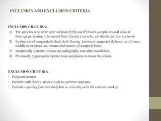
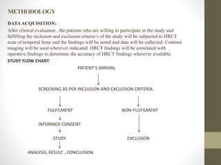
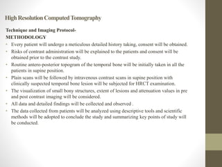
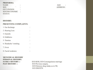
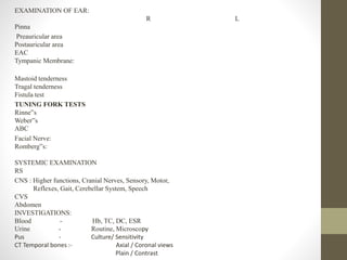
This document outlines a thesis protocol for evaluating temporal bone pathologies using high resolution computed tomography (HRCT). The introduction discusses the complex anatomy of the temporal bone and limitations of plain radiography. HRCT provides excellent spatial resolution to depict boundaries between ear cavities and assess extent of disease. The aim is to study HRCT's role in evaluating temporal bone pathologies. Objectives include assessing distributions of conditions like infections and tumors, evaluating involvement of middle ear and mastoid air cells, intracranial complications, and relationships with neurovascular structures. The methodology will involve prospective data collection from HRCT scans of referred patients which will then be analyzed.
1. “ROLE OF HIGH RESOLUTION COMPUTED TOMOGRAPHY IN
EVALUATION OF TEMPORAL BONE PATHOLOGIES.”
Thesis protocol for MD RADIOLOGY.
MGM Medical College and Hospital, Aurangabad.
Guided by DR. PRASANNA MISHRIKOTKAR
Associate Professor
Department of RADIOLOGY.
MGM Medical College and Hospital, Aurangabad.
Submitted by
DR. ANJALI DESHMUKH
Resident in MD RADIOLOGY
MGM Medical College and Hospital,Aurangabad.
2. INTRODUCTION
• The temporal bone is a complex anatomic structure that contains the organs of hearing and
balance.
• In addition, major vessels and nerves course through it and it also has a close proximity to the
brain.
• Temporal bone has direct contact with brainstem, cerebellum and temporal lobe of brain.
• Plain radiograph remains inexpensive tool of the study of temporal bone but has major
limitations due to complex anatomy and overlapping of various bony structures.
3. • High-resolution computed tomography (HRCT) offers excellent spatial and density
resolution using special algorithms.
• It provides information not only about bony outline but also soft tissue changes
making it possible to demonstrate the location and extent of disease as well as its
complications.
• Furthermore, coronal and axial CT scanning together has dramatically improved the
imaging of temporal bone.
• HRCT accurately depicts the boundaries between the external, middle and inner ear
cavities thereby localized the disease precisely and also greatly demarcate thin
boundary between temporal bone and intracranial compartment with exact details of
intracranial spread of primary temporal bone disease.
• Contrast media help to evaluate the vascularity and contrast enhancing characteristics
particularly in soft tissue lesions of temporal bone giving clues to the histopathology.
4. AIMANDOBJECTIVES
AIM
To study the role of Role of high resolution computed tomography in evaluation of
temporal bone pathologies.
OBJECTIVES
1. To evaluate pathologies of temporal bone such as congenital anomalies,
infections, trauma, neoplasms and their disease distribution.
2. To evaluate the extent of pathological process and sites of involvement of the
middle ear and the mastoid air cell system in these patients.
3. To study the intracranial complications.
4. To study the relationship of the tympanomastoid compartment to the adjacent
critical neurovascular structures .
5. REVIEWOFLITERATURE
1. Boyraz E et al (2009) conducted a study to show ability of CT temporal bones to detect
tympanosclerotic plaques on 19 tympanoplasty cases between January 2006 and May 2006.
The tympanosclerotic plaques obtained from surgical specimens were sent for
histopathological examination and preoperative temporal bone CT scans were evaluted. This
study showed that temporal bone CT scan is a valuable tool to diagnose the localize the
tympanosclerosis, in patients with chronic otitis media and conductive hearing loss. When
combined with clinical findings, CT scans can be useful for preoperative evaluation of
tympanosclerosis.18
2. Zelikovich EI (2004),et al used temporal bone CT to examine 87 patients with chronic otitis
media . The patients' age ranged from 2 to 74 years. The CT signs of chronic purulent otitis
media with and without cholesteatoma were identified. CT shows changes in the walls of the
middle ear cavity, including the roof and allows labyrinthine fistula and intracranial
complications to be detected .
6. 3. Study conducted by Sandeep Berry et al (1998) on 30 patients of unsafe chronic suppurative
otitis media with pre-operative CT scanning and surgical exploration of the middle ear and
mastoid, and comparison of CT findings with the surgical findings. The study showed that CT
scan was highly sensitive for soft tissue density mass in the tympanomastoid compartment.
They concluded that the CT scan of the temporal bone was best to depict pathology which is
not clinically evident.
4. The study conducted by Luchikhin LA et al on 30 patients with chronic otitis media (1995)
compared temporal bone computed tomography findings with the surgical findings . The study
showed that CT temporal bones provided excellent information on the pathological process
and was found to be of immense value before subjecting the patients for surgery
7. MATERIALSANDMETHOLDOLGY
STUDY SUBJECTS: Patients referred to the radiology department for HRCT evaluation of
temporal bone.
STUDY CENTER: This study will be carried out at Department of Radiology,MGM Medical
college & Hospital,Aurangabad.
STUDY DURATION: Study will be done for a period of 2 years after approval from ethical
committee.
STUDYDESIGN: Cross sectional descriptive study.
SAMPLE SIZE:
n=Z^2P[1-P]/d^2
P=Prevalance.
Z= 1.96 for 95% confidence interval.
d=Allowable error.
N=Sample size.
MINIMUM SAMPLE SIZE = Time Bound
8. INCLUSIONANDEXCLUSIONCRITERIA
INCLUSI0N CRITERIA:
1) The patients who were referred from OPD and IPD with complaints and clinical
findings pertaining to temporal bone disease ( earache, ear discharge, hearing loss).
2) Evaluation of congenitally deaf child, having known or suspected deformities of inner,
middle or external ear, trauma and tumors of temporal bone.
3) Incidentally detected lesions on radiography and other modalities.
4) Previously diagnosed temporal bone neoplasms to know the extent.
EXCLUSION CRITERIA :
• Pregnant women.
• Patients with electric device such as cochlear implants.
• Patients requiring contrast study but is clinically unfit for contrast workup.
9. METHODOLOGY
DATAACQUISITION:
After clinical evaluation , the patients who are willing to participate in the study and
fulfilling the inclusion and exclusion criteria’s of the study will be subjected to HRCT
scan of temporal bone and the findings will be noted and data will be collected. Contrast
imaging will be used wherever indicated. HRCT findings will be correlated with
operative findings to determine the accuracy of HRCT findings wherever available.
STUDY FLOW CHART:
PATIENT’S ARRIVAL
SCREENING AS PER INCLUSION AND EXCLUSION CRITERIA.
FULFILMENT NON-FULFILMENT
INFORMED CONSENT
STUDY EXCLUSION
ANALYSIS, RESULT , CONCLUSION
10. HighResolutionComputedTomography
Technique and Imaging Protocol-
METHODOLOGY
• Every patient will undergo a meticulous detailed history taking, consent will be obtained.
• Risks of contrast administration will be explained to the patients and consent will be
obtained prior to the contrast study.
• Routine antero-posterior topogram of the temporal bone will be initially taken in all the
patients in supine position.
• Plain scans will be followed by intravenous contrast scans in supine position with
clinically suspected temporal bone lesion will be subjected for HRCT examination.
• The visualization of small bony structures, extent of lesions and attenuation values in pre
and post contrast imaging will be considered.
• All data and detailed findings will be collected and observed .
• The data collected from patients will be analyzed using descriptive tools and scientific
methods will be adopted to conclude the study and summarizing key points of study will
be conducted.
11. ETHICAL CLEARANCE:
Application for ethical clearance will be submitted to the institutional ethical review board at
MGM Medical College and Hospital.
STATISTICALANALYSIS:
Data will be entered in Microsoft Excel and analyzed using SPSS version 24.Mean and SD
will be calculated for qualitative variables. Also data will be represented in form of visual
impression like bar-diagram etc.
Chi-square will be applied to check significant outcomes. ‘P’ value will be checked at 5%
level of significance.
12. PROFORMA
NAME : AGE :
SEX ` : ADDRESS :
OCCUPATION :
SOCIOECONOMIC :
STATUS
HISTORY:
PRESENTING COMPLAINTS:
1. Ear discharge :
2. Hearing Loss :
3. Earache :
4. Giddiness :
5. Tinnitus :
6. Headache/ vomiting :
7. Fever :
8. Facial weakness :
MENSTRUAL HISTORY
PERSONAL HISTORY:
FAMILY HISTORY :- H/O HOH, H/O Consanguinous marriage
PAST HISTORY :- H/O Previous surgery,
H/O Ototoxic drug intake as in TB.
H/O DM/HTN
13. EXAMINATION OF EAR:
R L
Pinna
Preauricular area
Postauricular area
EAC
Tympanic Membrane:
Mastoid tenderness
Tragal tenderness
Fistula test
TUNING FORK TESTS
Rinne‟s
Weber‟s
ABC
Facial Nerve:
Romberg‟s:
SYSTEMIC EXAMINATION
RS
CNS : Higher functions, Cranial Nerves, Sensory, Motor,
Reflexes, Gait, Cerebellar System, Speech
CVS
Abdomen
INVESTIGATIONS:
Blood - Hb, TC, DC, ESR
Urine - Routine, Microscopy
Pus - Culture/ Sensitivity
CT Temporal bones :- Axial / Coronal views
Plain / Contrast
14. REFERENCES
1. Abhijit Kishorkumar Sankhla, Neha Dubey. Assessment of Temporal Bone Diseases
by High Resolution Computed Tomography – Institution based Study.International
Journal of Contemporary Medicine Surgery and Radiology. 2019; 4(2): 87-90.
2. Sasmita Parida, Pravat Nallini Routray, Jayashree Mohanty, Snigdha Pattanaik. Role of
HRCT in Temporal Bone Diseases - A Study of 100 Cases.JK Science. 2018; 20 (1):34-
38.
3. Shadab Maqsood, Iqubal Hussain Dar, Shabir Ahmad Bhat. Role of high resolution
computed tomography in evaluation of temporal bone diseases. IAIM, 2018; 5(12):15-22.
4. Prakash S Handi, Mallikarjun N Patil, Nisha P.High resolution computed tomography of
temporal bone in the evaluation of otologic diseases.Int J Otorhinolaryngol Head Neck
Surg. 2018; 4(1):87-92.
5. Khrietouzo Dan Kire, Anish S, Beno Jefferson, I Venkatraman. Role of HRCT Temporal
Bone in Ear Pathologies. Journal of Dental and Medical Sciences.2017;16(12): 86-90.
6. Seema V, Raini KP, Thomas S, Mini MV, Daniel E. High Resolution Computed
Tomography in the Evaluation of Temporal Bone Cholesteatoma. J Med Sci and Clin Res.
2017; 5(08):26614-20.
7. Sneha Ankush More, Dilip L Lakhkar, Sushil Kachewar, Pramod Kumar S. High-
resolution Computed Tomographic Evaluation of Pathologies of Temporal Bone. Sch. J.
App. Med. Sci., 2017; 5(3B):770-79.
15. 8. Jyothi AC, Shrikrishna BH. Role of high resolution computed tomography in the
evaluation of temporal bone lesions: our experience. Int J Otorhinolaryngol Head Neck
Surg. 2016; 2(3):135-9.
9. Gaurano JL, Joharjy IA. Middle ear cholesteatoma: Characteristic CTfindings in 64
patients. Ann Saudi Med 2004; 24(6):442–47.
10. Chakeres DW, Augustyn MA. Temporal bone. In: Haaga JR., Lanzieri CF, Gilkeson RC.
CT and MR Imaging of the Whole Body. 4th ed. Ohio, Mosby, 2003: 495-552.
11. Som PM, Bergeron RT. Head and Neck Imaging, 2nd edition, St Louis, CV Mosby, 1991,
p. 1093-1108.
12. Howard JD, Elster AD, May JS. Temporal bone: Three dimensional CT. Radiology.
1990; 177(2):427-30.
13. Brogan M, Chakeres DW. Computed tomography and magnetic resonance imaging of
the normal anatomy of the temporal bone. Semin Ultrasound CT MR.1989; 1010:178-
94.
14. Tono T, Miyanaga S, Morimitsu T, Matsumoto I. Computed tomographic evaluation of
middle ear aeration following intact canal wall tympanoplasty. Auris Nasus Larynx.
1987; 14(3):123-30.
15. Jacklu J, Dillon WP, Schindler RA. Computed tomography in suppurative ear disease:a
correlation of surgical and radiographic findings. Laryngoscope. 1984; 94(6):746-52.