SlideShare a Scribd company logo
1 of 13
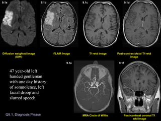
Q9.1. Diagnosis Please
Post-contrast Axial T1-wtd
image
T1-wtd image
FLAIR Image
Post-contrast coronal T1
wtd image
MRA Circle of Willis
Diffusion weighted image
(DWI)
Diffusion weighted image (DWI)
9.1a 9.1b 9.1c 9.1d
9.1e 9.1f
47 year-old left
handed gentleman
with one day history
of somnolence, left
facial droop and
slurred speech.
Q9.2. Diagnosis Please
Post-contrast Axial T1-wtd
image
T1-wtd image
FLAIR Image
(DWI) image
51 year-old patient multiple myeloma presented with acute onset of right side weakness leading to MRI of the brain 3 days later.
51 year-old patient with multiple myeloma presented
with acute onset of right sided weakness leading to MRI
of the brain 3 days later.
9.2a 9.2b 9.2c 9.2d
Q9.3. Diagnosis Please
Post-contrast Axial T1-wtd
image
T1-wtd image
FLAIR Image
Post-contrast coronal T1
wtd image
MR Angiography Neck
Diffusion weighted image
(DWI)
72 year-old left handed white man
with history of chest pain, 5 days
prior to MRI, which subsided.
Patient went to a gas station and
could not read the credit card and
signs in the gas station and was
unable to read the magazines in
the doctor’s office. Neurological
examination revealed alexia
without agraphia.
9.3a 9.3b 9.3c 9.3d
9.3e 9.3f
9.4a. Non-contrast CT Brain 9.4b. Non-contrast CT Brain
Q9.4. Diagnosis Please
9.4c. Non-contrast CT Brain
June 29, 2004 July 30, 2004
Patient is status post recent right temporal craniectomy for extensive
right middle fossa skull-based meningioma, with follow-up post-
operative CT images.
9.4a 9.4b 9.4c
Q9.5. Diagnosis Please
58 year-old male with history of renal cell carcinoma
presented with 3 weeks history of dressing apraxia consisting
of difficulty in performing routine tasks such as getting
dressed, tying his shoes, difficulty with recall and inferior
quadrantopsia with no focal motor deficits. Symptoms slowly
improved. Differential diagnosis: Stroke versus metastasis.
Post-contrast Axial T1-wtd
image
T1-wtd image
FLAIR Image
Post-contrast sagittal T1
wtd image
Diffusion weighted image
(DWI)
9.5a 9.5b 9.5c 9.5d
9.5e
Q9.6. Diagnosis Please
July 31, 2003
T1-wtd image
DW Image FLAIR Image Post-contrast Axial T1-wtd
image
9.6a 9.6b 9.6c 9.6d
December 31, 2003
DW Image FLAIR Image T1-wtd image Post-contrast Axial T1-wtd
image
9.6e 9.6f 9.6g 9.6h
73 year-old male with stage IV non-small cell carcinoma presented with 2 weeks history of
sudden onset of speech difficulty with difficulty in word finding, symptoms gradually
improved. Clinical diagnosis: Stroke versus metastasis.
Diffusion weighted image (DWI)
9.1a 9.1b 9.1c 9.1d 9.1e 9.1f
Diagnosis: Acute one day old infarction involving the right middle
cerebral artery (MCA) territory.
Acute infarction is seen as an area of increased signal intensity on
DWI (arrow in A), FLAIR image (arrow in B), with no evidence of
hemorrhage on T1-wtd image (C) and no enhancement on post
contrast images (D). Intravascular enhancement also an indication
of acute stroke is shown on coronal T1 weighted image (arrow in F).
MR angiography of circle of Willis demonstrates small caliber of
right Sylvian branches of MCA (arrows in E) when compared to the
normal side.
Post-contrast Axial T1-wtd
image
T1-wtd image
FLAIR Image
DWI
9.2a 9.2b 9.2c 9.2d
Diagnosis: Small acute 3 day old infarction involving the left
insular cortex, the territory of left MCA, best noted on diffusion
weighted image (arrow in A) and with enhancement (arrow in D).
9.3a 9.3b 9.3c 9.3d 9.3e 9.3f
Diagnosis: Acute infarction (5 day old) involving the left posterior
cerebral artery (PCA) territory.
The area of infarction is seen as an area of increased signal intensity
on DWI (arrow in A) and FLAIR image (arrow in B) involving the
left posterior temporal-occipital lobe. Mild enhancement seen on
post contrast images (arrow in D and F). Figure E represents
screening 2D time of flight MR angiogram of the neck vessels
revealing no obvious occlusion of major vessels in the neck.
Diagnosis: Acute infarction (5 day old) involving the left posterior cerebral artery (PCA) territory.
9.4a. Non-contrast CT Brain 9.4b. Non-contrast CT Brain 9.4c. Non-contrast CT Brain
June 29, 2004 July 30, 2004
Non-contrast CT brain on 8 days post-operatively demonstrated a
focal area of low attenuation within the right frontal lobe cortex
(arrow in A) and adjacent white matter representing acute infarct.
Scan done a month later (B & C) demonstrated a massive
acute/subacute infarct involving the right MCA territory (white
arrows in B & C) and ACA (anterior cerebral artery) territory
(yellow arrow in C) with mass effect and subfalcine herniation to
the left (fig. B).
Q9.5. Diagnosis Please
Post-contrast Axial T1-wtd
image
T1-wtd image
FLAIR Image
Post-contrast sagittal T1
wtd image
Diffusion weighted image
(DWI)
9.5a 9.5b 9.5c 9.5d
9.5e
Diagnosis: Subacute infarct 3 weeks old involving the
right middle cerebral (MCA) artery territory.
Abnormal T2 wtd hyperintensity involving the right posterior temporal-
parietal-occipital lobe with gyral thickening best noted on FLAIR image
(arrows in B). Subacute blood outlining the cortex is best seen on pre-
contrast T1-wtd image (arrows in C). There is no definite contrast
enhancement obscured by bright signal intensity of blood. The right MCA
territory infarct is also shown on sagittal post contrast T1-wtd image
(arrows in E). Diffusion weighted image reveals bright signal intensity
(arrows in A) involving the cortex from restricted diffusion, an important
sequence in the diagnosis of acute stroke.
Q9.6. Diagnosis Please
July 31, 2003
December 31, 2003
Diagnosis: Non-hemorrhagic subacute enhancing infarct (2 weeks old) involving
the left basal ganglia region. Subacute infarct is seen as an area of increased signal
intensity on FLAIR image (arrow in B) and bright signal intensity on DWI (arrow in
A). Enhancement of the infarct is shown on post contrast image (arrow in D).
A repeat MRI scan done 5 months later
showed resolution of infarct and no
evidence of bright signal intensity on
diffusion weighted image E.
5 months old infarct.
9.6a 9.6b 9.6c 9.6d
9.6e 9.6f 9.6g 9.6h
1a
2a
5b
6d
4b
6e
Imaging Findings of Stroke:
• MR imaging of the brain is far more sensitive than CT imaging to recognize acute
infarction.
• Diffusion wtd. pulse sequence (DW imaging) is the most sensitive MR sequence to
demonstrate stroke. This sequence is sensitive to restricted diffusion within the cell from
stroke-induced cytotoxic edema and the region of acute stroke is seen as an area of bright
signal on DWI (Figs. 1a, 2a). Cytotoxic edema can occur immediately after the initial insult
thus DWI images can reveal, the area of acute infarct immediately after the insult.
• Intravascular contrast enhancement, another sign of early stroke (Figure 1f).
• Sulcal effacement, gyral edema (Fig. 5b), loss of gray-white matter interface can occur
within 12 hours of stroke.
• Parenchymal contrast enhancement (Fig. 6d), mass effect (Fig. 4b) and hemorrhage can
occur within 1-7 days of insult.
Subacute infarct: (1 week to 8 weeks)
•Focal area of encephalomalacia
•Porencephalic dilatation of adjacent ventricle.
• Residual old blood products may be present.
Old Infarct:
• Contrast enhancement slowly decreases in time but can persist for 8 weeks, with
decreasing mass effect and abnormal signal intensity:
Acute Stroke (up to 7 days)

More Related Content

Similar to Znsnsnsnsnsnsnsnsnsnsnsnsnsnsnsnzmxmxmxmxmmxxm

Presentation1, radiological imaging of corpus callosum lesios.
Presentation1, radiological imaging of corpus callosum lesios.Presentation1, radiological imaging of corpus callosum lesios.
Presentation1, radiological imaging of corpus callosum lesios.
Abdellah Nazeer
 
Stroke and stroke mimics
Stroke and stroke mimicsStroke and stroke mimics
Stroke and stroke mimics
Nathaliazuos
 
Errores En Interpretacion De Tc Cráneo
Errores En Interpretacion De Tc CráneoErrores En Interpretacion De Tc Cráneo
Errores En Interpretacion De Tc Cráneo
rahterrazas
 
Thrombotic Occlusion Of The Common Carotid Artery
Thrombotic Occlusion Of The Common Carotid ArteryThrombotic Occlusion Of The Common Carotid Artery
Thrombotic Occlusion Of The Common Carotid Artery
MedicineAndHealthNeurolog
 
Marchiafava–Bignami disease (MBD)
Marchiafava–Bignami disease (MBD) Marchiafava–Bignami disease (MBD)
Marchiafava–Bignami disease (MBD)
Dr Surendra Khosya
 

Similar to Znsnsnsnsnsnsnsnsnsnsnsnsnsnsnsnzmxmxmxmxmmxxm (20)

imagingofspinaltrauMA
imagingofspinaltrauMAimagingofspinaltrauMA
imagingofspinaltrauMA
 
Casos Clínicos
Casos ClínicosCasos Clínicos
Casos Clínicos
 
Stroke
StrokeStroke
Stroke
 
Presentation2, radiological imaging of phakomatosis.
Presentation2, radiological imaging of phakomatosis.Presentation2, radiological imaging of phakomatosis.
Presentation2, radiological imaging of phakomatosis.
 
Presentation1, radiological imaging of corpus callosum lesios.
Presentation1, radiological imaging of corpus callosum lesios.Presentation1, radiological imaging of corpus callosum lesios.
Presentation1, radiological imaging of corpus callosum lesios.
 
Ariunaa spine trauma
Ariunaa spine traumaAriunaa spine trauma
Ariunaa spine trauma
 
Stroke and stroke mimics
Stroke and stroke mimicsStroke and stroke mimics
Stroke and stroke mimics
 
Diagnostic Imaging of Cerebral Trauma
Diagnostic Imaging of Cerebral TraumaDiagnostic Imaging of Cerebral Trauma
Diagnostic Imaging of Cerebral Trauma
 
First Clinical Results of Coronary CT Subtraction
First Clinical Results of Coronary CT SubtractionFirst Clinical Results of Coronary CT Subtraction
First Clinical Results of Coronary CT Subtraction
 
3. mri in acute stroke 2017 vietnam v2
3. mri in acute stroke 2017 vietnam v23. mri in acute stroke 2017 vietnam v2
3. mri in acute stroke 2017 vietnam v2
 
Clinical correlation and interpretation of Brain MRI by dr.Sagor
Clinical correlation and interpretation of Brain MRI by dr.SagorClinical correlation and interpretation of Brain MRI by dr.Sagor
Clinical correlation and interpretation of Brain MRI by dr.Sagor
 
Susceptibility Weighted Imaging (SWI)
Susceptibility Weighted Imaging (SWI)Susceptibility Weighted Imaging (SWI)
Susceptibility Weighted Imaging (SWI)
 
Role of magnetic resonance imaging in coronary artery disease MRCA part 7 Dr ...
Role of magnetic resonance imaging in coronary artery disease MRCA part 7 Dr ...Role of magnetic resonance imaging in coronary artery disease MRCA part 7 Dr ...
Role of magnetic resonance imaging in coronary artery disease MRCA part 7 Dr ...
 
4D-flow MRI of double aortic arch in a 14-year-old patient Authors: Nadya Al-...
4D-flow MRI of double aortic arch in a 14-year-old patient Authors: Nadya Al-...4D-flow MRI of double aortic arch in a 14-year-old patient Authors: Nadya Al-...
4D-flow MRI of double aortic arch in a 14-year-old patient Authors: Nadya Al-...
 
MRI in obstetric practice part 2
MRI in obstetric practice   part 2MRI in obstetric practice   part 2
MRI in obstetric practice part 2
 
Errores En Interpretacion De Tc Cráneo
Errores En Interpretacion De Tc CráneoErrores En Interpretacion De Tc Cráneo
Errores En Interpretacion De Tc Cráneo
 
Traumatic Brain Injury Pearls and Pitfalls (2014)
Traumatic Brain Injury Pearls and Pitfalls (2014)Traumatic Brain Injury Pearls and Pitfalls (2014)
Traumatic Brain Injury Pearls and Pitfalls (2014)
 
Thrombotic Occlusion Of The Common Carotid Artery
Thrombotic Occlusion Of The Common Carotid ArteryThrombotic Occlusion Of The Common Carotid Artery
Thrombotic Occlusion Of The Common Carotid Artery
 
23205056
2320505623205056
23205056
 
Marchiafava–Bignami disease (MBD)
Marchiafava–Bignami disease (MBD) Marchiafava–Bignami disease (MBD)
Marchiafava–Bignami disease (MBD)
 

More from ssuser227d6b

3 EABD- Utility.pptxjdjdjdjdjdjdjdjdkdkdkkd
3 EABD- Utility.pptxjdjdjdjdjdjdjdjdkdkdkkd3 EABD- Utility.pptxjdjdjdjdjdjdjdjdkdkdkkd
3 EABD- Utility.pptxjdjdjdjdjdjdjdjdkdkdkkd
ssuser227d6b
 
Topic -- The Power of Social Media and its effect on Youth.pptx
Topic --  The Power of Social Media and its effect on Youth.pptxTopic --  The Power of Social Media and its effect on Youth.pptx
Topic -- The Power of Social Media and its effect on Youth.pptx
ssuser227d6b
 
DOC-20230424-WA0010..pptx
DOC-20230424-WA0010..pptxDOC-20230424-WA0010..pptx
DOC-20230424-WA0010..pptx
ssuser227d6b
 
T32IntroMS_Newton2019.pptx
T32IntroMS_Newton2019.pptxT32IntroMS_Newton2019.pptx
T32IntroMS_Newton2019.pptx
ssuser227d6b
 
Vegetative Propagation (Cutting) KSD.pptx
Vegetative Propagation (Cutting) KSD.pptxVegetative Propagation (Cutting) KSD.pptx
Vegetative Propagation (Cutting) KSD.pptx
ssuser227d6b
 
presentation1-160607012326 (1).pdf
presentation1-160607012326 (1).pdfpresentation1-160607012326 (1).pdf
presentation1-160607012326 (1).pdf
ssuser227d6b
 
Sarcoidosis.ppt
Sarcoidosis.pptSarcoidosis.ppt
Sarcoidosis.ppt
ssuser227d6b
 
Sarcoidosis PK (2) (1).pptx
Sarcoidosis PK (2) (1).pptxSarcoidosis PK (2) (1).pptx
Sarcoidosis PK (2) (1).pptx
ssuser227d6b
 

More from ssuser227d6b (16)

2024-04-15-21-13-10.pptxnnnnnnnnnnnnnnnnnj
2024-04-15-21-13-10.pptxnnnnnnnnnnnnnnnnnj2024-04-15-21-13-10.pptxnnnnnnnnnnnnnnnnnj
2024-04-15-21-13-10.pptxnnnnnnnnnnnnnnnnnj
 
3 EABD- Utility.pptxjdjdjdjdjdjdjdjdkdkdkkd
3 EABD- Utility.pptxjdjdjdjdjdjdjdjdkdkdkkd3 EABD- Utility.pptxjdjdjdjdjdjdjdjdkdkdkkd
3 EABD- Utility.pptxjdjdjdjdjdjdjdjdkdkdkkd
 
Topic -- The Power of Social Media and its effect on Youth.pptx
Topic --  The Power of Social Media and its effect on Youth.pptxTopic --  The Power of Social Media and its effect on Youth.pptx
Topic -- The Power of Social Media and its effect on Youth.pptx
 
Kishan Deshmukh ppt 3.pptx
Kishan Deshmukh ppt 3.pptxKishan Deshmukh ppt 3.pptx
Kishan Deshmukh ppt 3.pptx
 
DOC-20230424-WA0008..pptx
DOC-20230424-WA0008..pptxDOC-20230424-WA0008..pptx
DOC-20230424-WA0008..pptx
 
DOC-20230424-WA0010..pptx
DOC-20230424-WA0010..pptxDOC-20230424-WA0010..pptx
DOC-20230424-WA0010..pptx
 
Presentation 01.pptx
Presentation 01.pptxPresentation 01.pptx
Presentation 01.pptx
 
Presentation 20.pptx
Presentation 20.pptxPresentation 20.pptx
Presentation 20.pptx
 
Thesis%20ppt%2003.pptx
Thesis%20ppt%2003.pptxThesis%20ppt%2003.pptx
Thesis%20ppt%2003.pptx
 
T32IntroMS_Newton2019.pptx
T32IntroMS_Newton2019.pptxT32IntroMS_Newton2019.pptx
T32IntroMS_Newton2019.pptx
 
Vegetative Propagation (Cutting) KSD.pptx
Vegetative Propagation (Cutting) KSD.pptxVegetative Propagation (Cutting) KSD.pptx
Vegetative Propagation (Cutting) KSD.pptx
 
Radiography of acquired Heart Disease.pptx
Radiography of acquired Heart Disease.pptxRadiography of acquired Heart Disease.pptx
Radiography of acquired Heart Disease.pptx
 
Anjali Deshmukh 2.pptx
Anjali Deshmukh 2.pptxAnjali Deshmukh 2.pptx
Anjali Deshmukh 2.pptx
 
presentation1-160607012326 (1).pdf
presentation1-160607012326 (1).pdfpresentation1-160607012326 (1).pdf
presentation1-160607012326 (1).pdf
 
Sarcoidosis.ppt
Sarcoidosis.pptSarcoidosis.ppt
Sarcoidosis.ppt
 
Sarcoidosis PK (2) (1).pptx
Sarcoidosis PK (2) (1).pptxSarcoidosis PK (2) (1).pptx
Sarcoidosis PK (2) (1).pptx
 

Recently uploaded

Internship Report].pdf iiwmoosmsosmshkssmk
Internship Report].pdf iiwmoosmsosmshkssmkInternship Report].pdf iiwmoosmsosmshkssmk
Internship Report].pdf iiwmoosmsosmshkssmk
SujalTamhane
 
Call Girls In Kengeri Satellite Town ☎ 7737669865 🥵 Book Your One night Stand
Call Girls In Kengeri Satellite Town ☎ 7737669865 🥵 Book Your One night StandCall Girls In Kengeri Satellite Town ☎ 7737669865 🥵 Book Your One night Stand
Call Girls In Kengeri Satellite Town ☎ 7737669865 🥵 Book Your One night Stand
amitlee9823
 
Vip Mumbai Call Girls Ghatkopar Call On 9920725232 With Body to body massage ...
Vip Mumbai Call Girls Ghatkopar Call On 9920725232 With Body to body massage ...Vip Mumbai Call Girls Ghatkopar Call On 9920725232 With Body to body massage ...
Vip Mumbai Call Girls Ghatkopar Call On 9920725232 With Body to body massage ...
amitlee9823
 
Chikkabanavara Call Girls: 🍓 7737669865 🍓 High Profile Model Escorts | Bangal...
Chikkabanavara Call Girls: 🍓 7737669865 🍓 High Profile Model Escorts | Bangal...Chikkabanavara Call Girls: 🍓 7737669865 🍓 High Profile Model Escorts | Bangal...
Chikkabanavara Call Girls: 🍓 7737669865 🍓 High Profile Model Escorts | Bangal...
amitlee9823
 
0425-GDSC-TMU.pdf0425-GDSC-TMU.pdf0425-GDSC-TMU.pdf0425-GDSC-TMU.pdf
0425-GDSC-TMU.pdf0425-GDSC-TMU.pdf0425-GDSC-TMU.pdf0425-GDSC-TMU.pdf0425-GDSC-TMU.pdf0425-GDSC-TMU.pdf0425-GDSC-TMU.pdf0425-GDSC-TMU.pdf
0425-GDSC-TMU.pdf0425-GDSC-TMU.pdf0425-GDSC-TMU.pdf0425-GDSC-TMU.pdf
ssuserded2d4
 
Call Girls Bidadi Just Call 👗 7737669865 👗 Top Class Call Girl Service Bangalore
Call Girls Bidadi Just Call 👗 7737669865 👗 Top Class Call Girl Service BangaloreCall Girls Bidadi Just Call 👗 7737669865 👗 Top Class Call Girl Service Bangalore
Call Girls Bidadi Just Call 👗 7737669865 👗 Top Class Call Girl Service Bangalore
amitlee9823
 
Call Girls Hosur Road Just Call 👗 7737669865 👗 Top Class Call Girl Service Ba...
Call Girls Hosur Road Just Call 👗 7737669865 👗 Top Class Call Girl Service Ba...Call Girls Hosur Road Just Call 👗 7737669865 👗 Top Class Call Girl Service Ba...
Call Girls Hosur Road Just Call 👗 7737669865 👗 Top Class Call Girl Service Ba...
amitlee9823
 
Call Girls Btm Layout Just Call 👗 7737669865 👗 Top Class Call Girl Service Ba...
Call Girls Btm Layout Just Call 👗 7737669865 👗 Top Class Call Girl Service Ba...Call Girls Btm Layout Just Call 👗 7737669865 👗 Top Class Call Girl Service Ba...
Call Girls Btm Layout Just Call 👗 7737669865 👗 Top Class Call Girl Service Ba...
amitlee9823
 
Call Girls Hosur Just Call 👗 7737669865 👗 Top Class Call Girl Service Bangalore
Call Girls Hosur Just Call 👗 7737669865 👗 Top Class Call Girl Service BangaloreCall Girls Hosur Just Call 👗 7737669865 👗 Top Class Call Girl Service Bangalore
Call Girls Hosur Just Call 👗 7737669865 👗 Top Class Call Girl Service Bangalore
amitlee9823
 
Call Girls Bidadi ☎ 7737669865☎ Book Your One night Stand (Bangalore)
Call Girls Bidadi ☎ 7737669865☎ Book Your One night Stand (Bangalore)Call Girls Bidadi ☎ 7737669865☎ Book Your One night Stand (Bangalore)
Call Girls Bidadi ☎ 7737669865☎ Book Your One night Stand (Bangalore)
amitlee9823
 

Recently uploaded (20)

Miletti Gabriela_Vision Plan for artist Jahzel.pdf
Miletti Gabriela_Vision Plan for artist Jahzel.pdfMiletti Gabriela_Vision Plan for artist Jahzel.pdf
Miletti Gabriela_Vision Plan for artist Jahzel.pdf
 
Pooja 9892124323, Call girls Services and Mumbai Escort Service Near Hotel Sa...
Pooja 9892124323, Call girls Services and Mumbai Escort Service Near Hotel Sa...Pooja 9892124323, Call girls Services and Mumbai Escort Service Near Hotel Sa...
Pooja 9892124323, Call girls Services and Mumbai Escort Service Near Hotel Sa...
 
CFO_SB_Career History_Multi Sector Experience
CFO_SB_Career History_Multi Sector ExperienceCFO_SB_Career History_Multi Sector Experience
CFO_SB_Career History_Multi Sector Experience
 
Internship Report].pdf iiwmoosmsosmshkssmk
Internship Report].pdf iiwmoosmsosmshkssmkInternship Report].pdf iiwmoosmsosmshkssmk
Internship Report].pdf iiwmoosmsosmshkssmk
 
Presentation for the country presentation
Presentation for the country presentationPresentation for the country presentation
Presentation for the country presentation
 
Call Girls In Kengeri Satellite Town ☎ 7737669865 🥵 Book Your One night Stand
Call Girls In Kengeri Satellite Town ☎ 7737669865 🥵 Book Your One night StandCall Girls In Kengeri Satellite Town ☎ 7737669865 🥵 Book Your One night Stand
Call Girls In Kengeri Satellite Town ☎ 7737669865 🥵 Book Your One night Stand
 
Vip Mumbai Call Girls Ghatkopar Call On 9920725232 With Body to body massage ...
Vip Mumbai Call Girls Ghatkopar Call On 9920725232 With Body to body massage ...Vip Mumbai Call Girls Ghatkopar Call On 9920725232 With Body to body massage ...
Vip Mumbai Call Girls Ghatkopar Call On 9920725232 With Body to body massage ...
 
Toxicokinetics studies.. (toxicokinetics evaluation in preclinical studies)
Toxicokinetics studies.. (toxicokinetics evaluation in preclinical studies)Toxicokinetics studies.. (toxicokinetics evaluation in preclinical studies)
Toxicokinetics studies.. (toxicokinetics evaluation in preclinical studies)
 
Chikkabanavara Call Girls: 🍓 7737669865 🍓 High Profile Model Escorts | Bangal...
Chikkabanavara Call Girls: 🍓 7737669865 🍓 High Profile Model Escorts | Bangal...Chikkabanavara Call Girls: 🍓 7737669865 🍓 High Profile Model Escorts | Bangal...
Chikkabanavara Call Girls: 🍓 7737669865 🍓 High Profile Model Escorts | Bangal...
 
0425-GDSC-TMU.pdf0425-GDSC-TMU.pdf0425-GDSC-TMU.pdf0425-GDSC-TMU.pdf
0425-GDSC-TMU.pdf0425-GDSC-TMU.pdf0425-GDSC-TMU.pdf0425-GDSC-TMU.pdf0425-GDSC-TMU.pdf0425-GDSC-TMU.pdf0425-GDSC-TMU.pdf0425-GDSC-TMU.pdf
0425-GDSC-TMU.pdf0425-GDSC-TMU.pdf0425-GDSC-TMU.pdf0425-GDSC-TMU.pdf
 
Call Girls Bidadi Just Call 👗 7737669865 👗 Top Class Call Girl Service Bangalore
Call Girls Bidadi Just Call 👗 7737669865 👗 Top Class Call Girl Service BangaloreCall Girls Bidadi Just Call 👗 7737669865 👗 Top Class Call Girl Service Bangalore
Call Girls Bidadi Just Call 👗 7737669865 👗 Top Class Call Girl Service Bangalore
 
Résumé (2 pager - 12 ft standard syntax)
Résumé (2 pager -  12 ft standard syntax)Résumé (2 pager -  12 ft standard syntax)
Résumé (2 pager - 12 ft standard syntax)
 
Call Girls Hosur Road Just Call 👗 7737669865 👗 Top Class Call Girl Service Ba...
Call Girls Hosur Road Just Call 👗 7737669865 👗 Top Class Call Girl Service Ba...Call Girls Hosur Road Just Call 👗 7737669865 👗 Top Class Call Girl Service Ba...
Call Girls Hosur Road Just Call 👗 7737669865 👗 Top Class Call Girl Service Ba...
 
Dubai Call Girls Kiki O525547819 Call Girls Dubai Koko
Dubai Call Girls Kiki O525547819 Call Girls Dubai KokoDubai Call Girls Kiki O525547819 Call Girls Dubai Koko
Dubai Call Girls Kiki O525547819 Call Girls Dubai Koko
 
Call Girls Btm Layout Just Call 👗 7737669865 👗 Top Class Call Girl Service Ba...
Call Girls Btm Layout Just Call 👗 7737669865 👗 Top Class Call Girl Service Ba...Call Girls Btm Layout Just Call 👗 7737669865 👗 Top Class Call Girl Service Ba...
Call Girls Btm Layout Just Call 👗 7737669865 👗 Top Class Call Girl Service Ba...
 
Call Girls Hosur Just Call 👗 7737669865 👗 Top Class Call Girl Service Bangalore
Call Girls Hosur Just Call 👗 7737669865 👗 Top Class Call Girl Service BangaloreCall Girls Hosur Just Call 👗 7737669865 👗 Top Class Call Girl Service Bangalore
Call Girls Hosur Just Call 👗 7737669865 👗 Top Class Call Girl Service Bangalore
 
Call Girls Bidadi ☎ 7737669865☎ Book Your One night Stand (Bangalore)
Call Girls Bidadi ☎ 7737669865☎ Book Your One night Stand (Bangalore)Call Girls Bidadi ☎ 7737669865☎ Book Your One night Stand (Bangalore)
Call Girls Bidadi ☎ 7737669865☎ Book Your One night Stand (Bangalore)
 
Solution Manual for First Course in Abstract Algebra A, 8th Edition by John B...
Solution Manual for First Course in Abstract Algebra A, 8th Edition by John B...Solution Manual for First Course in Abstract Algebra A, 8th Edition by John B...
Solution Manual for First Course in Abstract Algebra A, 8th Edition by John B...
 
Dubai Call Girls Starlet O525547819 Call Girls Dubai Showen Dating
Dubai Call Girls Starlet O525547819 Call Girls Dubai Showen DatingDubai Call Girls Starlet O525547819 Call Girls Dubai Showen Dating
Dubai Call Girls Starlet O525547819 Call Girls Dubai Showen Dating
 
Call Girls Alandi Road Call Me 7737669865 Budget Friendly No Advance Booking
Call Girls Alandi Road Call Me 7737669865 Budget Friendly No Advance BookingCall Girls Alandi Road Call Me 7737669865 Budget Friendly No Advance Booking
Call Girls Alandi Road Call Me 7737669865 Budget Friendly No Advance Booking
 

Znsnsnsnsnsnsnsnsnsnsnsnsnsnsnsnzmxmxmxmxmmxxm

  • 1. Q9.1. Diagnosis Please Post-contrast Axial T1-wtd image T1-wtd image FLAIR Image Post-contrast coronal T1 wtd image MRA Circle of Willis Diffusion weighted image (DWI) Diffusion weighted image (DWI) 9.1a 9.1b 9.1c 9.1d 9.1e 9.1f 47 year-old left handed gentleman with one day history of somnolence, left facial droop and slurred speech.
  • 2. Q9.2. Diagnosis Please Post-contrast Axial T1-wtd image T1-wtd image FLAIR Image (DWI) image 51 year-old patient multiple myeloma presented with acute onset of right side weakness leading to MRI of the brain 3 days later. 51 year-old patient multiple myeloma presented with acute onset of right side weakness leading to MRI of the brain 3 days later. 51 year-old patient with multiple myeloma presented with acute onset of right sided weakness leading to MRI of the brain 3 days later. 9.2a 9.2b 9.2c 9.2d
  • 3. Q9.3. Diagnosis Please Post-contrast Axial T1-wtd image T1-wtd image FLAIR Image Post-contrast coronal T1 wtd image MR Angiography Neck Diffusion weighted image (DWI) 72 year-old left handed white man with history of chest pain, 5 days prior to MRI, which subsided. Patient went to a gas station and could not read the credit card and signs in the gas station and was unable to read the magazines in the doctor’s office. Neurological examination revealed alexia without agraphia. 9.3a 9.3b 9.3c 9.3d 9.3e 9.3f
  • 4. 9.4a. Non-contrast CT Brain 9.4b. Non-contrast CT Brain Q9.4. Diagnosis Please 9.4c. Non-contrast CT Brain June 29, 2004 July 30, 2004 Patient is status post recent right temporal craniectomy for extensive right middle fossa skull-based meningioma, with follow-up post- operative CT images. 9.4a 9.4b 9.4c
  • 5. Q9.5. Diagnosis Please 58 year-old male with history of renal cell carcinoma presented with 3 weeks history of dressing apraxia consisting of difficulty in performing routine tasks such as getting dressed, tying his shoes, difficulty with recall and inferior quadrantopsia with no focal motor deficits. Symptoms slowly improved. Differential diagnosis: Stroke versus metastasis. Post-contrast Axial T1-wtd image T1-wtd image FLAIR Image Post-contrast sagittal T1 wtd image Diffusion weighted image (DWI) 9.5a 9.5b 9.5c 9.5d 9.5e
  • 6. Q9.6. Diagnosis Please July 31, 2003 T1-wtd image DW Image FLAIR Image Post-contrast Axial T1-wtd image 9.6a 9.6b 9.6c 9.6d December 31, 2003 DW Image FLAIR Image T1-wtd image Post-contrast Axial T1-wtd image 9.6e 9.6f 9.6g 9.6h 73 year-old male with stage IV non-small cell carcinoma presented with 2 weeks history of sudden onset of speech difficulty with difficulty in word finding, symptoms gradually improved. Clinical diagnosis: Stroke versus metastasis.
  • 7. Diffusion weighted image (DWI) 9.1a 9.1b 9.1c 9.1d 9.1e 9.1f Diagnosis: Acute one day old infarction involving the right middle cerebral artery (MCA) territory. Acute infarction is seen as an area of increased signal intensity on DWI (arrow in A), FLAIR image (arrow in B), with no evidence of hemorrhage on T1-wtd image (C) and no enhancement on post contrast images (D). Intravascular enhancement also an indication of acute stroke is shown on coronal T1 weighted image (arrow in F). MR angiography of circle of Willis demonstrates small caliber of right Sylvian branches of MCA (arrows in E) when compared to the normal side.
  • 8. Post-contrast Axial T1-wtd image T1-wtd image FLAIR Image DWI 9.2a 9.2b 9.2c 9.2d Diagnosis: Small acute 3 day old infarction involving the left insular cortex, the territory of left MCA, best noted on diffusion weighted image (arrow in A) and with enhancement (arrow in D).
  • 9. 9.3a 9.3b 9.3c 9.3d 9.3e 9.3f Diagnosis: Acute infarction (5 day old) involving the left posterior cerebral artery (PCA) territory. The area of infarction is seen as an area of increased signal intensity on DWI (arrow in A) and FLAIR image (arrow in B) involving the left posterior temporal-occipital lobe. Mild enhancement seen on post contrast images (arrow in D and F). Figure E represents screening 2D time of flight MR angiogram of the neck vessels revealing no obvious occlusion of major vessels in the neck. Diagnosis: Acute infarction (5 day old) involving the left posterior cerebral artery (PCA) territory.
  • 10. 9.4a. Non-contrast CT Brain 9.4b. Non-contrast CT Brain 9.4c. Non-contrast CT Brain June 29, 2004 July 30, 2004 Non-contrast CT brain on 8 days post-operatively demonstrated a focal area of low attenuation within the right frontal lobe cortex (arrow in A) and adjacent white matter representing acute infarct. Scan done a month later (B & C) demonstrated a massive acute/subacute infarct involving the right MCA territory (white arrows in B & C) and ACA (anterior cerebral artery) territory (yellow arrow in C) with mass effect and subfalcine herniation to the left (fig. B).
  • 11. Q9.5. Diagnosis Please Post-contrast Axial T1-wtd image T1-wtd image FLAIR Image Post-contrast sagittal T1 wtd image Diffusion weighted image (DWI) 9.5a 9.5b 9.5c 9.5d 9.5e Diagnosis: Subacute infarct 3 weeks old involving the right middle cerebral (MCA) artery territory. Abnormal T2 wtd hyperintensity involving the right posterior temporal- parietal-occipital lobe with gyral thickening best noted on FLAIR image (arrows in B). Subacute blood outlining the cortex is best seen on pre- contrast T1-wtd image (arrows in C). There is no definite contrast enhancement obscured by bright signal intensity of blood. The right MCA territory infarct is also shown on sagittal post contrast T1-wtd image (arrows in E). Diffusion weighted image reveals bright signal intensity (arrows in A) involving the cortex from restricted diffusion, an important sequence in the diagnosis of acute stroke.
  • 12. Q9.6. Diagnosis Please July 31, 2003 December 31, 2003 Diagnosis: Non-hemorrhagic subacute enhancing infarct (2 weeks old) involving the left basal ganglia region. Subacute infarct is seen as an area of increased signal intensity on FLAIR image (arrow in B) and bright signal intensity on DWI (arrow in A). Enhancement of the infarct is shown on post contrast image (arrow in D). A repeat MRI scan done 5 months later showed resolution of infarct and no evidence of bright signal intensity on diffusion weighted image E. 5 months old infarct. 9.6a 9.6b 9.6c 9.6d 9.6e 9.6f 9.6g 9.6h
  • 13. 1a 2a 5b 6d 4b 6e Imaging Findings of Stroke: • MR imaging of the brain is far more sensitive than CT imaging to recognize acute infarction. • Diffusion wtd. pulse sequence (DW imaging) is the most sensitive MR sequence to demonstrate stroke. This sequence is sensitive to restricted diffusion within the cell from stroke-induced cytotoxic edema and the region of acute stroke is seen as an area of bright signal on DWI (Figs. 1a, 2a). Cytotoxic edema can occur immediately after the initial insult thus DWI images can reveal, the area of acute infarct immediately after the insult. • Intravascular contrast enhancement, another sign of early stroke (Figure 1f). • Sulcal effacement, gyral edema (Fig. 5b), loss of gray-white matter interface can occur within 12 hours of stroke. • Parenchymal contrast enhancement (Fig. 6d), mass effect (Fig. 4b) and hemorrhage can occur within 1-7 days of insult. Subacute infarct: (1 week to 8 weeks) •Focal area of encephalomalacia •Porencephalic dilatation of adjacent ventricle. • Residual old blood products may be present. Old Infarct: • Contrast enhancement slowly decreases in time but can persist for 8 weeks, with decreasing mass effect and abnormal signal intensity: Acute Stroke (up to 7 days)