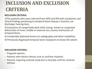
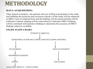
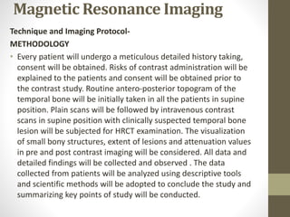
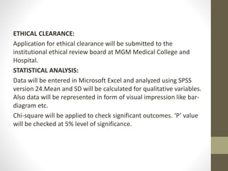
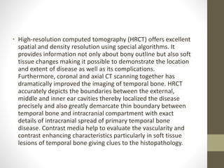
SlideShare a Scribd company logo
1 of 15
“ROLE OF HIGH RESOLUTION COMPUTED TOMOGRAPHY IN
EVALUATION OF TEMPORAL BONE PATHOLOGIES.”
Thesis protocol for MD RADIOLOGY.
MGM Medical College and Hospital, Aurangabad.
Guided by DR. PRASANNA MISHRIKOTKAR
Associate Professor
Department of RADIOLOGY.
MGM Medical College and Hospital, Aurangabad.
Submitted by
DR. ANJALI DESHMUKH
Resident in MD RADIOLOGY
MGM Medical College and Hospital,Aurangabad.
INTRODUCTION
• The temporal bone is a complex anatomic structure that contains
the organs of hearing and balance. In addition, major vessels and
nerves course through it and it also has a close proximity to the
brain. Temporal bone has direct contact with brainstem, cerebellum
and temporal lobe of brain. Before computed tomography (CT)
imaging modalities available for the evaluation of temporal bone
were plain radiograph, polytomography, angiography, and
cisternography. Plain radiograph remains inexpensive tool of the
study of temporal bone but has major limitations due to complex
anatomy and overlapping of various bony structures.
• High-resolution computed tomography (HRCT) offers excellent
spatial and density resolution using special algorithms. It
provides information not only about bony outline but also soft
tissue changes making it possible to demonstrate the location
and extent of disease as well as its complications.
Furthermore, coronal and axial CT scanning together has
dramatically improved the imaging of temporal bone. HRCT
accurately depicts the boundaries between the external,
middle and inner ear cavities thereby localized the disease
precisely and also greatly demarcate thin boundary between
temporal bone and intracranial compartment with exact
details of intracranial spread of primary temporal bone
disease. Contrast media help to evaluate the vascularity and
contrast enhancing characteristics particularly in soft tissue
lesions of temporal bone giving clues to the histopathology.
AIM AND OBJECTIVES
AIM
To study the role of Role of high resolution computed
tomography in evaluation of temporal bone pathologies.
OBJECTIVES
1. To evaluate pathologies of temporal bone such as congenital
anomales, infections, trauma, neoplasms and their disease
distribution.
2. To evaluate the extent of pathological process and sites of
involvement of the middle ear and the mastoid air cell system in
these patients.
3. To study the intracranial complications.
4. To study the relationship of the tympanomastoid
compartment to the adjacent , critical neurovascular structures .
REVIEW OF LITERATURE
• Zelikovich EI (2004),et al used temporal bone CT to examine 87
patients with chronic otitis media . The patients' age ranged from 2
to 74 years. The CT signs of chronic purulent otitis media with and
without cholesteatoma were identified. CT shows changes in the
walls of the middle ear cavity, including the roof and allows
labyrinthine fistula and intracranial complications to be detected .
• The study conducted by Luchikhin LA et al on 30 patients with
chronic otitis media (1995) compared temporal bone computed
tomography findings with the surgical findings . The study showed
that CT temporal bones provided excellent information on the
pathological process and was found to be of immense value before
subjecting the patients for surgery
• Boyraz E et al (2009) conducted a study to show ability of CT temporal
bones to detect tympanosclerotic plaques on 19 tympanoplasty cases
between January 2006 and May 2006. The tympanosclerotic plaques
obtained from surgical specimens were sent for histopathological
examination and preoperative temporal bone CT scans were evaluted.
This study showed that temporal bone CT scan is a valuable tool to
diagnose the localize the tympanosclerosis, in patients with chronic
otitis media and conductive hearing loss. When combined with
clinical findings, CT scans can be useful for preoperative evaluation of
tympanosclerosis.18
• Study conducted by Sandeep Berry et al (1998) on 30 patients of
unsafe chronic suppurative otitis media with pre-operative CT
scanning and surgical exploration of the middle ear and mastoid, and
comparison of CT findings with the surgical findings. The study
showed that CT scan was highly sensitive for soft tissue density mass
in the tympanomastoid compartment. They concluded that the CT
scan of the temporal bone was best to depict pathology which is not
clinically evident.
MATERIALS AND METHOLDOLGY
STUDY SUBJECTS: Patients referred to the radiology department for HRCT
evaluation of temporal bone.
STUDY CENTER: This study will be carried out at Department of
Radiology,MGM Medical college & Hospital,Aurangabad.
STUDY DURATION: Study will be done for a period of 2 years after approval
from ethical committee.
STUDYDESIGN: Cross sectional descriptive study.
SAMPLE SIZE:
n=Z^2P[1-P]/d^2
P=Prevalance.
Z= 1.96 for 95% confidence interval.
d=Allowable error.
N=Sample size.
MINIMUM SAMPLE SIZE = Time Bound
INCLUSION AND EXCLUSION
CRITERIA
INCLUSI0N CRITERIA:
1)The patients who were referred from OPD and IPD with complaints and
clinical findings pertaining to temporal bone disease ( earache, ear
discharge, hearing loss).
2) Evaluation of congenitally deaf child, having known or suspected
deformities of inner, middle or external ear, trauma and tumors of
temporal bone.
3) Incidentally detected lesions on radiography and other modalities.
4) Previously diagnosed temporal bone neoplasms to know the extent.
EXCLUSION CRITERIA :
• Pregnant women.
• Patients with electric device such as cochlear implants.
• Patients requiring contrast study but is clinically unfit for contrast
workup.
METHODOLOGY
DATA ACQUISITION:
After clinical evaluation , the patients who are willing to participate in the study
and fulfilling the inclusion and exclusion criteria’s of the study will be subjected
to HRCT scan of temporal bone and the findings will be noted and data will be
collected. Contrast imaging will be used wherever indicated. HRCT findings
will be correlated with operative findings to determine the accuracy of HRCT
findings wherever available.
STUDY FLOW CHART:
PATIENT’S ARRIVAL
SCREENING AS PER INCLUSION AND EXCLUSION CRITERIA.
FULFILMENT NON-FULFILMENT
INFORMED CONSENT
STUDY EXCLUSION
ANALYSIS, RESULT , CONCLUSION
Magnetic Resonance Imaging
Technique and Imaging Protocol-
METHODOLOGY
• Every patient will undergo a meticulous detailed history taking,
consent will be obtained. Risks of contrast administration will be
explained to the patients and consent will be obtained prior to
the contrast study. Routine antero-posterior topogram of the
temporal bone will be initially taken in all the patients in supine
position. Plain scans will be followed by intravenous contrast
scans in supine position with clinically suspected temporal bone
lesion will be subjected for HRCT examination. The visualization
of small bony structures, extent of lesions and attenuation values
in pre and post contrast imaging will be considered. All data and
detailed findings will be collected and observed . The data
collected from patients will be analyzed using descriptive tools
and scientific methods will be adopted to conclude the study and
summarizing key points of study will be conducted.
ETHICAL CLEARANCE:
Application for ethical clearance will be submitted to the
institutional ethical review board at MGM Medical College and
Hospital.
STATISTICAL ANALYSIS:
Data will be entered in Microsoft Excel and analyzed using SPSS
version 24.Mean and SD will be calculated for qualitative variables.
Also data will be represented in form of visual impression like bar-
diagram etc.
Chi-square will be applied to check significant outcomes. ‘P’ value
will be checked at 5% level of significance.
• PROFORMA
NAME : AGE :
SEX : ADDRESS :
OCCUPATION :
SOCIOECONOMIC :
STATUS
HISTORY:
PRESENTING COMPLAINTS:
1. Ear discharge : `Side, Onset, Duration, Amount, Type of discharge,
Aggravating / Relieving Factors, Response to
treatment
2. Hard of hearing : Side, Acute / Insidious, Degree Progressive
/continuous/
Intermittent , Whether handicapping, Family
history,
H/o trauma to ear,
H/o exposure to loud nice.
3. Earache : Duration, Side, Mild/Server, More on movement of
ear /
during mastication, Increase during ear discharge or
not
4. Giddiness : Duration, Intermittent / Continuous, Associated
with
posture, H/o intake of ototoxic drugs, Aggravating /
relieving factors.
5. Tinnitus : Side, Unilateral / Bilateral, Duration, Sudden /
Gradual,
• Progression – Severe / Static / Decreasing,
Continuous / intermittent/ Pulsatile/ Clicking,
Aggravating / Relieving factors.
6. Headache/ vomiting
7. Fever
8. Facial weakness
• MENSTRUAL HISTORY
PERSONAL HISTORY:
FAMILY HISTORY :- H/O HOH, H/O Consanguinous marriage
PAST HISTORY :- H/O Previous surgery,
H/O Ototoxic drug intake as in TB.
H/O DM/HTN
EXAMINATION OF EAR:
R L
Pinna
•
Preauricular area
Postauricular area
EAC
Tympanic Membrane:
•
Mastoid tenderness
Tragal tenderness
Fistula test
• TUNING FORK TESTS
Rinne‟s
Weber‟s
ABC
• Facial Nerve:
Romberg‟s:
Examination of Oral Cavity/ Oropharynx / Nose
Palate : Bulge/ Movements of soft palate, postnasal drip, tonsils,
posterior
pharyngeal
wall, Dental hygiene.
SYSTEMIC EXAMINATION
RS CNS : Higher functions, Cranial Nerves, Sensory,
Motor,
Reflexes, Gait, Cerebellar System, Speech
CVS
Abdomen
INVESTIGATIONS:
Blood - Hb, TC, DC, ESR
Urine - Routine, Microscopy
Pus - Culture/ Sensitivity
CT Temporal bones :- Axial / Coronal views
Plain / Contrast
•
REFERENCES
1. Brogan M, Chakeres DW. Computed tomography and magnetic resonance imaging of
the normal anatomy of the temporal bone. Semin Ultrasound CT MR.1989; 1010:178-
94.
2. Howard JD, Elster AD, May JS. Temporal bone: Three dimensional CT. Radiology. 1990;
177(2):427-30.
3. Chakeres DW, Augustyn MA. Temporal bone. In: Haaga JR., Lanzieri CF, Gilkeson RC.
CT and MR Imaging of the Whole Body. 4th ed. Ohio, Mosby, 2003: 495-552.
4. Tono T, Miyanaga S, Morimitsu T, Matsumoto I. Computed tomographic evaluation of
middle ear aeration following intact canal wall tympanoplasty. Auris Nasus Larynx. 1987;
14(3):123-30.
5.Jacklu J, Dillon WP, Schindler RA. Computed tomography in suppurative ear disease:a
correlation of surgical and radiographic findings. Laryngoscope. 1984; 94(6):746-52.
6. Som PM, Bergeron RT. Head and Neck Imaging, 2nd edition, St Louis, CV Mosby, 1991,
p. 1093-1108.
7. Jyothi AC, Shrikrishna BH. Role of high resolution computed tomography in the
evaluation of temporal bone lesions: our experience. Int J Otorhinolaryngol Head Neck
Surg. 2016; 2(3):135-9.
8. Seema V, Raini KP, Thomas S, Mini MV, Daniel E. High Resolution Computed Tomography in the
Evaluation of Temporal Bone Cholesteatoma. J Med Sci and Clin Res. 2017; 5(08):26614-20.
9. Sneha Ankush More, Dilip L Lakhkar, Sushil Kachewar, Pramod Kumar S. High-resolution Computed
Tomographic Evaluation of Pathologies of Temporal Bone. Sch. J. App. Med. Sci., 2017; 5(3B):770-79
10. Khrietouzo Dan Kire, Anish S, Beno Jefferson, I Venkatraman. Role of HRCT Temporal Bone in Ear
Pathologies. Journal of Dental and Medical Sciences.2017;16(12): 86-90
11. Abhijit Kishorkumar Sankhla, Neha Dubey. Assessment of Temporal Bone Diseases by
High Resolution Computed Tomography – Institution based Study.International Journal of
Contemporary Medicine Surgery and Radiology. 2019; 4(2): 87-90
12. Sasmita Parida, Pravat Nallini Routray, Jayashree Mohanty, Snigdha Pattanaik. Role of
HRCT in Temporal Bone Diseases - A Study of 100 Cases.JK Science. 2018; 20 (1):34-38
13. Shadab Maqsood, Iqubal Hussain Dar, Shabir Ahmad Bhat. Role of high resolution
computed tomography in evaluation of temporal bone diseases. IAIM, 2018; 5(12):15-22.
14. Prakash S Handi, Mallikarjun N Patil, Nisha P.High resolution computed tomography of
temporal bone in the evaluation of otologic diseases.Int J Otorhinolaryngol Head Neck
Surg. 2018; 4(1):87-92
15. Gaurano JL, Joharjy IA. Middle ear cholesteatoma: Characteristic CT findings in 64
patients. Ann Saudi Med 2004; 24(6):442–47.

More Related Content

Similar to DOC-20230424-WA0008..pptx

Correlation of the interdental and the interradicular bone loss
Correlation of the interdental and the interradicular bone lossCorrelation of the interdental and the interradicular bone loss
Correlation of the interdental and the interradicular bone lossDr Gauri Kapila
 
Temporal bone tumors staging and radiological assesment
Temporal bone tumors staging and radiological assesmentTemporal bone tumors staging and radiological assesment
Temporal bone tumors staging and radiological assesmenttamer ebaied
 
Arthi protoco1811l.
Arthi protoco1811l.Arthi protoco1811l.
Arthi protoco1811l.gosha30
 
Retrosternal SZISACON anaesthesia periop
Retrosternal SZISACON anaesthesia periopRetrosternal SZISACON anaesthesia periop
Retrosternal SZISACON anaesthesia periopUmaKumar14
 
Furlw vs 2 flap on ET function.pptx
Furlw vs 2 flap on ET function.pptxFurlw vs 2 flap on ET function.pptx
Furlw vs 2 flap on ET function.pptxlifestory4
 
Effects of rapid palatal expansion on the sagittal and vertical dimensions of...
Effects of rapid palatal expansion on the sagittal and vertical dimensions of...Effects of rapid palatal expansion on the sagittal and vertical dimensions of...
Effects of rapid palatal expansion on the sagittal and vertical dimensions of...EdwardHAngle
 
ENDOSCOPIC TRANSCANAL STAPEDOTOMY.pptx
ENDOSCOPIC TRANSCANAL STAPEDOTOMY.pptxENDOSCOPIC TRANSCANAL STAPEDOTOMY.pptx
ENDOSCOPIC TRANSCANAL STAPEDOTOMY.pptxGaganaNagaraj3
 
Role of CT Mastoids in the Diagnosis and Management of Chronic Inflammatory E...
Role of CT Mastoids in the Diagnosis and Management of Chronic Inflammatory E...Role of CT Mastoids in the Diagnosis and Management of Chronic Inflammatory E...
Role of CT Mastoids in the Diagnosis and Management of Chronic Inflammatory E...Dr.Juveria Majeed
 
International Organisation of Scientific Research
International Organisation of Scientific ResearchInternational Organisation of Scientific Research
International Organisation of Scientific ResearchDr.Juveria Majeed
 
Long-Term Outcomes of Endoscopic Treatment for Bile Duct Stones in Patients A...
Long-Term Outcomes of Endoscopic Treatment for Bile Duct Stones in Patients A...Long-Term Outcomes of Endoscopic Treatment for Bile Duct Stones in Patients A...
Long-Term Outcomes of Endoscopic Treatment for Bile Duct Stones in Patients A...JapaneseJournalofGas
 
Long-Term Outcomes of Endoscopic Treatment for Bile Duct Stones in Patients A...
Long-Term Outcomes of Endoscopic Treatment for Bile Duct Stones in Patients A...Long-Term Outcomes of Endoscopic Treatment for Bile Duct Stones in Patients A...
Long-Term Outcomes of Endoscopic Treatment for Bile Duct Stones in Patients A...JohnJulie1
 
4.- The tubarial salivary glands. A potential new organ at risk for radiother...
4.- The tubarial salivary glands. A potential new organ at risk for radiother...4.- The tubarial salivary glands. A potential new organ at risk for radiother...
4.- The tubarial salivary glands. A potential new organ at risk for radiother...ssusere6675a1
 
The use of low level laser in periodontal disease
The use of low level laser in periodontal diseaseThe use of low level laser in periodontal disease
The use of low level laser in periodontal diseaseJan Tunér
 

Similar to DOC-20230424-WA0008..pptx (18)

Correlation of the interdental and the interradicular bone loss
Correlation of the interdental and the interradicular bone lossCorrelation of the interdental and the interradicular bone loss
Correlation of the interdental and the interradicular bone loss
 
American Journal of Anesthesia & Clinical Research
American Journal of Anesthesia & Clinical ResearchAmerican Journal of Anesthesia & Clinical Research
American Journal of Anesthesia & Clinical Research
 
Temporal bone tumors staging and radiological assesment
Temporal bone tumors staging and radiological assesmentTemporal bone tumors staging and radiological assesment
Temporal bone tumors staging and radiological assesment
 
Arthi protoco1811l.
Arthi protoco1811l.Arthi protoco1811l.
Arthi protoco1811l.
 
Retrosternal SZISACON anaesthesia periop
Retrosternal SZISACON anaesthesia periopRetrosternal SZISACON anaesthesia periop
Retrosternal SZISACON anaesthesia periop
 
Furlw vs 2 flap on ET function.pptx
Furlw vs 2 flap on ET function.pptxFurlw vs 2 flap on ET function.pptx
Furlw vs 2 flap on ET function.pptx
 
NM
NMNM
NM
 
DARS
DARSDARS
DARS
 
Effects of rapid palatal expansion on the sagittal and vertical dimensions of...
Effects of rapid palatal expansion on the sagittal and vertical dimensions of...Effects of rapid palatal expansion on the sagittal and vertical dimensions of...
Effects of rapid palatal expansion on the sagittal and vertical dimensions of...
 
ENDOSCOPIC TRANSCANAL STAPEDOTOMY.pptx
ENDOSCOPIC TRANSCANAL STAPEDOTOMY.pptxENDOSCOPIC TRANSCANAL STAPEDOTOMY.pptx
ENDOSCOPIC TRANSCANAL STAPEDOTOMY.pptx
 
Role of CT Mastoids in the Diagnosis and Management of Chronic Inflammatory E...
Role of CT Mastoids in the Diagnosis and Management of Chronic Inflammatory E...Role of CT Mastoids in the Diagnosis and Management of Chronic Inflammatory E...
Role of CT Mastoids in the Diagnosis and Management of Chronic Inflammatory E...
 
International Organisation of Scientific Research
International Organisation of Scientific ResearchInternational Organisation of Scientific Research
International Organisation of Scientific Research
 
intrusion.pdf
intrusion.pdfintrusion.pdf
intrusion.pdf
 
Long-Term Outcomes of Endoscopic Treatment for Bile Duct Stones in Patients A...
Long-Term Outcomes of Endoscopic Treatment for Bile Duct Stones in Patients A...Long-Term Outcomes of Endoscopic Treatment for Bile Duct Stones in Patients A...
Long-Term Outcomes of Endoscopic Treatment for Bile Duct Stones in Patients A...
 
Long-Term Outcomes of Endoscopic Treatment for Bile Duct Stones in Patients A...
Long-Term Outcomes of Endoscopic Treatment for Bile Duct Stones in Patients A...Long-Term Outcomes of Endoscopic Treatment for Bile Duct Stones in Patients A...
Long-Term Outcomes of Endoscopic Treatment for Bile Duct Stones in Patients A...
 
4.- The tubarial salivary glands. A potential new organ at risk for radiother...
4.- The tubarial salivary glands. A potential new organ at risk for radiother...4.- The tubarial salivary glands. A potential new organ at risk for radiother...
4.- The tubarial salivary glands. A potential new organ at risk for radiother...
 
84th Publication- IJECSE- 2ND Name.pdf
84th Publication- IJECSE- 2ND Name.pdf84th Publication- IJECSE- 2ND Name.pdf
84th Publication- IJECSE- 2ND Name.pdf
 
The use of low level laser in periodontal disease
The use of low level laser in periodontal diseaseThe use of low level laser in periodontal disease
The use of low level laser in periodontal disease
 

More from ssuser227d6b

2024-04-15-21-13-10.pptxnnnnnnnnnnnnnnnnnj
2024-04-15-21-13-10.pptxnnnnnnnnnnnnnnnnnj2024-04-15-21-13-10.pptxnnnnnnnnnnnnnnnnnj
2024-04-15-21-13-10.pptxnnnnnnnnnnnnnnnnnjssuser227d6b
 
3 EABD- Utility.pptxjdjdjdjdjdjdjdjdkdkdkkd
3 EABD- Utility.pptxjdjdjdjdjdjdjdjdkdkdkkd3 EABD- Utility.pptxjdjdjdjdjdjdjdjdkdkdkkd
3 EABD- Utility.pptxjdjdjdjdjdjdjdjdkdkdkkdssuser227d6b
 
Topic -- The Power of Social Media and its effect on Youth.pptx
Topic --  The Power of Social Media and its effect on Youth.pptxTopic --  The Power of Social Media and its effect on Youth.pptx
Topic -- The Power of Social Media and its effect on Youth.pptxssuser227d6b
 
Znsnsnsnsnsnsnsnsnsnsnsnsnsnsnsnzmxmxmxmxmmxxm
ZnsnsnsnsnsnsnsnsnsnsnsnsnsnsnsnzmxmxmxmxmmxxmZnsnsnsnsnsnsnsnsnsnsnsnsnsnsnsnzmxmxmxmxmmxxm
Znsnsnsnsnsnsnsnsnsnsnsnsnsnsnsnzmxmxmxmxmmxxmssuser227d6b
 
Kishan Deshmukh ppt 3.pptx
Kishan Deshmukh ppt 3.pptxKishan Deshmukh ppt 3.pptx
Kishan Deshmukh ppt 3.pptxssuser227d6b
 
Presentation 20.pptx
Presentation 20.pptxPresentation 20.pptx
Presentation 20.pptxssuser227d6b
 
T32IntroMS_Newton2019.pptx
T32IntroMS_Newton2019.pptxT32IntroMS_Newton2019.pptx
T32IntroMS_Newton2019.pptxssuser227d6b
 
Vegetative Propagation (Cutting) KSD.pptx
Vegetative Propagation (Cutting) KSD.pptxVegetative Propagation (Cutting) KSD.pptx
Vegetative Propagation (Cutting) KSD.pptxssuser227d6b
 
Radiography of acquired Heart Disease.pptx
Radiography of acquired Heart Disease.pptxRadiography of acquired Heart Disease.pptx
Radiography of acquired Heart Disease.pptxssuser227d6b
 
Anjali Deshmukh 2.pptx
Anjali Deshmukh 2.pptxAnjali Deshmukh 2.pptx
Anjali Deshmukh 2.pptxssuser227d6b
 
presentation1-160607012326 (1).pdf
presentation1-160607012326 (1).pdfpresentation1-160607012326 (1).pdf
presentation1-160607012326 (1).pdfssuser227d6b
 
Sarcoidosis PK (2) (1).pptx
Sarcoidosis PK (2) (1).pptxSarcoidosis PK (2) (1).pptx
Sarcoidosis PK (2) (1).pptxssuser227d6b
 

More from ssuser227d6b (13)

2024-04-15-21-13-10.pptxnnnnnnnnnnnnnnnnnj
2024-04-15-21-13-10.pptxnnnnnnnnnnnnnnnnnj2024-04-15-21-13-10.pptxnnnnnnnnnnnnnnnnnj
2024-04-15-21-13-10.pptxnnnnnnnnnnnnnnnnnj
 
3 EABD- Utility.pptxjdjdjdjdjdjdjdjdkdkdkkd
3 EABD- Utility.pptxjdjdjdjdjdjdjdjdkdkdkkd3 EABD- Utility.pptxjdjdjdjdjdjdjdjdkdkdkkd
3 EABD- Utility.pptxjdjdjdjdjdjdjdjdkdkdkkd
 
Topic -- The Power of Social Media and its effect on Youth.pptx
Topic --  The Power of Social Media and its effect on Youth.pptxTopic --  The Power of Social Media and its effect on Youth.pptx
Topic -- The Power of Social Media and its effect on Youth.pptx
 
Znsnsnsnsnsnsnsnsnsnsnsnsnsnsnsnzmxmxmxmxmmxxm
ZnsnsnsnsnsnsnsnsnsnsnsnsnsnsnsnzmxmxmxmxmmxxmZnsnsnsnsnsnsnsnsnsnsnsnsnsnsnsnzmxmxmxmxmmxxm
Znsnsnsnsnsnsnsnsnsnsnsnsnsnsnsnzmxmxmxmxmmxxm
 
Kishan Deshmukh ppt 3.pptx
Kishan Deshmukh ppt 3.pptxKishan Deshmukh ppt 3.pptx
Kishan Deshmukh ppt 3.pptx
 
Presentation 20.pptx
Presentation 20.pptxPresentation 20.pptx
Presentation 20.pptx
 
T32IntroMS_Newton2019.pptx
T32IntroMS_Newton2019.pptxT32IntroMS_Newton2019.pptx
T32IntroMS_Newton2019.pptx
 
Vegetative Propagation (Cutting) KSD.pptx
Vegetative Propagation (Cutting) KSD.pptxVegetative Propagation (Cutting) KSD.pptx
Vegetative Propagation (Cutting) KSD.pptx
 
Radiography of acquired Heart Disease.pptx
Radiography of acquired Heart Disease.pptxRadiography of acquired Heart Disease.pptx
Radiography of acquired Heart Disease.pptx
 
Anjali Deshmukh 2.pptx
Anjali Deshmukh 2.pptxAnjali Deshmukh 2.pptx
Anjali Deshmukh 2.pptx
 
presentation1-160607012326 (1).pdf
presentation1-160607012326 (1).pdfpresentation1-160607012326 (1).pdf
presentation1-160607012326 (1).pdf
 
Sarcoidosis.ppt
Sarcoidosis.pptSarcoidosis.ppt
Sarcoidosis.ppt
 
Sarcoidosis PK (2) (1).pptx
Sarcoidosis PK (2) (1).pptxSarcoidosis PK (2) (1).pptx
Sarcoidosis PK (2) (1).pptx
 

Recently uploaded

如何办理(UCLA毕业证书)加州大学洛杉矶分校毕业证成绩单留信学历认证原版一比一
如何办理(UCLA毕业证书)加州大学洛杉矶分校毕业证成绩单留信学历认证原版一比一如何办理(UCLA毕业证书)加州大学洛杉矶分校毕业证成绩单留信学历认证原版一比一
如何办理(UCLA毕业证书)加州大学洛杉矶分校毕业证成绩单留信学历认证原版一比一ga6c6bdl
 
(MEGHA) Hinjewadi Call Girls Just Call 7001035870 [ Cash on Delivery ] Pune E...
(MEGHA) Hinjewadi Call Girls Just Call 7001035870 [ Cash on Delivery ] Pune E...(MEGHA) Hinjewadi Call Girls Just Call 7001035870 [ Cash on Delivery ] Pune E...
(MEGHA) Hinjewadi Call Girls Just Call 7001035870 [ Cash on Delivery ] Pune E...ranjana rawat
 
(办理学位证)加州州立大学北岭分校毕业证成绩单原版一比一
(办理学位证)加州州立大学北岭分校毕业证成绩单原版一比一(办理学位证)加州州立大学北岭分校毕业证成绩单原版一比一
(办理学位证)加州州立大学北岭分校毕业证成绩单原版一比一Fi sss
 
Call Girls Delhi {Rohini} 9711199012 high profile service
Call Girls Delhi {Rohini} 9711199012 high profile serviceCall Girls Delhi {Rohini} 9711199012 high profile service
Call Girls Delhi {Rohini} 9711199012 high profile servicerehmti665
 
如何办理伦敦大学伯贝克学院毕业证(BBK毕业证) 成绩单留信学历认证原版一比一
如何办理伦敦大学伯贝克学院毕业证(BBK毕业证) 成绩单留信学历认证原版一比一如何办理伦敦大学伯贝克学院毕业证(BBK毕业证) 成绩单留信学历认证原版一比一
如何办理伦敦大学伯贝克学院毕业证(BBK毕业证) 成绩单留信学历认证原版一比一ga6c6bdl
 
Russian Call Girls Kolkata Chhaya 🤌 8250192130 🚀 Vip Call Girls Kolkata
Russian Call Girls Kolkata Chhaya 🤌  8250192130 🚀 Vip Call Girls KolkataRussian Call Girls Kolkata Chhaya 🤌  8250192130 🚀 Vip Call Girls Kolkata
Russian Call Girls Kolkata Chhaya 🤌 8250192130 🚀 Vip Call Girls Kolkataanamikaraghav4
 
如何办理萨省大学毕业证(UofS毕业证)成绩单留信学历认证原版一比一
如何办理萨省大学毕业证(UofS毕业证)成绩单留信学历认证原版一比一如何办理萨省大学毕业证(UofS毕业证)成绩单留信学历认证原版一比一
如何办理萨省大学毕业证(UofS毕业证)成绩单留信学历认证原版一比一ga6c6bdl
 
Slim Call Girls Service Badshah Nagar * 9548273370 Naughty Call Girls Service...
Slim Call Girls Service Badshah Nagar * 9548273370 Naughty Call Girls Service...Slim Call Girls Service Badshah Nagar * 9548273370 Naughty Call Girls Service...
Slim Call Girls Service Badshah Nagar * 9548273370 Naughty Call Girls Service...nagunakhan
 
NO1 Qualified Best Black Magic Specialist Near Me Spiritual Healer Powerful L...
NO1 Qualified Best Black Magic Specialist Near Me Spiritual Healer Powerful L...NO1 Qualified Best Black Magic Specialist Near Me Spiritual Healer Powerful L...
NO1 Qualified Best Black Magic Specialist Near Me Spiritual Healer Powerful L...Amil baba
 
Real Sure (Call Girl) in I.G.I. Airport 8377087607 Hot Call Girls In Delhi NCR
Real Sure (Call Girl) in I.G.I. Airport 8377087607 Hot Call Girls In Delhi NCRReal Sure (Call Girl) in I.G.I. Airport 8377087607 Hot Call Girls In Delhi NCR
Real Sure (Call Girl) in I.G.I. Airport 8377087607 Hot Call Girls In Delhi NCRdollysharma2066
 
vip Krishna Nagar Call Girls 9999965857 Call or WhatsApp Now Book
vip Krishna Nagar Call Girls 9999965857 Call or WhatsApp Now Bookvip Krishna Nagar Call Girls 9999965857 Call or WhatsApp Now Book
vip Krishna Nagar Call Girls 9999965857 Call or WhatsApp Now Bookmanojkuma9823
 
Hifi Babe North Delhi Call Girl Service Fun Tonight
Hifi Babe North Delhi Call Girl Service Fun TonightHifi Babe North Delhi Call Girl Service Fun Tonight
Hifi Babe North Delhi Call Girl Service Fun TonightKomal Khan
 
如何办理(NUS毕业证书)新加坡国立大学毕业证成绩单留信学历认证原版一比一
如何办理(NUS毕业证书)新加坡国立大学毕业证成绩单留信学历认证原版一比一如何办理(NUS毕业证书)新加坡国立大学毕业证成绩单留信学历认证原版一比一
如何办理(NUS毕业证书)新加坡国立大学毕业证成绩单留信学历认证原版一比一ga6c6bdl
 
Russian Call Girls In South Delhi Delhi 9711199012 💋✔💕😘 Independent Escorts D...
Russian Call Girls In South Delhi Delhi 9711199012 💋✔💕😘 Independent Escorts D...Russian Call Girls In South Delhi Delhi 9711199012 💋✔💕😘 Independent Escorts D...
Russian Call Girls In South Delhi Delhi 9711199012 💋✔💕😘 Independent Escorts D...nagunakhan
 
Hifi Defence Colony Call Girls Service WhatsApp -> 9999965857 Available 24x7 ...
Hifi Defence Colony Call Girls Service WhatsApp -> 9999965857 Available 24x7 ...Hifi Defence Colony Call Girls Service WhatsApp -> 9999965857 Available 24x7 ...
Hifi Defence Colony Call Girls Service WhatsApp -> 9999965857 Available 24x7 ...srsj9000
 

Recently uploaded (20)

Low rate Call girls in Delhi Justdial | 9953330565
Low rate Call girls in Delhi Justdial | 9953330565Low rate Call girls in Delhi Justdial | 9953330565
Low rate Call girls in Delhi Justdial | 9953330565
 
young call girls in Khanpur,🔝 9953056974 🔝 escort Service
young call girls in  Khanpur,🔝 9953056974 🔝 escort Serviceyoung call girls in  Khanpur,🔝 9953056974 🔝 escort Service
young call girls in Khanpur,🔝 9953056974 🔝 escort Service
 
如何办理(UCLA毕业证书)加州大学洛杉矶分校毕业证成绩单留信学历认证原版一比一
如何办理(UCLA毕业证书)加州大学洛杉矶分校毕业证成绩单留信学历认证原版一比一如何办理(UCLA毕业证书)加州大学洛杉矶分校毕业证成绩单留信学历认证原版一比一
如何办理(UCLA毕业证书)加州大学洛杉矶分校毕业证成绩单留信学历认证原版一比一
 
(MEGHA) Hinjewadi Call Girls Just Call 7001035870 [ Cash on Delivery ] Pune E...
(MEGHA) Hinjewadi Call Girls Just Call 7001035870 [ Cash on Delivery ] Pune E...(MEGHA) Hinjewadi Call Girls Just Call 7001035870 [ Cash on Delivery ] Pune E...
(MEGHA) Hinjewadi Call Girls Just Call 7001035870 [ Cash on Delivery ] Pune E...
 
(办理学位证)加州州立大学北岭分校毕业证成绩单原版一比一
(办理学位证)加州州立大学北岭分校毕业证成绩单原版一比一(办理学位证)加州州立大学北岭分校毕业证成绩单原版一比一
(办理学位证)加州州立大学北岭分校毕业证成绩单原版一比一
 
Call Girls Delhi {Rohini} 9711199012 high profile service
Call Girls Delhi {Rohini} 9711199012 high profile serviceCall Girls Delhi {Rohini} 9711199012 high profile service
Call Girls Delhi {Rohini} 9711199012 high profile service
 
如何办理伦敦大学伯贝克学院毕业证(BBK毕业证) 成绩单留信学历认证原版一比一
如何办理伦敦大学伯贝克学院毕业证(BBK毕业证) 成绩单留信学历认证原版一比一如何办理伦敦大学伯贝克学院毕业证(BBK毕业证) 成绩单留信学历认证原版一比一
如何办理伦敦大学伯贝克学院毕业证(BBK毕业证) 成绩单留信学历认证原版一比一
 
9953330565 Low Rate Call Girls In Jahangirpuri Delhi NCR
9953330565 Low Rate Call Girls In Jahangirpuri  Delhi NCR9953330565 Low Rate Call Girls In Jahangirpuri  Delhi NCR
9953330565 Low Rate Call Girls In Jahangirpuri Delhi NCR
 
Russian Call Girls Kolkata Chhaya 🤌 8250192130 🚀 Vip Call Girls Kolkata
Russian Call Girls Kolkata Chhaya 🤌  8250192130 🚀 Vip Call Girls KolkataRussian Call Girls Kolkata Chhaya 🤌  8250192130 🚀 Vip Call Girls Kolkata
Russian Call Girls Kolkata Chhaya 🤌 8250192130 🚀 Vip Call Girls Kolkata
 
如何办理萨省大学毕业证(UofS毕业证)成绩单留信学历认证原版一比一
如何办理萨省大学毕业证(UofS毕业证)成绩单留信学历认证原版一比一如何办理萨省大学毕业证(UofS毕业证)成绩单留信学历认证原版一比一
如何办理萨省大学毕业证(UofS毕业证)成绩单留信学历认证原版一比一
 
young call girls in Gtb Nagar,🔝 9953056974 🔝 escort Service
young call girls in Gtb Nagar,🔝 9953056974 🔝 escort Serviceyoung call girls in Gtb Nagar,🔝 9953056974 🔝 escort Service
young call girls in Gtb Nagar,🔝 9953056974 🔝 escort Service
 
Slim Call Girls Service Badshah Nagar * 9548273370 Naughty Call Girls Service...
Slim Call Girls Service Badshah Nagar * 9548273370 Naughty Call Girls Service...Slim Call Girls Service Badshah Nagar * 9548273370 Naughty Call Girls Service...
Slim Call Girls Service Badshah Nagar * 9548273370 Naughty Call Girls Service...
 
NO1 Qualified Best Black Magic Specialist Near Me Spiritual Healer Powerful L...
NO1 Qualified Best Black Magic Specialist Near Me Spiritual Healer Powerful L...NO1 Qualified Best Black Magic Specialist Near Me Spiritual Healer Powerful L...
NO1 Qualified Best Black Magic Specialist Near Me Spiritual Healer Powerful L...
 
Real Sure (Call Girl) in I.G.I. Airport 8377087607 Hot Call Girls In Delhi NCR
Real Sure (Call Girl) in I.G.I. Airport 8377087607 Hot Call Girls In Delhi NCRReal Sure (Call Girl) in I.G.I. Airport 8377087607 Hot Call Girls In Delhi NCR
Real Sure (Call Girl) in I.G.I. Airport 8377087607 Hot Call Girls In Delhi NCR
 
vip Krishna Nagar Call Girls 9999965857 Call or WhatsApp Now Book
vip Krishna Nagar Call Girls 9999965857 Call or WhatsApp Now Bookvip Krishna Nagar Call Girls 9999965857 Call or WhatsApp Now Book
vip Krishna Nagar Call Girls 9999965857 Call or WhatsApp Now Book
 
Hifi Babe North Delhi Call Girl Service Fun Tonight
Hifi Babe North Delhi Call Girl Service Fun TonightHifi Babe North Delhi Call Girl Service Fun Tonight
Hifi Babe North Delhi Call Girl Service Fun Tonight
 
如何办理(NUS毕业证书)新加坡国立大学毕业证成绩单留信学历认证原版一比一
如何办理(NUS毕业证书)新加坡国立大学毕业证成绩单留信学历认证原版一比一如何办理(NUS毕业证书)新加坡国立大学毕业证成绩单留信学历认证原版一比一
如何办理(NUS毕业证书)新加坡国立大学毕业证成绩单留信学历认证原版一比一
 
Russian Call Girls In South Delhi Delhi 9711199012 💋✔💕😘 Independent Escorts D...
Russian Call Girls In South Delhi Delhi 9711199012 💋✔💕😘 Independent Escorts D...Russian Call Girls In South Delhi Delhi 9711199012 💋✔💕😘 Independent Escorts D...
Russian Call Girls In South Delhi Delhi 9711199012 💋✔💕😘 Independent Escorts D...
 
Hifi Defence Colony Call Girls Service WhatsApp -> 9999965857 Available 24x7 ...
Hifi Defence Colony Call Girls Service WhatsApp -> 9999965857 Available 24x7 ...Hifi Defence Colony Call Girls Service WhatsApp -> 9999965857 Available 24x7 ...
Hifi Defence Colony Call Girls Service WhatsApp -> 9999965857 Available 24x7 ...
 
CIVIL ENGINEERING
CIVIL ENGINEERINGCIVIL ENGINEERING
CIVIL ENGINEERING
 

DOC-20230424-WA0008..pptx

  • 1. “ROLE OF HIGH RESOLUTION COMPUTED TOMOGRAPHY IN EVALUATION OF TEMPORAL BONE PATHOLOGIES.” Thesis protocol for MD RADIOLOGY. MGM Medical College and Hospital, Aurangabad. Guided by DR. PRASANNA MISHRIKOTKAR Associate Professor Department of RADIOLOGY. MGM Medical College and Hospital, Aurangabad. Submitted by DR. ANJALI DESHMUKH Resident in MD RADIOLOGY MGM Medical College and Hospital,Aurangabad.
  • 2. INTRODUCTION • The temporal bone is a complex anatomic structure that contains the organs of hearing and balance. In addition, major vessels and nerves course through it and it also has a close proximity to the brain. Temporal bone has direct contact with brainstem, cerebellum and temporal lobe of brain. Before computed tomography (CT) imaging modalities available for the evaluation of temporal bone were plain radiograph, polytomography, angiography, and cisternography. Plain radiograph remains inexpensive tool of the study of temporal bone but has major limitations due to complex anatomy and overlapping of various bony structures.
  • 3. • High-resolution computed tomography (HRCT) offers excellent spatial and density resolution using special algorithms. It provides information not only about bony outline but also soft tissue changes making it possible to demonstrate the location and extent of disease as well as its complications. Furthermore, coronal and axial CT scanning together has dramatically improved the imaging of temporal bone. HRCT accurately depicts the boundaries between the external, middle and inner ear cavities thereby localized the disease precisely and also greatly demarcate thin boundary between temporal bone and intracranial compartment with exact details of intracranial spread of primary temporal bone disease. Contrast media help to evaluate the vascularity and contrast enhancing characteristics particularly in soft tissue lesions of temporal bone giving clues to the histopathology.
  • 4. AIM AND OBJECTIVES AIM To study the role of Role of high resolution computed tomography in evaluation of temporal bone pathologies. OBJECTIVES 1. To evaluate pathologies of temporal bone such as congenital anomales, infections, trauma, neoplasms and their disease distribution. 2. To evaluate the extent of pathological process and sites of involvement of the middle ear and the mastoid air cell system in these patients. 3. To study the intracranial complications. 4. To study the relationship of the tympanomastoid compartment to the adjacent , critical neurovascular structures .
  • 5. REVIEW OF LITERATURE • Zelikovich EI (2004),et al used temporal bone CT to examine 87 patients with chronic otitis media . The patients' age ranged from 2 to 74 years. The CT signs of chronic purulent otitis media with and without cholesteatoma were identified. CT shows changes in the walls of the middle ear cavity, including the roof and allows labyrinthine fistula and intracranial complications to be detected . • The study conducted by Luchikhin LA et al on 30 patients with chronic otitis media (1995) compared temporal bone computed tomography findings with the surgical findings . The study showed that CT temporal bones provided excellent information on the pathological process and was found to be of immense value before subjecting the patients for surgery
  • 6. • Boyraz E et al (2009) conducted a study to show ability of CT temporal bones to detect tympanosclerotic plaques on 19 tympanoplasty cases between January 2006 and May 2006. The tympanosclerotic plaques obtained from surgical specimens were sent for histopathological examination and preoperative temporal bone CT scans were evaluted. This study showed that temporal bone CT scan is a valuable tool to diagnose the localize the tympanosclerosis, in patients with chronic otitis media and conductive hearing loss. When combined with clinical findings, CT scans can be useful for preoperative evaluation of tympanosclerosis.18 • Study conducted by Sandeep Berry et al (1998) on 30 patients of unsafe chronic suppurative otitis media with pre-operative CT scanning and surgical exploration of the middle ear and mastoid, and comparison of CT findings with the surgical findings. The study showed that CT scan was highly sensitive for soft tissue density mass in the tympanomastoid compartment. They concluded that the CT scan of the temporal bone was best to depict pathology which is not clinically evident.
  • 7. MATERIALS AND METHOLDOLGY STUDY SUBJECTS: Patients referred to the radiology department for HRCT evaluation of temporal bone. STUDY CENTER: This study will be carried out at Department of Radiology,MGM Medical college & Hospital,Aurangabad. STUDY DURATION: Study will be done for a period of 2 years after approval from ethical committee. STUDYDESIGN: Cross sectional descriptive study. SAMPLE SIZE: n=Z^2P[1-P]/d^2 P=Prevalance. Z= 1.96 for 95% confidence interval. d=Allowable error. N=Sample size. MINIMUM SAMPLE SIZE = Time Bound
  • 8. INCLUSION AND EXCLUSION CRITERIA INCLUSI0N CRITERIA: 1)The patients who were referred from OPD and IPD with complaints and clinical findings pertaining to temporal bone disease ( earache, ear discharge, hearing loss). 2) Evaluation of congenitally deaf child, having known or suspected deformities of inner, middle or external ear, trauma and tumors of temporal bone. 3) Incidentally detected lesions on radiography and other modalities. 4) Previously diagnosed temporal bone neoplasms to know the extent. EXCLUSION CRITERIA : • Pregnant women. • Patients with electric device such as cochlear implants. • Patients requiring contrast study but is clinically unfit for contrast workup.
  • 9. METHODOLOGY DATA ACQUISITION: After clinical evaluation , the patients who are willing to participate in the study and fulfilling the inclusion and exclusion criteria’s of the study will be subjected to HRCT scan of temporal bone and the findings will be noted and data will be collected. Contrast imaging will be used wherever indicated. HRCT findings will be correlated with operative findings to determine the accuracy of HRCT findings wherever available. STUDY FLOW CHART: PATIENT’S ARRIVAL SCREENING AS PER INCLUSION AND EXCLUSION CRITERIA. FULFILMENT NON-FULFILMENT INFORMED CONSENT STUDY EXCLUSION ANALYSIS, RESULT , CONCLUSION
  • 10. Magnetic Resonance Imaging Technique and Imaging Protocol- METHODOLOGY • Every patient will undergo a meticulous detailed history taking, consent will be obtained. Risks of contrast administration will be explained to the patients and consent will be obtained prior to the contrast study. Routine antero-posterior topogram of the temporal bone will be initially taken in all the patients in supine position. Plain scans will be followed by intravenous contrast scans in supine position with clinically suspected temporal bone lesion will be subjected for HRCT examination. The visualization of small bony structures, extent of lesions and attenuation values in pre and post contrast imaging will be considered. All data and detailed findings will be collected and observed . The data collected from patients will be analyzed using descriptive tools and scientific methods will be adopted to conclude the study and summarizing key points of study will be conducted.
  • 11. ETHICAL CLEARANCE: Application for ethical clearance will be submitted to the institutional ethical review board at MGM Medical College and Hospital. STATISTICAL ANALYSIS: Data will be entered in Microsoft Excel and analyzed using SPSS version 24.Mean and SD will be calculated for qualitative variables. Also data will be represented in form of visual impression like bar- diagram etc. Chi-square will be applied to check significant outcomes. ‘P’ value will be checked at 5% level of significance.
  • 12. • PROFORMA NAME : AGE : SEX : ADDRESS : OCCUPATION : SOCIOECONOMIC : STATUS HISTORY: PRESENTING COMPLAINTS: 1. Ear discharge : `Side, Onset, Duration, Amount, Type of discharge, Aggravating / Relieving Factors, Response to treatment 2. Hard of hearing : Side, Acute / Insidious, Degree Progressive /continuous/ Intermittent , Whether handicapping, Family history, H/o trauma to ear, H/o exposure to loud nice. 3. Earache : Duration, Side, Mild/Server, More on movement of ear / during mastication, Increase during ear discharge or not 4. Giddiness : Duration, Intermittent / Continuous, Associated with posture, H/o intake of ototoxic drugs, Aggravating / relieving factors. 5. Tinnitus : Side, Unilateral / Bilateral, Duration, Sudden / Gradual, • Progression – Severe / Static / Decreasing, Continuous / intermittent/ Pulsatile/ Clicking, Aggravating / Relieving factors. 6. Headache/ vomiting 7. Fever 8. Facial weakness • MENSTRUAL HISTORY PERSONAL HISTORY: FAMILY HISTORY :- H/O HOH, H/O Consanguinous marriage PAST HISTORY :- H/O Previous surgery, H/O Ototoxic drug intake as in TB. H/O DM/HTN EXAMINATION OF EAR: R L Pinna • Preauricular area Postauricular area EAC Tympanic Membrane: • Mastoid tenderness Tragal tenderness Fistula test
  • 13. • TUNING FORK TESTS Rinne‟s Weber‟s ABC • Facial Nerve: Romberg‟s: Examination of Oral Cavity/ Oropharynx / Nose Palate : Bulge/ Movements of soft palate, postnasal drip, tonsils, posterior pharyngeal wall, Dental hygiene. SYSTEMIC EXAMINATION RS CNS : Higher functions, Cranial Nerves, Sensory, Motor, Reflexes, Gait, Cerebellar System, Speech CVS Abdomen INVESTIGATIONS: Blood - Hb, TC, DC, ESR Urine - Routine, Microscopy Pus - Culture/ Sensitivity CT Temporal bones :- Axial / Coronal views Plain / Contrast •
  • 14. REFERENCES 1. Brogan M, Chakeres DW. Computed tomography and magnetic resonance imaging of the normal anatomy of the temporal bone. Semin Ultrasound CT MR.1989; 1010:178- 94. 2. Howard JD, Elster AD, May JS. Temporal bone: Three dimensional CT. Radiology. 1990; 177(2):427-30. 3. Chakeres DW, Augustyn MA. Temporal bone. In: Haaga JR., Lanzieri CF, Gilkeson RC. CT and MR Imaging of the Whole Body. 4th ed. Ohio, Mosby, 2003: 495-552. 4. Tono T, Miyanaga S, Morimitsu T, Matsumoto I. Computed tomographic evaluation of middle ear aeration following intact canal wall tympanoplasty. Auris Nasus Larynx. 1987; 14(3):123-30. 5.Jacklu J, Dillon WP, Schindler RA. Computed tomography in suppurative ear disease:a correlation of surgical and radiographic findings. Laryngoscope. 1984; 94(6):746-52. 6. Som PM, Bergeron RT. Head and Neck Imaging, 2nd edition, St Louis, CV Mosby, 1991, p. 1093-1108. 7. Jyothi AC, Shrikrishna BH. Role of high resolution computed tomography in the evaluation of temporal bone lesions: our experience. Int J Otorhinolaryngol Head Neck Surg. 2016; 2(3):135-9.
  • 15. 8. Seema V, Raini KP, Thomas S, Mini MV, Daniel E. High Resolution Computed Tomography in the Evaluation of Temporal Bone Cholesteatoma. J Med Sci and Clin Res. 2017; 5(08):26614-20. 9. Sneha Ankush More, Dilip L Lakhkar, Sushil Kachewar, Pramod Kumar S. High-resolution Computed Tomographic Evaluation of Pathologies of Temporal Bone. Sch. J. App. Med. Sci., 2017; 5(3B):770-79 10. Khrietouzo Dan Kire, Anish S, Beno Jefferson, I Venkatraman. Role of HRCT Temporal Bone in Ear Pathologies. Journal of Dental and Medical Sciences.2017;16(12): 86-90 11. Abhijit Kishorkumar Sankhla, Neha Dubey. Assessment of Temporal Bone Diseases by High Resolution Computed Tomography – Institution based Study.International Journal of Contemporary Medicine Surgery and Radiology. 2019; 4(2): 87-90 12. Sasmita Parida, Pravat Nallini Routray, Jayashree Mohanty, Snigdha Pattanaik. Role of HRCT in Temporal Bone Diseases - A Study of 100 Cases.JK Science. 2018; 20 (1):34-38 13. Shadab Maqsood, Iqubal Hussain Dar, Shabir Ahmad Bhat. Role of high resolution computed tomography in evaluation of temporal bone diseases. IAIM, 2018; 5(12):15-22. 14. Prakash S Handi, Mallikarjun N Patil, Nisha P.High resolution computed tomography of temporal bone in the evaluation of otologic diseases.Int J Otorhinolaryngol Head Neck Surg. 2018; 4(1):87-92 15. Gaurano JL, Joharjy IA. Middle ear cholesteatoma: Characteristic CT findings in 64 patients. Ann Saudi Med 2004; 24(6):442–47.