2. 2
Epidemiology of SLE
Prevalence of SLE:
In U.S:18-24/100000
black female:7.9-10.5/100000
white female:4/100000
More common in urban than in rural areas
Female:male=1.4-5.8:1 (children)
8-13:1 (adult)
2:1 (older)
Onset age=65% between 16-55 y/o
20% < 16 y/o
15% > 55 y/o
Identical twin:30%
first degre relative:5%
Annual incidence of new case:6/100000 (low-risk group)
35/100000 (high risk group)
3. 3
Subacute Cutaneous Lupus
Erythematosus
• Widespread, non-scarring but often
photosensitive rash
• Annular or papulosquamous morphology
• Mild systemic disease common but renal
involvement rare
• Positive ANA in most patients, but anti-nDNA
uncommon
• Anti-Ro in two thirds patients
• HLA-DR3 present in the majority of patients
4. 4
Symptoms Percentage
Fatigue 80-100
Fever >80
Weight loss >60
Arthritis, arthralgia 95
Skin
Butterfly rash
Photosensitivity
Mucous membrane lesion
Alopecia
Raynaud’s phenomenon
Purpura
Urticaria
>80
>50
<58
27-41
<71
17-30
15
8
Renal
Nephrosis
50
18
Castrointestinal 38
Pulmonary
Pleruisy
Effusion
Pneumonia
0.9-98
45
24
29
Cardiac
Pericarditis
Murmurs
ECG changes
46
8-48
23
34-70
Lymphadenopathy 50
Splenomegaly 10-20
Hepatomegaly 25
Central nervous system
Functional
Psychosis
Convulsions
25-75
most
5-52
15-20
Table 61-1. FREQUENCY OF CLINICAL SYMPTOMS IN SLE AT ANY TIME
5. 5
Table 9-3. THE SPECTRUM OF ANAs
Chromatin
Anti-native DNA
Anti-single-stranded DNA
Anti-Z DNA
Anti-centromere
Anti-Ku
Anti-HMG proteins
Anti-topoisomerase I (Scl-70 antigen)
Anti-topoisomerase II
Anti-PBC 95K
Anti-lamins
Nucleolar Components
Anti-RNA polymerase I
Anti-Th
Anti-Us (fibrillarin)
Anti-Pm/Scl
Anti-NOR-90
Other Cellular Components
Anti-unclear pore complexes
Anti-centrosomes
Anti-midbody
Anti-spindle
Anti-Mi
Anti-Su
Nuclear Ribonucleoproteins
Anti-U1 RNP
Anti-Sm
Anti-Ro
Anti-La
Anti-U2 RNP
Anti-U4 U6 RNP
Anti-U5 RNP
Anti-5S rRNAprotein
Cytoplasmic Components
Anti-Jo-1 (tRNAhistidyl synthetase)
Anti-tRNAalanyl synthetase
Anti-tRNAthreonyl synthetase
Anti-tRNAglycyl synthetase
Anti-signal recognition particle (SRP)
Anti-ribosomes
13. 13
Table 61-2. MUSCULOSKELETAL MANIFESTATIONS IN SLE
SLE RA
Arthralgia Common Common
Arthritis Common Deforming
Symmetry Yes Yes
Joints involved PIP > MCP > wrist > knee MCp > wrist > knee
Synovial hypertrophy Rare Common
Synovial membrane abnormality Minimal Proliferative
Synovial fluid Transudate Exudate
Subcutaneous nodules Rare 35%
Erosions Very rare Common
Morning stiffness Minutes Hours
Myalgia Common Common
Myositis Rare Uncommon
Osteoporosis Variable Common
Avascular necrosis 5-50% Uncommon
Deforming arthritis
Swan neck
Ulnar deviation
Uncommon
10%
5%
Common
Common
Common
MCP, Metacarpophalangeal joint; PIP, proximal interphalangeal joint.
14. 14
Possible causes of leukopenia in SLE
• Immune destruction
• Marrow suppression
• Hypersplenism
• Drugs
15. 15
Possible causes of anemia in SLE
• Anemia of chronic disease
• Auto-immune hemolytic anemia
• Hypoplastic anemia
• Blood loss due to thrombocytopenia or NSAID use
• Hypersplenism
• Anemia of renal failure
16. 16
AN APPROACH TO THE MANAGEMENT OF LUPUS THROMBOCYTOPENIA
• Confirm diagnosis by examining peripheral smear and bone marrow examination, and determine
severity
• Rule out drug effects and discontinue all but absolutely essential drugs
• Rule out thrombotic throbocytopenic purpura (may be indicated by anemia with pronounced
reticulocytosis and fragmented erythrocytes in the peripheral smear)
• Rule out infection:
viral:HIV, HBV, CMV
bacterial:subacute bacterial endocarditis, gram-negative sepsis
• Look for evidence of lupus activity in other organs; beware of major organ involvement
• Use prednisone 0.25-1.0mg/kg/day for 3-4 weeks if platelets < 50.000/mm3 (unless otherwise
indicated for other manifestations of lupus); taper after 3-4 weeks
The goal is a stable platelet count > 50.000/mm3
• If prednisone fails or unable to tape, consider danazol (400-800mg/day),-globulin or
splenectomy
• In patients refractory to these modalities or patients with major organ involvement, use monthly
pulses of cyclophosphamide for at least 6 months
Fig. 7.14 An approach to the management of lupus thrombocytopenia.
17. 17
Lupus lymphadeenitis (I)
• The prevalence of lymphadenopathy range from 12-
59% of lupus patients
• The most common sites are cervical (43%), mesenteric
(21%), axillary (18%) and inguinal (17%). Unusual sites
such as hilar, mediastinal and retroperitoneal were also
reported
• The pathognomonic pathologic feature of lupus
lymphadenitis, the hematoxylin body, was described by
Ginzler and Fox in 1940, which stain with periodic acid-
Schiff and Feulgen methods, are coalescent amorphic
aggregates of deeply basophilic material found within
areas of lymph node necrosis.
• The hematoxylin bodies are highly specific for SLE, are
also found in glomeruli, endocardium and spleen
18. 18
Lupus lymphadeenitis (II)
• Other lymph node features include paracortical focl of
necrosis marked infiltration by histiocytes, lymphocytes and
plasma cells and the preesence of imunoblasts. Neutrophils,
eosinophils and granulomata are conspiculously absent.
• Both lupus lymphadenitis and KFD are characterized by of
histiocytic and immunoblastic infiltrates. A prominent plasma
cell component strongly suggests lupus lymphadenitis. When
present, hematoxylin bodies are virtually diagnostic of SLE.
• The clinical feature, building on these pathologic
resemblances, supports a link between KFD and SLE.
Perhaps KFD and SLE share a common inclting event, such
as exposure to an enviromental or infectious agent, that can
produce either disorder. Alternatively, KFD may be an
antoimmune-midiated necrotizing lymphadenitis that can
remain self-limited or develop into SLE
19. 19
Primary Respiratory System Involvement in Systemic Lupus Erythematosus
Upper airway disease
Epiglottitis
Subglottic stenosis
Vocal cord paralysis
Laryngeal edema or ulceration
Inflammatory mass lesions or nodules
Cricoarytenoid arthritis
Necrotizing vasculitis
Parenchymal disease
Acute lupus pneumonitis
Alveolar hemorrhage syndrome
Chronic lupus pneumonitis or interstitial lung disease
Lymphocytic interstitial pneumonia or pseudolymphoma
Bronchiolitis obliterans with or without organizing pneumonia
Respiratory muscle disease
Shrinking lung syndrome
Pleural disease
Pleuritis with or without effusion
Vascular diaease
Pulmonary hypertension
Pulmonary embolism
Acute reversible hypoxemia
23. 23
Characteristic Clinicopathologic Correlations in the Major Classes of Lupus Nephritis
Normal or minimal disease
Mesangial lupus nephritis
Clinical:low-grade hematuria, proteinuria, normal renal function
Pathology:increased mesangial cells, matrix, and immune complexes; patent glomerular
capillary loops
Focal proliferative lupus nephritis
Clinical:nephritic urine sediment, variable but usually nonnephrotic proteinuria
Pathology:segmental proliferation, necrosis, crescents compromising capillary loops in <50%
of glomeruli; mesangial and subendothelial immune complex deposits
Diffuse proliferative lupus nephritis
Clinical :nephritic urine sediment, nephrotic syndrome, hypertension, variable renal insufficiency
Pathology:global proliferation, necrosis in >50% of glomeruli; variable sclerosis, atrophy, and
fibrosis; mesangial, subendothelial, and subepithelial immune complex deposits
Membranous nephropathy
Clinical:nephrotic syndrome
Pathology:diffuse capillary loop thickening; subepithelial immune deposits
24. 24
Morphological Classification of Lupus Nephritis
(modified WHO Classification)
Class Biopsy finding
I Normal glomerule
II Pure messngial alteration
III Focal proliferative glomerulonephritis
IV Diffuse proliferative glomerulonephritis
V Menbranous glomerulopathy
VI Advanced glomerulosclerosis
25. 25
INDICES OF ACTIVITY AND CHRONICITY IN LUPUS NEPHRITIS*
Activity Index (Range 0 to 24)
Glomerular hypercillularity
Leukocyte exudation
Karyorrhexis/fibrinoid necrosis
Cellular crescents
Hyaline thrombi
Tubulointerstitial inflammation
Chronicity index (Range 0 to 12)
Glomerular lesions
Glomerular sclerosis
Fibrous crescents
Tubulointerstitial lesions
Tubular atrophy
Interstitial fibrosis
*Individual lesions are scored 0 to 3+ (absent, mild, moderate, severe). Indices are composite scores for
individual lesions in each category of activity or chronicity. Necrosis/karyorrhexis and cellular crescents
are weighted by a factor of 2.
26. 26
INDICATIONS FOR RENAL BIOPSY IN LUPUS NEPHRITIS
Proteinuria of >1g/day
The threshold is conventionally 1-2g/day
Less proteinuria does not preclude biopsy if it occurs in the context of major serologic
abnormalities, especially hypocomplementemia
At the other extreme, the presence of full-bolwn nephrotic and nephritic syndromes may make
renal biopsy unnecessary
Progressive azotemia
Decreasing renal function in associtaion with active urinary sediment is an indication for biopsy
in order to assess the extent of crescents and necrosis which would warrant very aggressive
therapy
Ambiguity or inconsistency of data
Lupus nephritis of indeterminate duration, severity and potential responsiveness warrants the
establishment of a fresh baseline database including determination of class,activity and
chronicity indices
Overlapping clinical features
Situations where clinical and laboratory data are compatible with different classes of lupus
nephritis, for which different approaches to management are warranted
Redirection of therapy
Partially treated or incompletely responsive lupus nephritis for which a change in therapeutic
plan is deemed appropriate
27. 27
SLE and ESRD (1)
• 5-22% of SLE patients progress to ESRD requiring H/D
• In USA, Iupus nephropathy accounting for 1.4% off all
ESRD
• Decreased clinical and serological lupus activity
following ESRD. Some theories had:
1.Depressed cellular and humoral immunity
2.Lack of mediators produced by the kidney
3.Removal of lupus factors by dialysis itself
4.Nature end point in SLE
• Survival of lupus patients on dialysis versus non-SLE
dialysis patients:no significant
28. 28
SLE and ESRD (2)
• Renal transplant graft survival of lupus versus non-lupus
patients:no difference
• Lupus patients have slightly better outcome with LR
rather than CAD grafts
• The transplantation time following dialysis need at least
3 months
• Recurrence of transplanted allograft is often similar to
histologic or immunofluorescent type as in the origin
kidney, but it is a rare event.
29. 29
NEUROPSYCHIATRIC MANIFESTATIONS IN SYSTEMIC LUPUS ERYTHEMATOSUS
Central Nervous System
Diffuse manifestations (35%-60%) Seizures (15%-35%)
Organic brain syndromes
Organic amnestic/cognitive dysfunction
Dementia
Altered consciousness
Grand mal
Focal
Temporal lobe
Petit mal
Psychiatric
Psychosis
Organic mood/anxiety syndromes
Other
Headaches
Aseptic meningitis
Pseudotumor cerebri
Normal pressure hydrocephalus
Focal manifestations (10%-35%)
Cranial neuropathies
Cerebrovascular accidents/strokes
Transverse myelltis
Movement disorders
Peripheral Nervous System
Peripheral Neuropathies (10%-20%) Other
Sensory polyneuropathy
Mononeruitis multiplex
Chronic, relapsing polyneruopathy
Guillien-Barre syndrome
Autonomic noruopathy
Myasthenial gravis
Eaton-Lambert syndrome
31. 31
Pathogenesis of Neuropsychiatric Events in Patients with SLE
•Primary events
Vascular occlusion from immune-complex-mediated or antibody (for example,
antiphospholipid) –mediated vasculopathy, vasculitis, leukoagglutination, or
thrombosis.
Cerebral dysfunction from antibodies to brain tissue† (antineruonal,
antiribosomal P protein) or cytokines (interleukin-6, interferon-).
•Secondary events
Infection (meningitis, abscess, discitis)
Cerebrovascular accidents due to accelerated atherosclerosis
Hypertensive encephalopathy
Metabolic encephalopathy (uremia, electrolyte imbalance, fever, hypoxia)
Hypercoagulable state due to the nephrotic syndrome
Drugs (glucocorticoids, nonsteroidal anti-inflammatory agents, trimethoprim and
sulfamethoxazole, hydroxychloroquine, azathioprine).
† Intrathecal production or entrance through a blood-brain barrier disturbed by
vascular injury.
32. 32
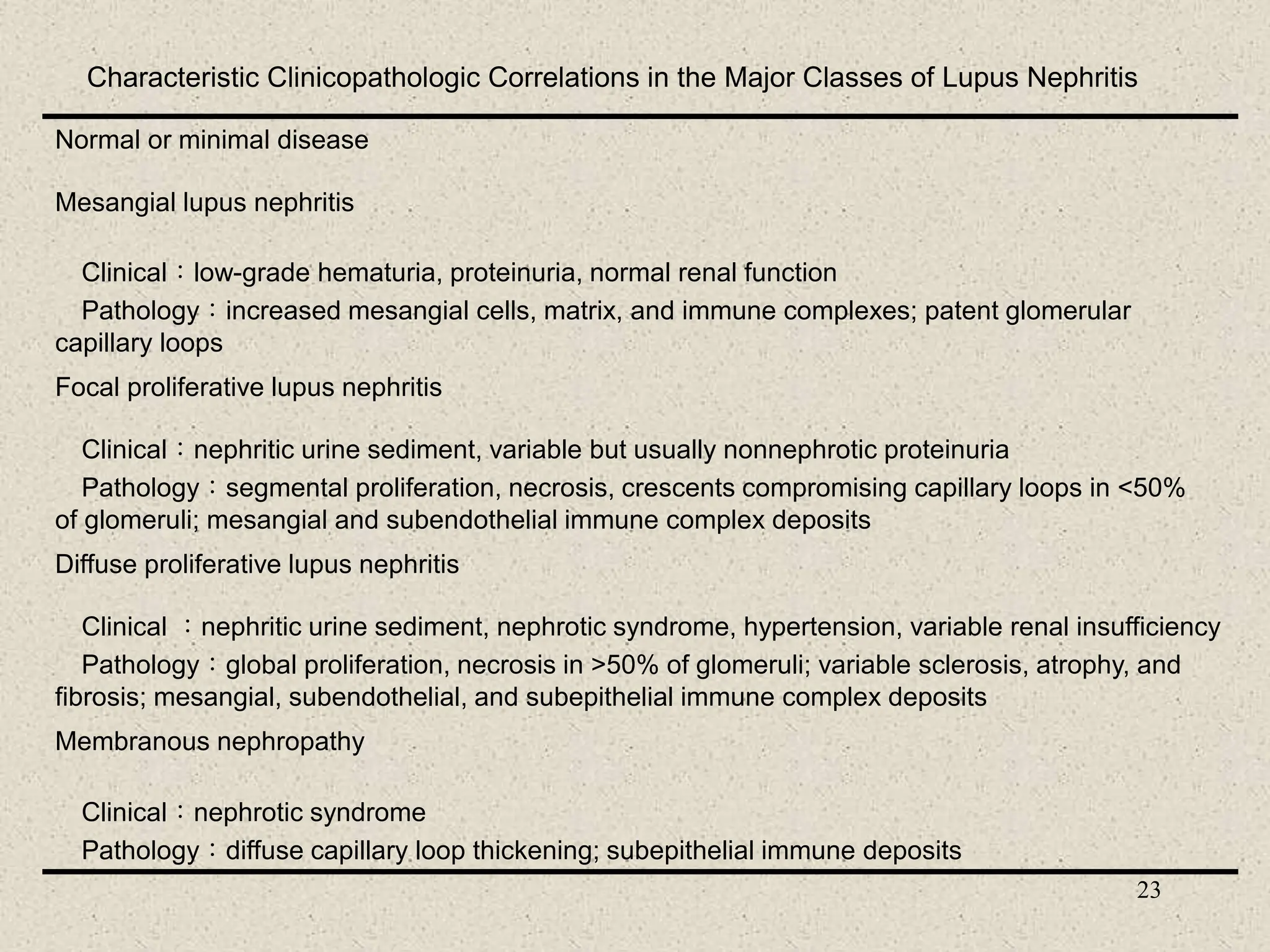
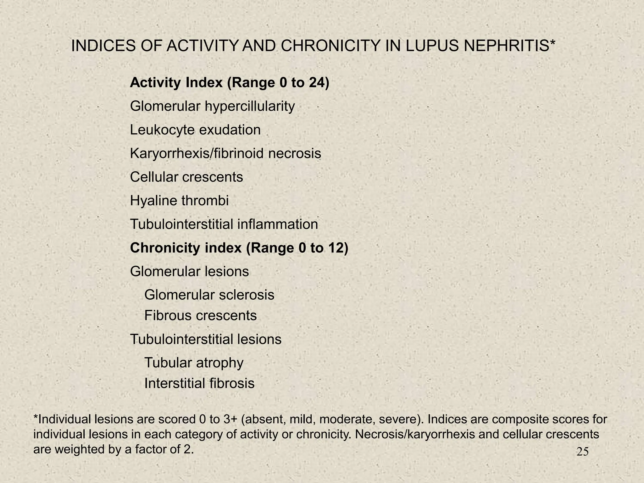
FREQUENCY OF ABNORMAL LABORATORY TESTS COMMONLY USED IN THE
EVALUATION OF NEUROPSYCHIATRIC LUPUS ERYTHEMATOSUS
Test Frequency of
Abnormal Test Result
Range (%)
Comment
Serologic
Antimeuronal antibodies
Antineurofilament antibodies
Antiribosomal-P antibodies
Antiphospholipid antibodies
30-92
58
45-90
45-80
Diffuse manifestations
Psychosis/depression
Focal manifestations, strokes
Cerebrospinal fluid
Routine
Pleocytosis
Increased protein]
Low glucose
6-34
22-50
3-8
Rule out infection and NSAID meningitis
Nonspecific
Rule out infection, transerse myelitis
Special
Antineuronal antibodies (lgG)
Elevated Q albumin
Elevated lgG/lgM index
Oligoclonal bands (2 bands)
90
8-33
25-66
20-82
Diffuse manifestations, present in 40%
with focal manifestations
Break in blood-brain barrier
Diffuse manifestations
33. 33
Pathologic studies in NPLE
Several autopsy series agree on several important points
There is no pathognomonic lesion that NPLE causes in
the brain that is diagnostically specific like the wire loop
lesion observed in lupus nephritis.
Active vasculitis is rare, bland vasculopathy (vascular
hyalinization, perivascular imflammation, and endothelial
proliferation associated with microinfarcts and
hemorrhage is the most common pathologic abnormalities
seen.)
Clinical manifestations may not be readily explained by
pathologic findings, some NPLE patients, particularly
those with diffuse neuropsychiatric manifestations may
have normal or unremarkable brain pathology.
34. 34
Proactive and preventive strategies in
addition to lupus therapies(1)
• Patients education programs, eliminate patient
nonadherence
• Monitor vital signs, update physical examination, and
have laboratory work done
• Adhere to a general conditioning exercise program to
minimize osteoporosis and muscle atrophy
• Cognitive therapy for lupus “fog”;biofeedback for
Raynaud’s phenomenon
• Counseling and stress management
• Physical and occupational therapy, ergonomic work
station evaluation
35. 35
Proactive and preventive strategies in
addition to lupus therapies(2)
• Aggressive proactive management of blood pressure,
blood sugars, serum lipids, and weight. Smoking
cessation.
• Yearly bone densitometry and osteoporosis
prevention measures.
• Annual electrocardiogram and chest x-ray
• Prompt evaluation of all fevers
• Periodic screening with carotid duplex scanning,
treadmill, or stress testing; screening for, and
prophylactic management of, antiphospholipid
antibodies.
36. 36
Therapies for lupus patients with
skin lesions(1)
• General
– Avoid sun: clothing, sunscreens, avoid hot part of
day with most UV-B light, camouflage cosmetics
– Stop smoking (so antimalarials works better)
– Thiazides and sulfonylureals may exacerbate skin
disease
37. 37
Therapies for lupus patients with
skin lesions(2)
• Routine therapy
– Topical steroids, intralesional steroids
– Hydroxychloroquine
– Oral corticosteroids
– Dapsone for bullous lesions
38. 38
Therapies for lupus patients with
skin lesions(3)
• Advanced therapy for resistant causes
– Subacute cutaneous lupus: mycophenylate
mofetil, retinoids, or cyclosporine
– Discoid lesions: chloroquine, clofazimine,
thalidomide, or cyclosporine
– Lupus profundus: dapsone
– Chronic lesions over 50% of body: topical
nitrogen mustard, BCNU, or tacrolimus
– Vasculitis: may need immunosuppressives
39. 39
Therapy for lupus patients with arthritis
(no internal organ involvement)
• First line: NSAIDs
• Cyclooxygenase-2 specific inhibitors
(but may induce thrombotic risk in
patients with antiphospholipid antibodies)
• Low dose hydroxychloroquine(200mg
twice a day)
40. 40
Indications of high dose corticosteroid
therapy in lupus patients
• Severe lupus nephritis
• CNS lupus with severe manifestations
• Autoimmune thrombocytopenia with
extremely low platelet counts
(e.g.<30000/mm3)
• Autoimmune hemolytic anemia
• Acute pneumonitis caused by SLE.
• Others: severe vasculitis with visceral organ
involvement, serious complications from
serositis (pleuritis, pericarditis, or peritonitis)
41. 41
Life-Threatening Manifestations of SLE:
Responses to glucocorticoids(1)
• Manifestations often responsive to glucocorticoids
– Vasculitis
– Severe dermatitis of subacute cutaneous lupus
erythematosus or SLE
– Polyarthritis
– Polyserositis—pericarditis, pleurisy, peritonitis
– Myocarditis
– Lupus pneumonitis
43. 43
Life-Threatening Manifestations of SLE:
Responses to glucocorticoids(3)
• Manifestations not often responsive to
glucocorticoids
– Thrombosis—includes strokes
– Glomerulonephritis—scarred end-stage renal
disease, pure membranous glomerulonephritis
– Resistant thrombocytopenia or hemolytic
anemia—occurs in a minority of patients; consider
splenectomy, cytotoxics, danazol, or
cyclosporine/neoral therapies
– Psychosis related to conditions other than SLE,
such as glucocorticoid therapy
44. 44
Therapy for patients with lupus nephritis
• Previously untreated patients with active
lupus nephritis or severe manifestations
( decreased renal function and /or high-
grade proteinuria)
– First line: high doses of corticosteroids
(about 1mg/kg/day)
– Cytotoxic drugs or other
immunosuppressive drugs
45. 45
The indications of cytotoxic drugs use in
the treatment of lupus nephritis
• Active and severe GN despite treatment with
high dose prednisone
• Responded to corticosteroids but require an
unacceptably high dose to maintain a
response.
• Unacceptable side effects from
corticosteroids.
• Chronic damage on a renal biopsy and other
indicators of a poor prognosis.
47. 47
The management of organ-threatening lupus
• Existing immunosuppressive therapies:
cyclophosphamide, mycophenolate mofetil,
cyclosporine A, fludarabine, cladribine (2-CDA)
• Apheresis
• Intravenous immunoglobulin
• Various biologic agents: BlyS inhibitor, CTLA-4Ig,
LL2IgG
• Stem cell transplantation
48. 48
Use of Cytotoxic Drugs in SLE :
Azathioprine
• requires 6–12 months to work well
• 1–3 mg/kg/day(initial dose)
• 1–2 mg/kg/day(maintenance dose)
• Advantage:probably reduces flares, reduces
renal
scarring, reduces glucocorticoid dose
requirement
• Side effects: Bone marrow suppression,
leukopenia, infection(herpes zoster), infertility,
malignancy, early menopause, hepatic
damage, nausea
49. 49
Use of Cytotoxic Drugs in SLE:
Cyclophosphamide
• requires 2–16 weeks to work well
• Initial dose:1-3 mg/kg/day orally or 8–20
mg/kg intravenously once a month
plus mesna
• Maintenance dose:0.5–2 mg/kg/day orally or 8–
20mg/kg intravenously every 4–12 wks plus mesna
• Adverse effects:probably reduces flares, reduces
renal scarring, reduces glucocorticoid dose
requirement
• Adverse effects: marrow suppression, leukopenia,
infection, infertility, malignancy,
menopause,cystitis,nausea
50. 50
The treatment in lupus patients with
autoimmune thrombocytopenia
• Splenectomy
• Danazol
• Immunosuppressive or cytotoxic drugs:
azathioprine, cyclophosphamide
• Intravenous immunoglobulin(IVIG)
51. 51
Other management principles in the
treatment of lupus patients(1)
• Thrombosis
-Anticoagulation
• Recurrent fetal loss with antiphospholipid
-Heparin in low dose or low-molecular-weight
heparin with or without aspirin
-If heparin ineffective or not tolerated, use low-dose
aspirin alone
-Glucocorticoids plus aspirin in moderate to high
dose may be used but is controversial
• Thrombocytopenia or hemolytic anemia
-Intravenous gamma globulin, splenectomy, danazol,
cyclosporine, cytotoxic drugs
52. 52
Other management principles in the
treatment of lupus patients(2)
• Seizures without other serious manifestations
-Anticonvulsants
• Behavior disorders or psychosis without other
serious manifestations:
-Psychoactive drugs, neuroleptics
• Pure membranous glomerulonephritis:
-Limited trials of immunosuppressives or no specific
treatment
53. 53
Other management principles in the
treatment of lupus patients(3)
• Avoid possible disease triggers-sulfa
antibiotics, sun, high estrogen-containing
birth control pills,alfalfa sprouts
• Prevent atherosclerosis
• Prevent osteoporosis
• Prevent infection
• Prevent progression of renal disease
• Prevent clots in patients with antiphospholipid
antibodies
• Treat fatigue.