SlideShare a Scribd company logo
Role of DNA & RNA in Protein Synthesis
DNA
DNA, is a double stranded nucleic acid consisting of deoxyribose sugar and carries the genetic
instructions used in the development and functioning of all known living organisms.
RNA
RNA is a single stranded molecule consisting of ribose sugar and it is transcribed (synthesized)
from DNA by enzymes called RNA polymerases. RNA acts as a messenger between DNA and
the protein synthesis complexes known as ribosomes.
Protein
Proteins are one of the vital biomolecules of life. These compounds perform a variety of essential
processes to sustain an organism's survival, which include clotting of blood, transporting oxygen,
contracting muscles and catalyzing chemical reactions. The building blocks of proteins are called
amino acids.
A typical α-amino acid consists of an amino group (-NH2), carboxyl group (-COOH) and an R
group. Amino acids in a protein chain are linked by a peptide bond. Usually, proteins have an N-
terminal end carrying a free amino group and C-terminus with -COOH.
Protein Synthesis
The synthesis of proteins starts with transcribing the instructions in DNA into mRNA.
The mRNA is then carried out of the cell's nucleus into the cytoplasm, specifically
into structures called ribosomes.
Protein production occurs in ribosomes containing rRNA.
The tRNA transports the amino acids to the ribosomes.
The code sequence in mRNA is then translated and specific proteins are synthesized
by stringing amino acids together.
The production or synthesis of polypeptide chains (proteins) includes two phases:
Transcription & Translation
Protein synthesis in prokaryotes and eukaryotes
Eukaryotes
In eukaryotes, mRNA is synthesized in the nucleus from pre-messenger RNA (pre mRNA)
molecules, and then shipped to the cytoplasm, where translation occurs.
Prokaryotes
In prokaryotes, transcription and translation occur in the same cellular compartment — the
cytosol. Ribosomes are the site of translation
Transcription
Transcription is the process through which a DNA sequence is enzymatically copied by an
RNA polymerase to produce a complementary RNA(mRNA). In other words, it is the transfer
of genetic information from DNA into RNA.
During transcription, only one strand of a DNA molecule is transcribed; this strands is called
antisense strand or template strand and the RNA so produced is termed as sense RNA.
The other strand of the DNA duplex is know as coding strand or sense strand.
As in DNA replication, transcription proceeds in the 5' → 3' direction (i.e. the old polymer is
read in the 3' → 5' direction and the new, complementary fragments are generated in the 5' →
3' direction).
Transcription, is catalyzed by DNA-directed RNA polymerase, or simply RNA polymerase.
Process of Transcription
Direction of Transcription
The DNA splits into two strands:
 Template strand: it is used to synthesize RNA.
 Coding Strand: (Informational strand): it is not used to synthesize RNA.
Transcription proceeds from the 3’ end to the 5’ end of the template.
Transcription Process
Transcription begins with the attachment of RNA polymerase holoenzyme to the promoter of
transcription unit and it ends when the core enzyme reaches the terminator site and dissociates
from the DNA .
The transcription process is divided into the following steps:
(1) initiation (2) elongation (3) termination.
Transcription Initiation
The initiation phase begins with the binding of RNA polymerase holoenzyme to the
promoter and ends when the holoenzyme leaves the promoter.
The initially the holoenzyme binds to the promoter DNA about 70-80 bp extending from -
55 to +20
The holoenzyme now induces ‘melting’, i.e. strand separation, in a <17 bp region that
includes the right end of the -10 sequence and extends just beyond the start point.
The holoenzyme now begins to transcribe the antisense strand, RNA synthesis begins at the
start point.
The nucleotides used are riboside 5’-triphosphates and RNA synthesis progresses in 5ꞌ-3ꞌ
direction. The enzyme permits the correct riboside to align opposite the deoxyribotides of
antisense strand.
When the first two riboside have been aligned, the enzyme catalyzes the formation of
phosphodiester bond between them, the diribotide so formed remains associated with the
template DNA strand and the enzyme. The holoenzyme sequentially adds the subsequent
riboside to the growing RNA chain.
The RNA chain remains as RNA∙DNA hybrid for ~2 to 3 nucleotides at its 3-end, i.e., the
growing end , But ~25 nucleotide at the growing end of the RNA chain are associated with
the template DNA and /or the enzyme.
Initiation in Prokaryotes
Elongation
The elongation phase begins when the RNA polymerase leaves the promoter region and
continues transcription of the template strand. As the core enzyme moves along the DNA
duplex, the regions downstream of the start point progressively become single-stranded, the
core enzyme sequentially adds the correct ribotides to the 3ꞌ-end of RNA chain. As the RNA
chain elongates, its 5ꞌ-region progressively separates from the template strand. The sense and
antisense strands of the DNA become progressively free as the transcription proceeds; these
single strands progressively reassociate to form double helix.Thus elongation involves
progressive disruption of the double helix to generate transient single-stranded regions; the
separated strands form a transcription bubble within which the template strand is transcribed.
The transcription bubble moves progressively downstream of the start point and disappears
when transcription terminates.
Termination
When the core enzyme reaches terminator site,
(1) No further ribotides are added to the RNA chain.
(2)The RNA chain dissociates from the template strand of DNA.
(3) the separated DNA strands reassociate to form a double helix. As a result, the
Transcription bubble disappears.
(4) the core enzyme dissociates from DNA.These events constitute the termination phase
of transcription.
The termination sites in prokaryotes have been classified into the following two groups:
(1) rho – independent terminators,
(2) rho – dependent terminators.
Rho-Dependent Terminators
At these terminators, a polypeptide called rho-factor is required for transcription termination.
Most likely, rho-factor binds the RNA transcript at a recognition site (-50-90 bases long) that is
located upstream of the termination site.
Rho-Independent Terminator
Terminator of transcription at these termination sites does not require rho factor; therefore, they
are called intrinsic terminators.
The RNA transcript from such terminators forms a typical hairpin loop, which is followed by a
run of – 6U residues.
The poly – U region probably signals the core enzyme to leave the DNA duplex.
Transcription in eukaryotes
RNA Polymerase
All eukaryotes possess three type of RNA polymerases called RNA polymerase I,II and III .
RNA polymerase I is located in nucleolus, and is responsible for transcription of genes for
rRNA, it is responsible for 50-70% of the activity in eukaryotes.
RNA polymerase II is located in the nucleoplasm, constitutes 20-40% of total activity and
transcribes all the genes that produce mRNA.
RNA polymerase III also occurs in nucleoplasm, provides ~10% of total polymerase
activity, and transcribes tRNA and other small RNA genes.
Process of transcription
In eukaryotes transcription unit generally contains a single gene.Transcription termination
occurs beyond the end of coding region.Transcription by RNA polymerase II is briefly
described below.
Transcription Complex
The complex of proteins formed at the promoter that ultimately leads to transcription
initiation is called transcription complex.
Transcription Initiation
Transcription begins at the start point, which is usually an A.
A closed binary complex is formed when RNA polymerase II binds the promoter.
The complex is then converted into an open binary complex in which the two strands of promoter
DNA become separated locally.
RNA polymerase now begins transcription at the start point, and presumably, the transcription factors
are released around this stage.
The basal transcription factors are able to initiate transcription at a low level.
Transcription Termination
It is likely that the transcription units have rather long terminator regions with multiple terminator
sites.
In some transcription units, termination occurs 1,000 bp downstream of the site corresponding to the
3' -end of mature mRNAs. In such cases, the 3' -ends of mRNAs are generated by cleavage following
transcription.
mRNA Processing
 The process of modification, mainly through cleavage and /or splicing of primary RNA
transcripts so as to produce functional mRNA molecules from them is called RNA
processing.
 RNA processing is carried out by ribonucleases, the enzymes that cleave RNA.
 They are of two types ., Exoribonucleases and Endoribonucleases.
 Exoribonucleases remove one base at a time from one end of RNA molecule, all know
enzymes trim RNA from the 3' end But Endoribonucleases produce internal cuts in
RNA molecules.
 Eukaryotic transcripts (pre-mRNA) contains exons (coding/expressed sequences) and
introns (non coding/ unexpressed sequences)
 All the introns are excised from the primary transcript and all the exons of a gene are
joined together in the proper order; this process is called splicing.
Translation
The mRNA carries genetic information encoded as a ribonucleotide sequence from the
chromosomes to the ribosomes.
The ribonucleotide are "read" by translational machinery in a sequence of nucleotide triplets
called codons. Each of those triplets codes for a specific amino acid.
Translation is the production of proteins by decoding mRNA produced in transcription.
The ribosome and tRNA molecules translate this code to produce proteins.
The ribosome is a multi subunit structure containing rRNA and proteins. It is the "factory"
where amino acids are assembled into proteins.
Genetic code
The number and the sequence of bases in mRNA specifying an amino acid is known as codon.
The set of bases in a tRNA that base-pairs with a codon of an mRNA is known as anticodon.
The sequence of bases in an anticodon is exactly the opposite of and complementary to that
present in the codon. For example the codon 5’AUG 3’ has the anticodon 3’ UAC 5’.
The set of all the codons that specify the 20 amino acids is termed as the genetic code.
Properties of the genetic code:
The code is universal: All prokaryotic and eukaryotic organisms use the same codon to
specify each amino acid.
The code is triplet: Three nucleotides make one codon. 61 of them code for amino acids and
3 viz. , UAA, UAG and UGA are nonsense codons or chain termination codons.
The code is degenerate: There are 64 codons available for 20 amino acids.
Most amino acids are encoded by two or more codons.
The code is non ambiguous: Each codon specifies only one of the 20 amino
acids. None of the codons code for two or more amino acids. The only
exception is the AUG codon which codes for formylmethionine in prokaryotes
at the initiation site. While at other positions it specifies methionine.
The code is non overlapping: A base in mRNA is not used for two different
codons.
The code is commaless: There is no special signal or commas between codons.
Ribosome and Ribosomal RNA (rRNA)
Ribosomal RNA (rRNA) occurs in association with proteins and is organized into almost
spherical bodies of about 200 A° diameter called ribosomes.
Ribosomes of prokaryotes are of 70 S size and are composed of 60% rRNA and 40%
protein. They dissociate into a smaller 30 S subunit and a larger 50 S subunit.
The 30S subunit of prokaryotic ribosomes has a single 16S rRNA molecule, which is
associated with 21 different ‘S’ proteins. The larger 50S subunit has a 23S rRNA and a 5S
rRNA molecule complexed with 31 different ‘L’ proteins. The basic structure of ribosomes
is due to their rRNA, and the proteins play a secondary role.
The cytoplasmic ribosomes of eukaryotes are of the size 80S and contain 40% rRNA and
60% protein. They consist of a 40S and a 60S subunit.
The 40 S subunit of eukaryotic ribosomes has a single 18S rRNA molecule and 33 different
proteins. The larger 60S subunit has one molecule each of 28S, 5.8S and 5S rRNA and 49
different proteins.
Functional sites on Ribosomes
A complete ribosome particle has two distinct and adjacent sites for the attachment of
aminoacyl-tRNAs (amino acid carrying tRNA), an aminoacyl attachment site (site A) and a
peptidyl site (site P).
An aminoacyl-tRNA first attaches to site A, the kind of aminoacyl-tRNA being determined
by the sequence of mRNA (codon) attached to the site A. the aminoacyl-tRNA, along with
the mRNA codon, now moves to the site P, making the site A available for the attachment of
a new aminoacyl-tRNA.
In addition, the ribosome has following sites as well:
1.mRNA site 2.peptidyl transferase site 3.EF-Tu site 4.EF-G site
5. 5 S RNA site 6.exit site
Transfer RNA (tRNA)
The existence of tRNA was demonstrated by Hoagland and coworkers in 1957.
Transfer RNA (tRNA) is a class of RNA of small size of 3S and generally, it has 76 to 95
nucleotides.
tRNA molecules have a high proportion of unusual bases. The unusual bases present in
tRNA are produced after transcription by modifications of the usual bases, A,U,G and C.
Further, tRNA molecules show considerable double helical structure, and more than 50% of
the bases in tRNA are paired.
Structure of tRNA
A ‘clover leaf model’ of tRNA structure has been proposed to account for its structural and
functional properties. The three nucleotides at the 5 -end are CCA in all the types of tRNA
molecules. This segment is the amino acid acceptor region. The amino acid binds to one of
the OH groups of the ribose of the terminal adenine.The tRNA molecule has three loops:
DHU loop, anticodon loop and thymine loop. Sometimes an extra loop, having a variable
number of nucleotides, is also present between the anticodon and thymine loops.
Formation of aminoacyl-tRNA
The amino acids are attached to tRNA molecules through a two step reaction process. Both
the reactions are catalysed by the same enzyme, aminoacyl tRNA synthetase.
The carboxyl group of the amino acid reacts with one of the –OH groups of the ribose of
the terminal adenine nucleotide to produce aminoacyl-tRNA .
1.first step: It consist of amino acid activation, in which the amino acid molecule reacts
with and ATP molecule to yield an aminoacyl-AMP (aminoacyl adenylate) molecule.
2.Second step: The amino acid form aminoacyl-AMP molecule is then transferred to a
tRNA molecule that is specific for the amino acid, and AMP is released.
The aminoacyl-AMP complex remains tightly bound to the enzyme aminoacyl-tRNA
synthetase during the entire reaction, i.e., both the steps.
The attachment of an amino acid to the concerned tRNA to yield aminoacyl-tRNA is called
charging of tRNA.
Recognition of codons
Recognition of specific codons of the mRNA is the function of tRNA molecule. This
recognition is due to the anticodon that base pairs with the mRNA codon.
Almost as a rule, A in the first place of anticodons (corresponding to the third base of
mRNA codon) is modified to I (inosine); I base pairs with any one of U,C and A.
Similarly, U in the first position of anticodon is always modified the modified U may base
pair with A,G and, in some cases with even U.
Translation
Translation begins near the 5-end of mRNA and progresses toward its 3 -end. In terms
of the polypeptide being synthesized, it starts with the N-terminal end of the
polypeptide, while the C-terminal amino acid is the last to be added to the chain.
The process of translation may be divided into the following 3 steps:
(1) Initiation,
(2) Elongation and
(3) Termination
Initiation
Initiation comprises all the events that precede the formation of the first peptide bond.
It
includes the following events:
 binding of the smaller subunit of ribosome to mRNA and
 binding of the first or initiator aminoacyl-tRNA to the P site of ribosome; this
completes the formation of initiation complex. Initiation is a relatively slow step in
translation.
Elongation
Elongation includes all the reactions from the formation of the first peptide bond to that
of the last peptide bond of the polypeptide chain.
Amino acids are added one at a time to the growing peptide chain.
It is the most rapid step of translation: in bacteria it proceeds at the rate of 15 amino
acids/second at 370C, while in eukaryotes its rate is 2 amino acids/second.
Elongation
The elongation step in bacteria is briefly described below. The complete initiation
complex has the following features:
ribosome is complete and ready for translation,
P site of ribosome is occupied by the initiator aminoacyl-tRNA (fmet-tRNAf) and
The A site is free.
The P site of ribosome must be occupied by peptidyl-tRNA (tRNA carrying a peptide
for the A site to be in a proper conformation to allow entry to an amino-acyl-tRNA.
Termination
Termination describes those events that occur after the formation of the last peptide bond
of the polypeptide, they culminate in the separation of the ribosome from mRNA.
Termination is signaled by the nonsense codons UAG, UAA and UGA, and is mediated by
certain proteins called release factors.
As a result of this the following three simultaneous events occur:
(1)the polypeptide chain detaches from the tRNA located at P site,
(2) followed by immediate release of tRNA from the P site and
(3) the release of ribosome from the mRNA.
The Translation Process
End product -protein
The end products of protein synthesis is a primary structure of a protein
A sequence of amino acid bonded together by peptide bonds
Point Mutation
A point mutation is a type of mutation in DNA or RNA, the cells genetic material, in which
one single nucleotide base is added, deleted or changed. DNA and RNA are made up of many
nucleotides. There are five different molecules that can make up nitrogenous bases on
nucleotides: cytosine, guanine, adenine, thymine (in DNA) and uracil (in RNA), abbreviated
C, G, A, T, and U. The specific sequence of nucleotides encodes all the information for
carrying out all cell processes. In general, a mutation is when a gene is altered through a
change in DNA structure; this may refer even to entire sections of chromosomes. A point
mutation is specifically when only one nucleotide base is changed in some way, although
multiple point mutations can occur in one strand of DNA or RNA.
Effects of point mutation
Sickle-Cell Anemia
Sickle-cell anemia is a recessive disorder caused by a single substitution in the gene that creates
hemoglobin, which carries oxygen in the blood. Normally, glutamic acid is produced in the chain, but
the substitution causes valine to be produced at that spot instead. When people have two copies of this
mutation, it results in thin sickle-shaped blood cells that sometimes cannot carry oxygen properly.
About 80% of people with sickle-cell disease are in sub-Saharan Africa, where being a carrier for
sickle-cell anemia (having only one copy of the gene, not two) actually helps protect against malaria.
It is also found in other parts of the world such as India and the Middle East, and affects about 1 in
500 African Americans. Symptoms include anemia, obstruction of blood vessels, and chest pain, and
it is treated with folic acid, blood transfusions, bone marrow transplants, and certain prescription
drugs.
Tay-Sachs
Tay-Sachs disease is another recessive disorder caused by point mutations. Different
mutations can cause this disorder, but they are all found on the HEXA gene
on chromosome 15. Tay-Sachs causes nerve cells to deteriorate over time, which in turn
results in the decline of physical and mental functioning. Both child and adult-onset forms of
the disease occur, and children with the disease usually die before the age of four. About 1 in
320,000 newborns in the United States develop Tay-Sachs. It occurs in higher frequencies in
Ashkenazi Jews, Cajuns, and French Canadians (about 1 in 3500 in these populations),
although the mutations associated with the disease are different in each population. There is
currently no treatment or cure.
Inheritence of Cystic Fibrosis
Cystic fibrosis is the most common inherited disease with a fatal outcome in industrialised
nations, and is becoming increasingly prevalent as survival is prolonged. Thirty years ago
most of these patients died in infancy but today about 36% reach adulthood. On the basis of
epidemiological analysis we can expect children born with cystic fibrosis in 1990 to have a
life expectancy of 40 years—in other words, more than 90% will reach adulthood.This will
entail costly and time consuming therapy. As in other chronic pulmonary diseases, lung
function parameters such as forced expiratory volume in one second (FEV1) are poor
predictors of the degree of disability.
Inheritence of Cystic Fibrosis
There are more than 30,000 Americans with cystic fibrosis (CF), the most common
autosomal recessive genetic disorder of Caucasians. CF patients born in 1970 had only a 50%
chance of reaching age 15, but in little more than two decades the chances of reaching that
age had increased to 95% . An increased understanding of normal airway physiology and the
pathophysiology of CF has spawned new treatments that have driven this impressive increase
in patient survival.
Inheritence of gender
The human genome is organized into 23 pairs of chromosomes (22 pairs of autosomes and
one pair of sex chromosomes), with each parent contributing one chromosome per pair.
The X and Y chromosomes, also known as the sex chromosomes, determine the biological
sex of an
individual: females inherit an X chromosome from the father for a XX genotype, while males
inherit a Y chromosome from the father for a XY genotype (mothers only pass on X
chromosomes). The presence or absence of the Y chromosome is critical because it contains
the genes necessary to override the biological default female development.
Inheritence of Sex linked
Sex linked is a trait in which a gene is located on a sex chromosome. In humans, the term
generally refers to traits that are influenced by genes on the X chromosome. This is because
the X chromosome is large and contains many more genes than the smaller Y chromosome.
In a sex-linked disease, it is usually males who are affected because they have a single copy
of X chromosome that carries the mutation. In females, the effect of the mutation may be
masked by the second healthy copy of the X chromosome.
These are traits that are found on either one of the chromosomes that determine sex, or the
sex chromosomes. And in humans this is the X or the Y chromosomes. And so some of the
more familiar sex-linked traits are hemophilia, red-green color blindness, congenital night
blindness, some high blood pressure genes, Duchenne muscular dystrophy, and also Fragile
X syndrome. So what's also very interesting is that you can imagine that for individuals who
are XY or males, having these different mutations on the genes, on the X chromosome, is
particularly problematic, because unlike females, there are not two X chromosomes that give
you the potential of carrying a normal gene on the X chromosome. Which is why in many
cases you'll see that males are more often afflicted with these sex-linked disorders.
Sex-linked disorder
The existence of X-linked disorders in humans has been recognized for many centuries,
based on lessons in religious texts and observations of specific human families (e.g., color
blindness or Daltonism). Our modern concepts of Mendelian (including X-linked)
inheritance originated just after the turn of the last century. Early concepts of dominance
and recessiveness were first used in conjunction with autosomal traits,andthen applied to
‘‘sex’’-linked traits to distinguish X-linked recessive and X-linked dominant inheritance.
The former was defined as vertical transmission in which carrierwomenpass the disorder to
affected sons, while the latter was defined as vertical transmission in which daughters of
affected malesare always affected, transmitting the disorder to offspring of both sexes.
X-linked disorder
X-linked disorders can be either dominant or recessive. X-linked dominant disorders are the
result of a mutation to the X chromosome that can affect either males or females. However,
some disorders, including Fragile X syndrome, affect males more severely than females,
likely because males do not have a second, normal copy of the X chromosome. Fragile X
syndrome is characterized by a wide range of developmental problems, including learning
disabilities. X-linked hypophosphatemia is another X-linked dominant condition that
manifests in a vitamin-D-resistant form of Rickets.
Y-linked disorder
Y chromosome mutations are called “Y-linked” and only affect males since they alone carry
a copy of that chromosome. Mutations to the relatively small Y chromosome can impact
male sexual function and secondary sex characteristics. Y-chromosome infertility is a
disorder that affects sperm production, caused by deletions to the azoospermia factor (AZF)
regions of the Y chromosome. In general, Y-linked disorders are only passed from father to
son; however, because affected males typically do not father children without assisted
reproductive technologies, Y-chromosome infertility is not typically passed on to offspring.
Role of chromosomal abnormalities in an inherited disease
Many human genetic disorders result from unbalanced chromosome abnormalities, in
which there is a net gain or loss of genetic material. Such imbalances often disrupt large
numbers of dosage-sensitive, developmentally important genes and result in specific and
complex phenotypes. Alternately, some chromosomal syndromes may be caused by a
deletion or duplication of a single gene with pleiotropic effects. Traditionally, chromosome
abnormalities were identified by visual inspection of the chromosomes under a
microscope. The use of molecular cytogenetic technologies, such as fluorescence in situ
hybridization and microarrays, has allowed for the identification of cryptic or
submicroscopic imbalances, which are not visible under the light microscope.
Microarrays have allowed for the identification of numerous new syndromes through a
genotype-first approach in which patients with the same or overlapping genomic alterations
are identified and then the phenotypes are described. Because many chromosomal alterations
are large and encompass numerous genes, the ascertainment of individuals with overlapping
deletions and varying clinical features may allow researchers to narrow the region in which
to search for candidate genes
Down Syndrome
Down syndrome (sometimes called Down’s syndrome) is a condition in which a child is born
with an extra copy of their 21st chromosome — hence its other name, trisomy 21. This
causes physical and mental developmental delays and disabilities
Many of the disabilities are lifelong, and they can also shorten life expectancy. However,
people with Down syndrome can live healthy and fulfilling lives.
What causes Down syndrome?
In all cases of reproduction, both parents pass their genes on to their children. These genes
are carried in chromosomes. When the baby’s cells develop, each cell is supposed to receive
23 pairs of chromosomes, for 46 chromosomes total. Half of the chromosomes are from the
mother, and half are from the father.
In children with Down syndrome, one of the chromosomes doesn’t separate properly. The
baby ends up with three copies, or an extra partial copy, of chromosome 21, instead of two.
This extra chromosome causes problems as the brain and physical features develop.
According to the National Down Syndrome Society (NDSS), about 1 in 700 babies in the
United States is born with Down syndrome. It’s the most common genetic disorder in the
United States.
References
 Massaia, A. and Xue, Y., 2017. Human Y chromosome copy number variation in the
next generation sequencing era and beyond. Human Genetics, 136(5), pp.591-603.
 Theisen, A. and Shaffer, L.G., 2010. Disorders caused by chromosome
abnormalities. The application of clinical genetics, 3, p.159
 MacIntyre, D.J., Blackwood, D.H.R., Porteous, D.J., Pickard, B.S. and Muir, W.J.,
2003. Chromosomal abnormalities and mental illness. Molecular Psychiatry, 8(3),
pp.275-287
 Dobyns, W.B., Filauro, A., Tomson, B.N., Chan, A.S., Ho, A.W., Ting, N.T.,
Oosterwijk, J.C. and Ober, C., 2004. Inheritance of most X‐linked traits is not
dominant or recessive, just X‐linked. American journal of medical genetics Part
A, 129(2), pp.136-143
 Staab, D., Wenninger, K., Gebert, N., Rupprath, K., Bisson, S., Trettin, M., Paul,
K.D., Keller, K.M. and Wahn, U., 1998. Quality of life in patients with cystic fibrosis
and their parents: what is important besides disease severity?. Thorax, 53(9),
pp.727-731
 Clunes, M.T. and Boucher, R.C., 2007. Cystic fibrosis: the mechanisms of
pathogenesis of an inherited lung disorder. Drug Discovery Today: Disease
Mechanisms, 4(2), pp.63-72
 Taliaferro, W.H. and Huck, J.G., 1923. The inheritance of sickle-cell anaemia in
man. Genetics, 8(6), p.594.
THANK YOU

More Related Content

What's hot

structure types and function of RNA
structure types and function of RNAstructure types and function of RNA
structure types and function of RNA
adnandinmohammed
 
Protein synthesis
Protein synthesis Protein synthesis
Protein synthesis
Samson Sakala Jnr
 
Reverse Transcription of RNA
Reverse Transcription of RNAReverse Transcription of RNA
Reverse Transcription of RNA
UNIVERSITI MALAYSIA SABAH
 
Translation
TranslationTranslation
Translation
Nandy 91
 
DNA Replication
DNA ReplicationDNA Replication
DNA Replication
Dr. A.D.Naveen Kumar
 
RNA: Nature, type and Structure
RNA: Nature, type and StructureRNA: Nature, type and Structure
RNA: Nature, type and Structure
Virendra Singh Shekhawat
 
Dna replication in prokaryotes
Dna replication in prokaryotesDna replication in prokaryotes
Dna replication in prokaryotes
Ashish Pratim Mahanta
 
RNA- STRUCTURE AND FUNCTIONS
RNA- STRUCTURE AND FUNCTIONSRNA- STRUCTURE AND FUNCTIONS
RNA- STRUCTURE AND FUNCTIONS
SushrutMohapatra
 
Types of RNA
Types of RNATypes of RNA
Types of RNA
Kayeen Vadakkan
 
Rna structure
Rna structureRna structure
Rna structure
Praveen Garg
 
Transcription
Transcription Transcription
Transcription
ranjani n
 
Transcription II- Post transcriptional modifications and inhibitors of Transc...
Transcription II- Post transcriptional modifications and inhibitors of Transc...Transcription II- Post transcriptional modifications and inhibitors of Transc...
Transcription II- Post transcriptional modifications and inhibitors of Transc...
Namrata Chhabra
 
Transcription in Pro- & eukaryotes
Transcription in Pro- & eukaryotesTranscription in Pro- & eukaryotes
Transcription in Pro- & eukaryotes
NurulhasanKhatri
 
REPLICATION
REPLICATIONREPLICATION
REPLICATION
YESANNA
 
Transcription process
Transcription processTranscription process
Transcription process
Touheed Ovi
 
Post Transcriptional Modifications
Post Transcriptional ModificationsPost Transcriptional Modifications
Post Transcriptional Modifications
Srishti Pandey
 
DNA and RNA types and functions
DNA and RNA types and functionsDNA and RNA types and functions
DNA and RNA types and functions
subhananthini jeyamurugan
 
Spliceosome
SpliceosomeSpliceosome
Spliceosome
EmaSushan
 
Genetic code features and character
Genetic code features and characterGenetic code features and character
Genetic code features and character
AnuKiruthika
 
Transcription in prokaryotes
Transcription in prokaryotesTranscription in prokaryotes
Transcription in prokaryotes
Microbiology
 

What's hot (20)

structure types and function of RNA
structure types and function of RNAstructure types and function of RNA
structure types and function of RNA
 
Protein synthesis
Protein synthesis Protein synthesis
Protein synthesis
 
Reverse Transcription of RNA
Reverse Transcription of RNAReverse Transcription of RNA
Reverse Transcription of RNA
 
Translation
TranslationTranslation
Translation
 
DNA Replication
DNA ReplicationDNA Replication
DNA Replication
 
RNA: Nature, type and Structure
RNA: Nature, type and StructureRNA: Nature, type and Structure
RNA: Nature, type and Structure
 
Dna replication in prokaryotes
Dna replication in prokaryotesDna replication in prokaryotes
Dna replication in prokaryotes
 
RNA- STRUCTURE AND FUNCTIONS
RNA- STRUCTURE AND FUNCTIONSRNA- STRUCTURE AND FUNCTIONS
RNA- STRUCTURE AND FUNCTIONS
 
Types of RNA
Types of RNATypes of RNA
Types of RNA
 
Rna structure
Rna structureRna structure
Rna structure
 
Transcription
Transcription Transcription
Transcription
 
Transcription II- Post transcriptional modifications and inhibitors of Transc...
Transcription II- Post transcriptional modifications and inhibitors of Transc...Transcription II- Post transcriptional modifications and inhibitors of Transc...
Transcription II- Post transcriptional modifications and inhibitors of Transc...
 
Transcription in Pro- & eukaryotes
Transcription in Pro- & eukaryotesTranscription in Pro- & eukaryotes
Transcription in Pro- & eukaryotes
 
REPLICATION
REPLICATIONREPLICATION
REPLICATION
 
Transcription process
Transcription processTranscription process
Transcription process
 
Post Transcriptional Modifications
Post Transcriptional ModificationsPost Transcriptional Modifications
Post Transcriptional Modifications
 
DNA and RNA types and functions
DNA and RNA types and functionsDNA and RNA types and functions
DNA and RNA types and functions
 
Spliceosome
SpliceosomeSpliceosome
Spliceosome
 
Genetic code features and character
Genetic code features and characterGenetic code features and character
Genetic code features and character
 
Transcription in prokaryotes
Transcription in prokaryotesTranscription in prokaryotes
Transcription in prokaryotes
 

Similar to Role of DNA and A in Protein synthesis

Biochem synthesis of rna(june.23.2010)
Biochem   synthesis of rna(june.23.2010)Biochem   synthesis of rna(june.23.2010)
Biochem synthesis of rna(june.23.2010)MBBS IMS MSU
 
Gene expression: Translation and Transcription
Gene expression: Translation and TranscriptionGene expression: Translation and Transcription
Gene expression: Translation and TranscriptionCyra Mae Soreda
 
molecular biology.pptx
molecular biology.pptxmolecular biology.pptx
molecular biology.pptx
HiraNoor47
 
Protein synthesis mechanism with reference of Translation and Transcription d...
Protein synthesis mechanism with reference of Translation and Transcription d...Protein synthesis mechanism with reference of Translation and Transcription d...
Protein synthesis mechanism with reference of Translation and Transcription d...
muhammad aleem ijaz
 
Role of DNA and RNA in Protein Synthesis
Role of DNA and RNA in Protein SynthesisRole of DNA and RNA in Protein Synthesis
Role of DNA and RNA in Protein Synthesis
CharupriyaChauhan1
 
lecture 3 Gene expression pptx
lecture 3 Gene expression           pptxlecture 3 Gene expression           pptx
lecture 3 Gene expression pptx
HanySaid33
 
Transcription
TranscriptionTranscription
Transcriptionaljeirou
 
protein synthesis
protein synthesisprotein synthesis
protein synthesis
Nawfal Aldujaily
 
Transcription &translation
Transcription &translationTranscription &translation
Transcription &translation
Prince Avi
 
Physiology-brief about RNA and its types
Physiology-brief about RNA and its typesPhysiology-brief about RNA and its types
Physiology-brief about RNA and its types
DrHamza Nafees
 
GENE EXPRESSION AND IT’S REGULATION.pptx
GENE EXPRESSION AND IT’S REGULATION.pptxGENE EXPRESSION AND IT’S REGULATION.pptx
GENE EXPRESSION AND IT’S REGULATION.pptx
yogesh532361
 
Protein synthesis
Protein synthesisProtein synthesis
Protein synthesisFiza Khan
 
Geneexpression
GeneexpressionGeneexpression
Geneexpression
RajeshwariTiwari2
 
Gene expression concept and analysis
Gene expression concept and analysisGene expression concept and analysis
Gene expression concept and analysis
Noha Lotfy Ibrahim
 
Gene Expression_AA1.ppt
Gene Expression_AA1.pptGene Expression_AA1.ppt
Gene Expression_AA1.ppt
midolyon1990gmailcom
 
Molecular Genetics Part II
Molecular Genetics Part IIMolecular Genetics Part II
Molecular Genetics Part IIJolie Yu
 
Provide an in depth description of biological information transfer (.pdf
Provide an in depth description of biological information transfer (.pdfProvide an in depth description of biological information transfer (.pdf
Provide an in depth description of biological information transfer (.pdf
MALASADHNANI
 
DNA- Transcription and Tranlation, RNA, Ribosomes and membrane proteins.pptx
DNA- Transcription and Tranlation, RNA, Ribosomes and membrane proteins.pptxDNA- Transcription and Tranlation, RNA, Ribosomes and membrane proteins.pptx
DNA- Transcription and Tranlation, RNA, Ribosomes and membrane proteins.pptx
LaibaSaher
 

Similar to Role of DNA and A in Protein synthesis (20)

Biochem synthesis of rna(june.23.2010)
Biochem   synthesis of rna(june.23.2010)Biochem   synthesis of rna(june.23.2010)
Biochem synthesis of rna(june.23.2010)
 
Gene expression: Translation and Transcription
Gene expression: Translation and TranscriptionGene expression: Translation and Transcription
Gene expression: Translation and Transcription
 
molecular biology.pptx
molecular biology.pptxmolecular biology.pptx
molecular biology.pptx
 
Protein synthesis mechanism with reference of Translation and Transcription d...
Protein synthesis mechanism with reference of Translation and Transcription d...Protein synthesis mechanism with reference of Translation and Transcription d...
Protein synthesis mechanism with reference of Translation and Transcription d...
 
Role of DNA and RNA in Protein Synthesis
Role of DNA and RNA in Protein SynthesisRole of DNA and RNA in Protein Synthesis
Role of DNA and RNA in Protein Synthesis
 
lecture 3 Gene expression pptx
lecture 3 Gene expression           pptxlecture 3 Gene expression           pptx
lecture 3 Gene expression pptx
 
Transcription
TranscriptionTranscription
Transcription
 
protein synthesis
protein synthesisprotein synthesis
protein synthesis
 
4,transcription
4,transcription4,transcription
4,transcription
 
Transcription &translation
Transcription &translationTranscription &translation
Transcription &translation
 
Physiology-brief about RNA and its types
Physiology-brief about RNA and its typesPhysiology-brief about RNA and its types
Physiology-brief about RNA and its types
 
GENE EXPRESSION AND IT’S REGULATION.pptx
GENE EXPRESSION AND IT’S REGULATION.pptxGENE EXPRESSION AND IT’S REGULATION.pptx
GENE EXPRESSION AND IT’S REGULATION.pptx
 
Protein synthesis
Protein synthesisProtein synthesis
Protein synthesis
 
Geneexpression
GeneexpressionGeneexpression
Geneexpression
 
Gene expression concept and analysis
Gene expression concept and analysisGene expression concept and analysis
Gene expression concept and analysis
 
Gene Expression_AA1.ppt
Gene Expression_AA1.pptGene Expression_AA1.ppt
Gene Expression_AA1.ppt
 
Molecular Genetics Part II
Molecular Genetics Part IIMolecular Genetics Part II
Molecular Genetics Part II
 
Provide an in depth description of biological information transfer (.pdf
Provide an in depth description of biological information transfer (.pdfProvide an in depth description of biological information transfer (.pdf
Provide an in depth description of biological information transfer (.pdf
 
Ig
IgIg
Ig
 
DNA- Transcription and Tranlation, RNA, Ribosomes and membrane proteins.pptx
DNA- Transcription and Tranlation, RNA, Ribosomes and membrane proteins.pptxDNA- Transcription and Tranlation, RNA, Ribosomes and membrane proteins.pptx
DNA- Transcription and Tranlation, RNA, Ribosomes and membrane proteins.pptx
 

More from Syed Khawar Abbas Asad

Human Population Growyh
Human Population GrowyhHuman Population Growyh
Human Population Growyh
Syed Khawar Abbas Asad
 
Environmental
EnvironmentalEnvironmental
Environmental
Syed Khawar Abbas Asad
 
Anatomy and physiology of respiration
Anatomy and physiology of respirationAnatomy and physiology of respiration
Anatomy and physiology of respiration
Syed Khawar Abbas Asad
 
Marfan syndrome
Marfan syndromeMarfan syndrome
Marfan syndrome
Syed Khawar Abbas Asad
 
What is hibernation
What is hibernationWhat is hibernation
What is hibernation
Syed Khawar Abbas Asad
 
Toxoplasmosis
ToxoplasmosisToxoplasmosis
Toxoplasmosis
Syed Khawar Abbas Asad
 
Wheat,its types ,importance,characteristics
Wheat,its types ,importance,characteristicsWheat,its types ,importance,characteristics
Wheat,its types ,importance,characteristics
Syed Khawar Abbas Asad
 
Importance of the maintenance of biodiversity
Importance of the maintenance of biodiversityImportance of the maintenance of biodiversity
Importance of the maintenance of biodiversity
Syed Khawar Abbas Asad
 
Social organization
Social organizationSocial organization
Social organization
Syed Khawar Abbas Asad
 
Computer networks
Computer networks Computer networks
Computer networks
Syed Khawar Abbas Asad
 

More from Syed Khawar Abbas Asad (10)

Human Population Growyh
Human Population GrowyhHuman Population Growyh
Human Population Growyh
 
Environmental
EnvironmentalEnvironmental
Environmental
 
Anatomy and physiology of respiration
Anatomy and physiology of respirationAnatomy and physiology of respiration
Anatomy and physiology of respiration
 
Marfan syndrome
Marfan syndromeMarfan syndrome
Marfan syndrome
 
What is hibernation
What is hibernationWhat is hibernation
What is hibernation
 
Toxoplasmosis
ToxoplasmosisToxoplasmosis
Toxoplasmosis
 
Wheat,its types ,importance,characteristics
Wheat,its types ,importance,characteristicsWheat,its types ,importance,characteristics
Wheat,its types ,importance,characteristics
 
Importance of the maintenance of biodiversity
Importance of the maintenance of biodiversityImportance of the maintenance of biodiversity
Importance of the maintenance of biodiversity
 
Social organization
Social organizationSocial organization
Social organization
 
Computer networks
Computer networks Computer networks
Computer networks
 

Recently uploaded

2024.06.01 Introducing a competency framework for languag learning materials ...
2024.06.01 Introducing a competency framework for languag learning materials ...2024.06.01 Introducing a competency framework for languag learning materials ...
2024.06.01 Introducing a competency framework for languag learning materials ...
Sandy Millin
 
Phrasal Verbs.XXXXXXXXXXXXXXXXXXXXXXXXXX
Phrasal Verbs.XXXXXXXXXXXXXXXXXXXXXXXXXXPhrasal Verbs.XXXXXXXXXXXXXXXXXXXXXXXXXX
Phrasal Verbs.XXXXXXXXXXXXXXXXXXXXXXXXXX
MIRIAMSALINAS13
 
special B.ed 2nd year old paper_20240531.pdf
special B.ed 2nd year old paper_20240531.pdfspecial B.ed 2nd year old paper_20240531.pdf
special B.ed 2nd year old paper_20240531.pdf
Special education needs
 
The Art Pastor's Guide to Sabbath | Steve Thomason
The Art Pastor's Guide to Sabbath | Steve ThomasonThe Art Pastor's Guide to Sabbath | Steve Thomason
The Art Pastor's Guide to Sabbath | Steve Thomason
Steve Thomason
 
GIÁO ÁN DẠY THÊM (KẾ HOẠCH BÀI BUỔI 2) - TIẾNG ANH 8 GLOBAL SUCCESS (2 CỘT) N...
GIÁO ÁN DẠY THÊM (KẾ HOẠCH BÀI BUỔI 2) - TIẾNG ANH 8 GLOBAL SUCCESS (2 CỘT) N...GIÁO ÁN DẠY THÊM (KẾ HOẠCH BÀI BUỔI 2) - TIẾNG ANH 8 GLOBAL SUCCESS (2 CỘT) N...
GIÁO ÁN DẠY THÊM (KẾ HOẠCH BÀI BUỔI 2) - TIẾNG ANH 8 GLOBAL SUCCESS (2 CỘT) N...
Nguyen Thanh Tu Collection
 
Cambridge International AS A Level Biology Coursebook - EBook (MaryFosbery J...
Cambridge International AS  A Level Biology Coursebook - EBook (MaryFosbery J...Cambridge International AS  A Level Biology Coursebook - EBook (MaryFosbery J...
Cambridge International AS A Level Biology Coursebook - EBook (MaryFosbery J...
AzmatAli747758
 
Additional Benefits for Employee Website.pdf
Additional Benefits for Employee Website.pdfAdditional Benefits for Employee Website.pdf
Additional Benefits for Employee Website.pdf
joachimlavalley1
 
Thesis Statement for students diagnonsed withADHD.ppt
Thesis Statement for students diagnonsed withADHD.pptThesis Statement for students diagnonsed withADHD.ppt
Thesis Statement for students diagnonsed withADHD.ppt
EverAndrsGuerraGuerr
 
Students, digital devices and success - Andreas Schleicher - 27 May 2024..pptx
Students, digital devices and success - Andreas Schleicher - 27 May 2024..pptxStudents, digital devices and success - Andreas Schleicher - 27 May 2024..pptx
Students, digital devices and success - Andreas Schleicher - 27 May 2024..pptx
EduSkills OECD
 
Instructions for Submissions thorugh G- Classroom.pptx
Instructions for Submissions thorugh G- Classroom.pptxInstructions for Submissions thorugh G- Classroom.pptx
Instructions for Submissions thorugh G- Classroom.pptx
Jheel Barad
 
The Challenger.pdf DNHS Official Publication
The Challenger.pdf DNHS Official PublicationThe Challenger.pdf DNHS Official Publication
The Challenger.pdf DNHS Official Publication
Delapenabediema
 
Unit 8 - Information and Communication Technology (Paper I).pdf
Unit 8 - Information and Communication Technology (Paper I).pdfUnit 8 - Information and Communication Technology (Paper I).pdf
Unit 8 - Information and Communication Technology (Paper I).pdf
Thiyagu K
 
TESDA TM1 REVIEWER FOR NATIONAL ASSESSMENT WRITTEN AND ORAL QUESTIONS WITH A...
TESDA TM1 REVIEWER  FOR NATIONAL ASSESSMENT WRITTEN AND ORAL QUESTIONS WITH A...TESDA TM1 REVIEWER  FOR NATIONAL ASSESSMENT WRITTEN AND ORAL QUESTIONS WITH A...
TESDA TM1 REVIEWER FOR NATIONAL ASSESSMENT WRITTEN AND ORAL QUESTIONS WITH A...
EugeneSaldivar
 
The approach at University of Liverpool.pptx
The approach at University of Liverpool.pptxThe approach at University of Liverpool.pptx
The approach at University of Liverpool.pptx
Jisc
 
CLASS 11 CBSE B.St Project AIDS TO TRADE - INSURANCE
CLASS 11 CBSE B.St Project AIDS TO TRADE - INSURANCECLASS 11 CBSE B.St Project AIDS TO TRADE - INSURANCE
CLASS 11 CBSE B.St Project AIDS TO TRADE - INSURANCE
BhavyaRajput3
 
Sectors of the Indian Economy - Class 10 Study Notes pdf
Sectors of the Indian Economy - Class 10 Study Notes pdfSectors of the Indian Economy - Class 10 Study Notes pdf
Sectors of the Indian Economy - Class 10 Study Notes pdf
Vivekanand Anglo Vedic Academy
 
Template Jadual Bertugas Kelas (Boleh Edit)
Template Jadual Bertugas Kelas (Boleh Edit)Template Jadual Bertugas Kelas (Boleh Edit)
Template Jadual Bertugas Kelas (Boleh Edit)
rosedainty
 
Operation Blue Star - Saka Neela Tara
Operation Blue Star   -  Saka Neela TaraOperation Blue Star   -  Saka Neela Tara
Operation Blue Star - Saka Neela Tara
Balvir Singh
 
How to Split Bills in the Odoo 17 POS Module
How to Split Bills in the Odoo 17 POS ModuleHow to Split Bills in the Odoo 17 POS Module
How to Split Bills in the Odoo 17 POS Module
Celine George
 
Mule 4.6 & Java 17 Upgrade | MuleSoft Mysore Meetup #46
Mule 4.6 & Java 17 Upgrade | MuleSoft Mysore Meetup #46Mule 4.6 & Java 17 Upgrade | MuleSoft Mysore Meetup #46
Mule 4.6 & Java 17 Upgrade | MuleSoft Mysore Meetup #46
MysoreMuleSoftMeetup
 

Recently uploaded (20)

2024.06.01 Introducing a competency framework for languag learning materials ...
2024.06.01 Introducing a competency framework for languag learning materials ...2024.06.01 Introducing a competency framework for languag learning materials ...
2024.06.01 Introducing a competency framework for languag learning materials ...
 
Phrasal Verbs.XXXXXXXXXXXXXXXXXXXXXXXXXX
Phrasal Verbs.XXXXXXXXXXXXXXXXXXXXXXXXXXPhrasal Verbs.XXXXXXXXXXXXXXXXXXXXXXXXXX
Phrasal Verbs.XXXXXXXXXXXXXXXXXXXXXXXXXX
 
special B.ed 2nd year old paper_20240531.pdf
special B.ed 2nd year old paper_20240531.pdfspecial B.ed 2nd year old paper_20240531.pdf
special B.ed 2nd year old paper_20240531.pdf
 
The Art Pastor's Guide to Sabbath | Steve Thomason
The Art Pastor's Guide to Sabbath | Steve ThomasonThe Art Pastor's Guide to Sabbath | Steve Thomason
The Art Pastor's Guide to Sabbath | Steve Thomason
 
GIÁO ÁN DẠY THÊM (KẾ HOẠCH BÀI BUỔI 2) - TIẾNG ANH 8 GLOBAL SUCCESS (2 CỘT) N...
GIÁO ÁN DẠY THÊM (KẾ HOẠCH BÀI BUỔI 2) - TIẾNG ANH 8 GLOBAL SUCCESS (2 CỘT) N...GIÁO ÁN DẠY THÊM (KẾ HOẠCH BÀI BUỔI 2) - TIẾNG ANH 8 GLOBAL SUCCESS (2 CỘT) N...
GIÁO ÁN DẠY THÊM (KẾ HOẠCH BÀI BUỔI 2) - TIẾNG ANH 8 GLOBAL SUCCESS (2 CỘT) N...
 
Cambridge International AS A Level Biology Coursebook - EBook (MaryFosbery J...
Cambridge International AS  A Level Biology Coursebook - EBook (MaryFosbery J...Cambridge International AS  A Level Biology Coursebook - EBook (MaryFosbery J...
Cambridge International AS A Level Biology Coursebook - EBook (MaryFosbery J...
 
Additional Benefits for Employee Website.pdf
Additional Benefits for Employee Website.pdfAdditional Benefits for Employee Website.pdf
Additional Benefits for Employee Website.pdf
 
Thesis Statement for students diagnonsed withADHD.ppt
Thesis Statement for students diagnonsed withADHD.pptThesis Statement for students diagnonsed withADHD.ppt
Thesis Statement for students diagnonsed withADHD.ppt
 
Students, digital devices and success - Andreas Schleicher - 27 May 2024..pptx
Students, digital devices and success - Andreas Schleicher - 27 May 2024..pptxStudents, digital devices and success - Andreas Schleicher - 27 May 2024..pptx
Students, digital devices and success - Andreas Schleicher - 27 May 2024..pptx
 
Instructions for Submissions thorugh G- Classroom.pptx
Instructions for Submissions thorugh G- Classroom.pptxInstructions for Submissions thorugh G- Classroom.pptx
Instructions for Submissions thorugh G- Classroom.pptx
 
The Challenger.pdf DNHS Official Publication
The Challenger.pdf DNHS Official PublicationThe Challenger.pdf DNHS Official Publication
The Challenger.pdf DNHS Official Publication
 
Unit 8 - Information and Communication Technology (Paper I).pdf
Unit 8 - Information and Communication Technology (Paper I).pdfUnit 8 - Information and Communication Technology (Paper I).pdf
Unit 8 - Information and Communication Technology (Paper I).pdf
 
TESDA TM1 REVIEWER FOR NATIONAL ASSESSMENT WRITTEN AND ORAL QUESTIONS WITH A...
TESDA TM1 REVIEWER  FOR NATIONAL ASSESSMENT WRITTEN AND ORAL QUESTIONS WITH A...TESDA TM1 REVIEWER  FOR NATIONAL ASSESSMENT WRITTEN AND ORAL QUESTIONS WITH A...
TESDA TM1 REVIEWER FOR NATIONAL ASSESSMENT WRITTEN AND ORAL QUESTIONS WITH A...
 
The approach at University of Liverpool.pptx
The approach at University of Liverpool.pptxThe approach at University of Liverpool.pptx
The approach at University of Liverpool.pptx
 
CLASS 11 CBSE B.St Project AIDS TO TRADE - INSURANCE
CLASS 11 CBSE B.St Project AIDS TO TRADE - INSURANCECLASS 11 CBSE B.St Project AIDS TO TRADE - INSURANCE
CLASS 11 CBSE B.St Project AIDS TO TRADE - INSURANCE
 
Sectors of the Indian Economy - Class 10 Study Notes pdf
Sectors of the Indian Economy - Class 10 Study Notes pdfSectors of the Indian Economy - Class 10 Study Notes pdf
Sectors of the Indian Economy - Class 10 Study Notes pdf
 
Template Jadual Bertugas Kelas (Boleh Edit)
Template Jadual Bertugas Kelas (Boleh Edit)Template Jadual Bertugas Kelas (Boleh Edit)
Template Jadual Bertugas Kelas (Boleh Edit)
 
Operation Blue Star - Saka Neela Tara
Operation Blue Star   -  Saka Neela TaraOperation Blue Star   -  Saka Neela Tara
Operation Blue Star - Saka Neela Tara
 
How to Split Bills in the Odoo 17 POS Module
How to Split Bills in the Odoo 17 POS ModuleHow to Split Bills in the Odoo 17 POS Module
How to Split Bills in the Odoo 17 POS Module
 
Mule 4.6 & Java 17 Upgrade | MuleSoft Mysore Meetup #46
Mule 4.6 & Java 17 Upgrade | MuleSoft Mysore Meetup #46Mule 4.6 & Java 17 Upgrade | MuleSoft Mysore Meetup #46
Mule 4.6 & Java 17 Upgrade | MuleSoft Mysore Meetup #46
 

Role of DNA and A in Protein synthesis

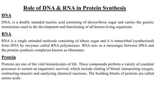
  • 1. Role of DNA & RNA in Protein Synthesis DNA DNA, is a double stranded nucleic acid consisting of deoxyribose sugar and carries the genetic instructions used in the development and functioning of all known living organisms. RNA RNA is a single stranded molecule consisting of ribose sugar and it is transcribed (synthesized) from DNA by enzymes called RNA polymerases. RNA acts as a messenger between DNA and the protein synthesis complexes known as ribosomes. Protein Proteins are one of the vital biomolecules of life. These compounds perform a variety of essential processes to sustain an organism's survival, which include clotting of blood, transporting oxygen, contracting muscles and catalyzing chemical reactions. The building blocks of proteins are called amino acids.
  • 2. A typical α-amino acid consists of an amino group (-NH2), carboxyl group (-COOH) and an R group. Amino acids in a protein chain are linked by a peptide bond. Usually, proteins have an N- terminal end carrying a free amino group and C-terminus with -COOH.
  • 3. Protein Synthesis The synthesis of proteins starts with transcribing the instructions in DNA into mRNA. The mRNA is then carried out of the cell's nucleus into the cytoplasm, specifically into structures called ribosomes. Protein production occurs in ribosomes containing rRNA. The tRNA transports the amino acids to the ribosomes. The code sequence in mRNA is then translated and specific proteins are synthesized by stringing amino acids together. The production or synthesis of polypeptide chains (proteins) includes two phases: Transcription & Translation
  • 4. Protein synthesis in prokaryotes and eukaryotes Eukaryotes In eukaryotes, mRNA is synthesized in the nucleus from pre-messenger RNA (pre mRNA) molecules, and then shipped to the cytoplasm, where translation occurs. Prokaryotes In prokaryotes, transcription and translation occur in the same cellular compartment — the cytosol. Ribosomes are the site of translation
  • 5. Transcription Transcription is the process through which a DNA sequence is enzymatically copied by an RNA polymerase to produce a complementary RNA(mRNA). In other words, it is the transfer of genetic information from DNA into RNA. During transcription, only one strand of a DNA molecule is transcribed; this strands is called antisense strand or template strand and the RNA so produced is termed as sense RNA. The other strand of the DNA duplex is know as coding strand or sense strand. As in DNA replication, transcription proceeds in the 5' → 3' direction (i.e. the old polymer is read in the 3' → 5' direction and the new, complementary fragments are generated in the 5' → 3' direction). Transcription, is catalyzed by DNA-directed RNA polymerase, or simply RNA polymerase.
  • 6. Process of Transcription Direction of Transcription The DNA splits into two strands:  Template strand: it is used to synthesize RNA.  Coding Strand: (Informational strand): it is not used to synthesize RNA. Transcription proceeds from the 3’ end to the 5’ end of the template.
  • 7. Transcription Process Transcription begins with the attachment of RNA polymerase holoenzyme to the promoter of transcription unit and it ends when the core enzyme reaches the terminator site and dissociates from the DNA . The transcription process is divided into the following steps: (1) initiation (2) elongation (3) termination. Transcription Initiation The initiation phase begins with the binding of RNA polymerase holoenzyme to the promoter and ends when the holoenzyme leaves the promoter. The initially the holoenzyme binds to the promoter DNA about 70-80 bp extending from - 55 to +20
  • 8. The holoenzyme now induces ‘melting’, i.e. strand separation, in a <17 bp region that includes the right end of the -10 sequence and extends just beyond the start point. The holoenzyme now begins to transcribe the antisense strand, RNA synthesis begins at the start point. The nucleotides used are riboside 5’-triphosphates and RNA synthesis progresses in 5ꞌ-3ꞌ direction. The enzyme permits the correct riboside to align opposite the deoxyribotides of antisense strand.
  • 9. When the first two riboside have been aligned, the enzyme catalyzes the formation of phosphodiester bond between them, the diribotide so formed remains associated with the template DNA strand and the enzyme. The holoenzyme sequentially adds the subsequent riboside to the growing RNA chain. The RNA chain remains as RNA∙DNA hybrid for ~2 to 3 nucleotides at its 3-end, i.e., the growing end , But ~25 nucleotide at the growing end of the RNA chain are associated with the template DNA and /or the enzyme.
  • 11. Elongation The elongation phase begins when the RNA polymerase leaves the promoter region and continues transcription of the template strand. As the core enzyme moves along the DNA duplex, the regions downstream of the start point progressively become single-stranded, the core enzyme sequentially adds the correct ribotides to the 3ꞌ-end of RNA chain. As the RNA chain elongates, its 5ꞌ-region progressively separates from the template strand. The sense and antisense strands of the DNA become progressively free as the transcription proceeds; these single strands progressively reassociate to form double helix.Thus elongation involves progressive disruption of the double helix to generate transient single-stranded regions; the separated strands form a transcription bubble within which the template strand is transcribed. The transcription bubble moves progressively downstream of the start point and disappears when transcription terminates.
  • 12. Termination When the core enzyme reaches terminator site, (1) No further ribotides are added to the RNA chain. (2)The RNA chain dissociates from the template strand of DNA. (3) the separated DNA strands reassociate to form a double helix. As a result, the Transcription bubble disappears. (4) the core enzyme dissociates from DNA.These events constitute the termination phase of transcription. The termination sites in prokaryotes have been classified into the following two groups: (1) rho – independent terminators, (2) rho – dependent terminators.
  • 13. Rho-Dependent Terminators At these terminators, a polypeptide called rho-factor is required for transcription termination. Most likely, rho-factor binds the RNA transcript at a recognition site (-50-90 bases long) that is located upstream of the termination site. Rho-Independent Terminator Terminator of transcription at these termination sites does not require rho factor; therefore, they are called intrinsic terminators. The RNA transcript from such terminators forms a typical hairpin loop, which is followed by a run of – 6U residues. The poly – U region probably signals the core enzyme to leave the DNA duplex.
  • 14. Transcription in eukaryotes RNA Polymerase All eukaryotes possess three type of RNA polymerases called RNA polymerase I,II and III . RNA polymerase I is located in nucleolus, and is responsible for transcription of genes for rRNA, it is responsible for 50-70% of the activity in eukaryotes. RNA polymerase II is located in the nucleoplasm, constitutes 20-40% of total activity and transcribes all the genes that produce mRNA. RNA polymerase III also occurs in nucleoplasm, provides ~10% of total polymerase activity, and transcribes tRNA and other small RNA genes.
  • 15. Process of transcription In eukaryotes transcription unit generally contains a single gene.Transcription termination occurs beyond the end of coding region.Transcription by RNA polymerase II is briefly described below. Transcription Complex The complex of proteins formed at the promoter that ultimately leads to transcription initiation is called transcription complex.
  • 16.
  • 17. Transcription Initiation Transcription begins at the start point, which is usually an A. A closed binary complex is formed when RNA polymerase II binds the promoter. The complex is then converted into an open binary complex in which the two strands of promoter DNA become separated locally. RNA polymerase now begins transcription at the start point, and presumably, the transcription factors are released around this stage. The basal transcription factors are able to initiate transcription at a low level. Transcription Termination It is likely that the transcription units have rather long terminator regions with multiple terminator sites. In some transcription units, termination occurs 1,000 bp downstream of the site corresponding to the 3' -end of mature mRNAs. In such cases, the 3' -ends of mRNAs are generated by cleavage following transcription.
  • 18. mRNA Processing  The process of modification, mainly through cleavage and /or splicing of primary RNA transcripts so as to produce functional mRNA molecules from them is called RNA processing.  RNA processing is carried out by ribonucleases, the enzymes that cleave RNA.  They are of two types ., Exoribonucleases and Endoribonucleases.  Exoribonucleases remove one base at a time from one end of RNA molecule, all know enzymes trim RNA from the 3' end But Endoribonucleases produce internal cuts in RNA molecules.  Eukaryotic transcripts (pre-mRNA) contains exons (coding/expressed sequences) and introns (non coding/ unexpressed sequences)  All the introns are excised from the primary transcript and all the exons of a gene are joined together in the proper order; this process is called splicing.
  • 19. Translation The mRNA carries genetic information encoded as a ribonucleotide sequence from the chromosomes to the ribosomes. The ribonucleotide are "read" by translational machinery in a sequence of nucleotide triplets called codons. Each of those triplets codes for a specific amino acid. Translation is the production of proteins by decoding mRNA produced in transcription. The ribosome and tRNA molecules translate this code to produce proteins. The ribosome is a multi subunit structure containing rRNA and proteins. It is the "factory" where amino acids are assembled into proteins.
  • 20. Genetic code The number and the sequence of bases in mRNA specifying an amino acid is known as codon. The set of bases in a tRNA that base-pairs with a codon of an mRNA is known as anticodon. The sequence of bases in an anticodon is exactly the opposite of and complementary to that present in the codon. For example the codon 5’AUG 3’ has the anticodon 3’ UAC 5’. The set of all the codons that specify the 20 amino acids is termed as the genetic code. Properties of the genetic code: The code is universal: All prokaryotic and eukaryotic organisms use the same codon to specify each amino acid. The code is triplet: Three nucleotides make one codon. 61 of them code for amino acids and 3 viz. , UAA, UAG and UGA are nonsense codons or chain termination codons.
  • 21. The code is degenerate: There are 64 codons available for 20 amino acids. Most amino acids are encoded by two or more codons. The code is non ambiguous: Each codon specifies only one of the 20 amino acids. None of the codons code for two or more amino acids. The only exception is the AUG codon which codes for formylmethionine in prokaryotes at the initiation site. While at other positions it specifies methionine. The code is non overlapping: A base in mRNA is not used for two different codons. The code is commaless: There is no special signal or commas between codons.
  • 22. Ribosome and Ribosomal RNA (rRNA) Ribosomal RNA (rRNA) occurs in association with proteins and is organized into almost spherical bodies of about 200 A° diameter called ribosomes. Ribosomes of prokaryotes are of 70 S size and are composed of 60% rRNA and 40% protein. They dissociate into a smaller 30 S subunit and a larger 50 S subunit. The 30S subunit of prokaryotic ribosomes has a single 16S rRNA molecule, which is associated with 21 different ‘S’ proteins. The larger 50S subunit has a 23S rRNA and a 5S rRNA molecule complexed with 31 different ‘L’ proteins. The basic structure of ribosomes is due to their rRNA, and the proteins play a secondary role. The cytoplasmic ribosomes of eukaryotes are of the size 80S and contain 40% rRNA and 60% protein. They consist of a 40S and a 60S subunit. The 40 S subunit of eukaryotic ribosomes has a single 18S rRNA molecule and 33 different proteins. The larger 60S subunit has one molecule each of 28S, 5.8S and 5S rRNA and 49 different proteins.
  • 23. Functional sites on Ribosomes A complete ribosome particle has two distinct and adjacent sites for the attachment of aminoacyl-tRNAs (amino acid carrying tRNA), an aminoacyl attachment site (site A) and a peptidyl site (site P). An aminoacyl-tRNA first attaches to site A, the kind of aminoacyl-tRNA being determined by the sequence of mRNA (codon) attached to the site A. the aminoacyl-tRNA, along with the mRNA codon, now moves to the site P, making the site A available for the attachment of a new aminoacyl-tRNA. In addition, the ribosome has following sites as well: 1.mRNA site 2.peptidyl transferase site 3.EF-Tu site 4.EF-G site 5. 5 S RNA site 6.exit site
  • 24.
  • 25. Transfer RNA (tRNA) The existence of tRNA was demonstrated by Hoagland and coworkers in 1957. Transfer RNA (tRNA) is a class of RNA of small size of 3S and generally, it has 76 to 95 nucleotides. tRNA molecules have a high proportion of unusual bases. The unusual bases present in tRNA are produced after transcription by modifications of the usual bases, A,U,G and C. Further, tRNA molecules show considerable double helical structure, and more than 50% of the bases in tRNA are paired. Structure of tRNA A ‘clover leaf model’ of tRNA structure has been proposed to account for its structural and functional properties. The three nucleotides at the 5 -end are CCA in all the types of tRNA molecules. This segment is the amino acid acceptor region. The amino acid binds to one of the OH groups of the ribose of the terminal adenine.The tRNA molecule has three loops: DHU loop, anticodon loop and thymine loop. Sometimes an extra loop, having a variable number of nucleotides, is also present between the anticodon and thymine loops.
  • 26.
  • 27. Formation of aminoacyl-tRNA The amino acids are attached to tRNA molecules through a two step reaction process. Both the reactions are catalysed by the same enzyme, aminoacyl tRNA synthetase. The carboxyl group of the amino acid reacts with one of the –OH groups of the ribose of the terminal adenine nucleotide to produce aminoacyl-tRNA . 1.first step: It consist of amino acid activation, in which the amino acid molecule reacts with and ATP molecule to yield an aminoacyl-AMP (aminoacyl adenylate) molecule. 2.Second step: The amino acid form aminoacyl-AMP molecule is then transferred to a tRNA molecule that is specific for the amino acid, and AMP is released. The aminoacyl-AMP complex remains tightly bound to the enzyme aminoacyl-tRNA synthetase during the entire reaction, i.e., both the steps. The attachment of an amino acid to the concerned tRNA to yield aminoacyl-tRNA is called charging of tRNA.
  • 28. Recognition of codons Recognition of specific codons of the mRNA is the function of tRNA molecule. This recognition is due to the anticodon that base pairs with the mRNA codon. Almost as a rule, A in the first place of anticodons (corresponding to the third base of mRNA codon) is modified to I (inosine); I base pairs with any one of U,C and A. Similarly, U in the first position of anticodon is always modified the modified U may base pair with A,G and, in some cases with even U.
  • 29. Translation Translation begins near the 5-end of mRNA and progresses toward its 3 -end. In terms of the polypeptide being synthesized, it starts with the N-terminal end of the polypeptide, while the C-terminal amino acid is the last to be added to the chain. The process of translation may be divided into the following 3 steps: (1) Initiation, (2) Elongation and (3) Termination
  • 30. Initiation Initiation comprises all the events that precede the formation of the first peptide bond. It includes the following events:  binding of the smaller subunit of ribosome to mRNA and  binding of the first or initiator aminoacyl-tRNA to the P site of ribosome; this completes the formation of initiation complex. Initiation is a relatively slow step in translation.
  • 31. Elongation Elongation includes all the reactions from the formation of the first peptide bond to that of the last peptide bond of the polypeptide chain. Amino acids are added one at a time to the growing peptide chain. It is the most rapid step of translation: in bacteria it proceeds at the rate of 15 amino acids/second at 370C, while in eukaryotes its rate is 2 amino acids/second.
  • 32. Elongation The elongation step in bacteria is briefly described below. The complete initiation complex has the following features: ribosome is complete and ready for translation, P site of ribosome is occupied by the initiator aminoacyl-tRNA (fmet-tRNAf) and The A site is free. The P site of ribosome must be occupied by peptidyl-tRNA (tRNA carrying a peptide for the A site to be in a proper conformation to allow entry to an amino-acyl-tRNA.
  • 33. Termination Termination describes those events that occur after the formation of the last peptide bond of the polypeptide, they culminate in the separation of the ribosome from mRNA. Termination is signaled by the nonsense codons UAG, UAA and UGA, and is mediated by certain proteins called release factors. As a result of this the following three simultaneous events occur: (1)the polypeptide chain detaches from the tRNA located at P site, (2) followed by immediate release of tRNA from the P site and (3) the release of ribosome from the mRNA.
  • 35. End product -protein The end products of protein synthesis is a primary structure of a protein A sequence of amino acid bonded together by peptide bonds
  • 36. Point Mutation A point mutation is a type of mutation in DNA or RNA, the cells genetic material, in which one single nucleotide base is added, deleted or changed. DNA and RNA are made up of many nucleotides. There are five different molecules that can make up nitrogenous bases on nucleotides: cytosine, guanine, adenine, thymine (in DNA) and uracil (in RNA), abbreviated C, G, A, T, and U. The specific sequence of nucleotides encodes all the information for carrying out all cell processes. In general, a mutation is when a gene is altered through a change in DNA structure; this may refer even to entire sections of chromosomes. A point mutation is specifically when only one nucleotide base is changed in some way, although multiple point mutations can occur in one strand of DNA or RNA.
  • 37. Effects of point mutation Sickle-Cell Anemia Sickle-cell anemia is a recessive disorder caused by a single substitution in the gene that creates hemoglobin, which carries oxygen in the blood. Normally, glutamic acid is produced in the chain, but the substitution causes valine to be produced at that spot instead. When people have two copies of this mutation, it results in thin sickle-shaped blood cells that sometimes cannot carry oxygen properly. About 80% of people with sickle-cell disease are in sub-Saharan Africa, where being a carrier for sickle-cell anemia (having only one copy of the gene, not two) actually helps protect against malaria. It is also found in other parts of the world such as India and the Middle East, and affects about 1 in 500 African Americans. Symptoms include anemia, obstruction of blood vessels, and chest pain, and it is treated with folic acid, blood transfusions, bone marrow transplants, and certain prescription drugs.
  • 38. Tay-Sachs Tay-Sachs disease is another recessive disorder caused by point mutations. Different mutations can cause this disorder, but they are all found on the HEXA gene on chromosome 15. Tay-Sachs causes nerve cells to deteriorate over time, which in turn results in the decline of physical and mental functioning. Both child and adult-onset forms of the disease occur, and children with the disease usually die before the age of four. About 1 in 320,000 newborns in the United States develop Tay-Sachs. It occurs in higher frequencies in Ashkenazi Jews, Cajuns, and French Canadians (about 1 in 3500 in these populations), although the mutations associated with the disease are different in each population. There is currently no treatment or cure.
  • 39. Inheritence of Cystic Fibrosis Cystic fibrosis is the most common inherited disease with a fatal outcome in industrialised nations, and is becoming increasingly prevalent as survival is prolonged. Thirty years ago most of these patients died in infancy but today about 36% reach adulthood. On the basis of epidemiological analysis we can expect children born with cystic fibrosis in 1990 to have a life expectancy of 40 years—in other words, more than 90% will reach adulthood.This will entail costly and time consuming therapy. As in other chronic pulmonary diseases, lung function parameters such as forced expiratory volume in one second (FEV1) are poor predictors of the degree of disability.
  • 40. Inheritence of Cystic Fibrosis There are more than 30,000 Americans with cystic fibrosis (CF), the most common autosomal recessive genetic disorder of Caucasians. CF patients born in 1970 had only a 50% chance of reaching age 15, but in little more than two decades the chances of reaching that age had increased to 95% . An increased understanding of normal airway physiology and the pathophysiology of CF has spawned new treatments that have driven this impressive increase in patient survival.
  • 41. Inheritence of gender The human genome is organized into 23 pairs of chromosomes (22 pairs of autosomes and one pair of sex chromosomes), with each parent contributing one chromosome per pair. The X and Y chromosomes, also known as the sex chromosomes, determine the biological sex of an individual: females inherit an X chromosome from the father for a XX genotype, while males inherit a Y chromosome from the father for a XY genotype (mothers only pass on X chromosomes). The presence or absence of the Y chromosome is critical because it contains the genes necessary to override the biological default female development.
  • 42. Inheritence of Sex linked Sex linked is a trait in which a gene is located on a sex chromosome. In humans, the term generally refers to traits that are influenced by genes on the X chromosome. This is because the X chromosome is large and contains many more genes than the smaller Y chromosome. In a sex-linked disease, it is usually males who are affected because they have a single copy of X chromosome that carries the mutation. In females, the effect of the mutation may be masked by the second healthy copy of the X chromosome.
  • 43.
  • 44. These are traits that are found on either one of the chromosomes that determine sex, or the sex chromosomes. And in humans this is the X or the Y chromosomes. And so some of the more familiar sex-linked traits are hemophilia, red-green color blindness, congenital night blindness, some high blood pressure genes, Duchenne muscular dystrophy, and also Fragile X syndrome. So what's also very interesting is that you can imagine that for individuals who are XY or males, having these different mutations on the genes, on the X chromosome, is particularly problematic, because unlike females, there are not two X chromosomes that give you the potential of carrying a normal gene on the X chromosome. Which is why in many cases you'll see that males are more often afflicted with these sex-linked disorders.
  • 45. Sex-linked disorder The existence of X-linked disorders in humans has been recognized for many centuries, based on lessons in religious texts and observations of specific human families (e.g., color blindness or Daltonism). Our modern concepts of Mendelian (including X-linked) inheritance originated just after the turn of the last century. Early concepts of dominance and recessiveness were first used in conjunction with autosomal traits,andthen applied to ‘‘sex’’-linked traits to distinguish X-linked recessive and X-linked dominant inheritance. The former was defined as vertical transmission in which carrierwomenpass the disorder to affected sons, while the latter was defined as vertical transmission in which daughters of affected malesare always affected, transmitting the disorder to offspring of both sexes.
  • 46. X-linked disorder X-linked disorders can be either dominant or recessive. X-linked dominant disorders are the result of a mutation to the X chromosome that can affect either males or females. However, some disorders, including Fragile X syndrome, affect males more severely than females, likely because males do not have a second, normal copy of the X chromosome. Fragile X syndrome is characterized by a wide range of developmental problems, including learning disabilities. X-linked hypophosphatemia is another X-linked dominant condition that manifests in a vitamin-D-resistant form of Rickets.
  • 47. Y-linked disorder Y chromosome mutations are called “Y-linked” and only affect males since they alone carry a copy of that chromosome. Mutations to the relatively small Y chromosome can impact male sexual function and secondary sex characteristics. Y-chromosome infertility is a disorder that affects sperm production, caused by deletions to the azoospermia factor (AZF) regions of the Y chromosome. In general, Y-linked disorders are only passed from father to son; however, because affected males typically do not father children without assisted reproductive technologies, Y-chromosome infertility is not typically passed on to offspring.
  • 48. Role of chromosomal abnormalities in an inherited disease Many human genetic disorders result from unbalanced chromosome abnormalities, in which there is a net gain or loss of genetic material. Such imbalances often disrupt large numbers of dosage-sensitive, developmentally important genes and result in specific and complex phenotypes. Alternately, some chromosomal syndromes may be caused by a deletion or duplication of a single gene with pleiotropic effects. Traditionally, chromosome abnormalities were identified by visual inspection of the chromosomes under a microscope. The use of molecular cytogenetic technologies, such as fluorescence in situ hybridization and microarrays, has allowed for the identification of cryptic or submicroscopic imbalances, which are not visible under the light microscope.
  • 49. Microarrays have allowed for the identification of numerous new syndromes through a genotype-first approach in which patients with the same or overlapping genomic alterations are identified and then the phenotypes are described. Because many chromosomal alterations are large and encompass numerous genes, the ascertainment of individuals with overlapping deletions and varying clinical features may allow researchers to narrow the region in which to search for candidate genes
  • 50. Down Syndrome Down syndrome (sometimes called Down’s syndrome) is a condition in which a child is born with an extra copy of their 21st chromosome — hence its other name, trisomy 21. This causes physical and mental developmental delays and disabilities Many of the disabilities are lifelong, and they can also shorten life expectancy. However, people with Down syndrome can live healthy and fulfilling lives.
  • 51. What causes Down syndrome? In all cases of reproduction, both parents pass their genes on to their children. These genes are carried in chromosomes. When the baby’s cells develop, each cell is supposed to receive 23 pairs of chromosomes, for 46 chromosomes total. Half of the chromosomes are from the mother, and half are from the father. In children with Down syndrome, one of the chromosomes doesn’t separate properly. The baby ends up with three copies, or an extra partial copy, of chromosome 21, instead of two. This extra chromosome causes problems as the brain and physical features develop. According to the National Down Syndrome Society (NDSS), about 1 in 700 babies in the United States is born with Down syndrome. It’s the most common genetic disorder in the United States.
  • 52. References  Massaia, A. and Xue, Y., 2017. Human Y chromosome copy number variation in the next generation sequencing era and beyond. Human Genetics, 136(5), pp.591-603.  Theisen, A. and Shaffer, L.G., 2010. Disorders caused by chromosome abnormalities. The application of clinical genetics, 3, p.159  MacIntyre, D.J., Blackwood, D.H.R., Porteous, D.J., Pickard, B.S. and Muir, W.J., 2003. Chromosomal abnormalities and mental illness. Molecular Psychiatry, 8(3), pp.275-287
  • 53.  Dobyns, W.B., Filauro, A., Tomson, B.N., Chan, A.S., Ho, A.W., Ting, N.T., Oosterwijk, J.C. and Ober, C., 2004. Inheritance of most X‐linked traits is not dominant or recessive, just X‐linked. American journal of medical genetics Part A, 129(2), pp.136-143  Staab, D., Wenninger, K., Gebert, N., Rupprath, K., Bisson, S., Trettin, M., Paul, K.D., Keller, K.M. and Wahn, U., 1998. Quality of life in patients with cystic fibrosis and their parents: what is important besides disease severity?. Thorax, 53(9), pp.727-731  Clunes, M.T. and Boucher, R.C., 2007. Cystic fibrosis: the mechanisms of pathogenesis of an inherited lung disorder. Drug Discovery Today: Disease Mechanisms, 4(2), pp.63-72  Taliaferro, W.H. and Huck, J.G., 1923. The inheritance of sickle-cell anaemia in man. Genetics, 8(6), p.594.