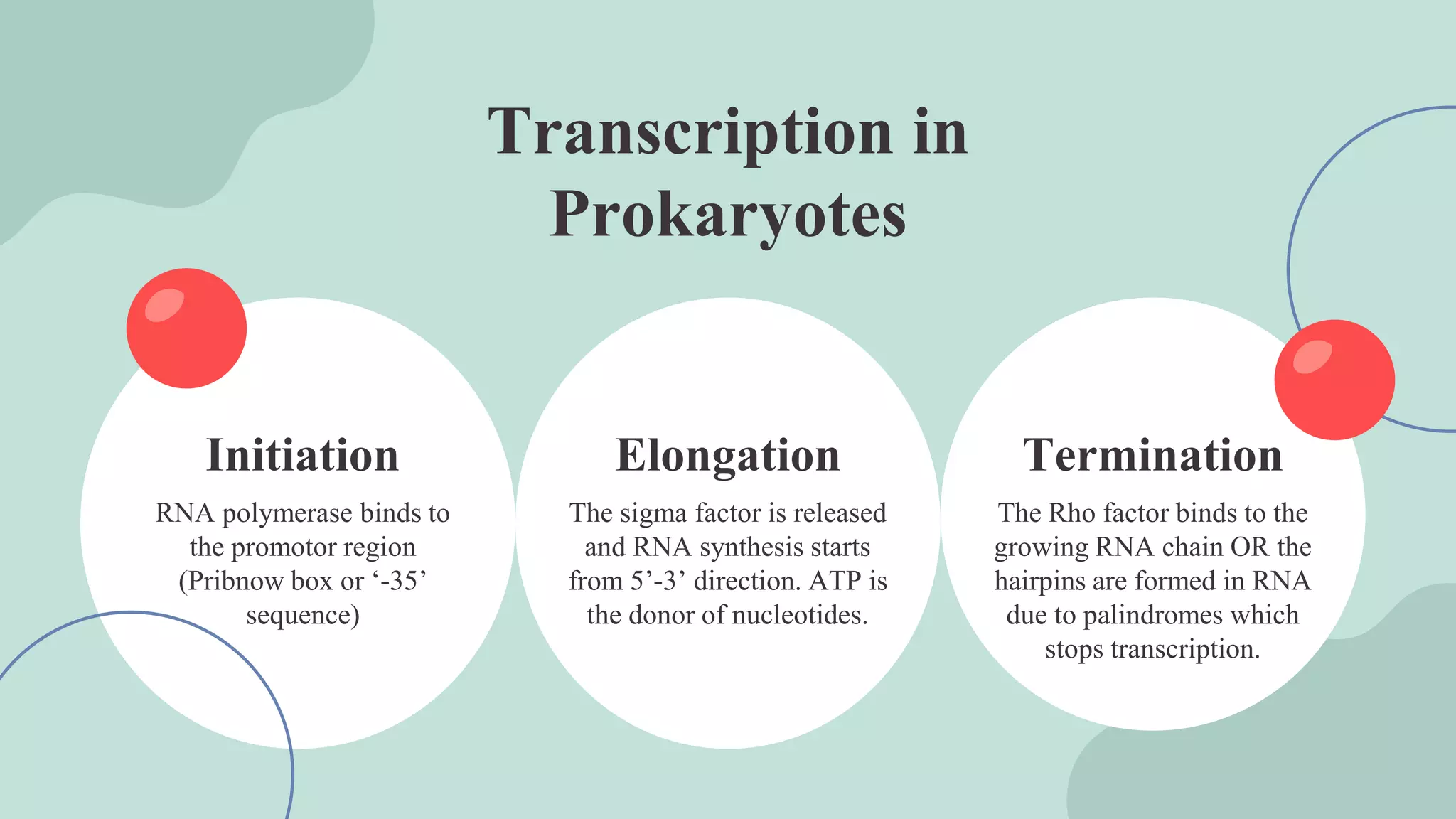
Department: Biochemistry
Submitted to: Dr- Ayesha Mushtaq
Dated: 6- 07 - 2023
STRUCTURAL
MECHANISM OF
PROTEIN
SYNTHESIS
BY: Laiba Saher
Biosynthesis of
Proteins
RNA Structure
Ribosomes Structure
Membrane Proteins
Structure
Table of Contents
01
Transcription, Translation, DNA
Structure.
02
You can describe the
topic of the section here
03 04
Detailed description of functions
and structures of mRNA, tRNA,
rRNA.
Detailed description of structure
and functions of Ribosomes.
Structural mechanism
of protein synthesis
With description of DNA
Structure, Transcription and
Translation
By. Laiba Saher
01
• DNA is a molecule which is carrier of
genetic information in nearly all
organisms.
• First discovered by the German
biochemist Frederich Miescher in the year
1869.
• Erwin Chargaff, James Watson, Francis
Crick, Maurice Wilkins and Rosalind
Franklin, discovered the structure of DNA
in the year 1953.
DNA
DNA: Structure
The human genome nearly consist of 3x10⁹ base pairs
With around 20,000 genes on 23 chromosomes
It is made up of subunits- nucleotides
Nucleotides= Ribose sugar+ base +phosphate
Sugar and phosphate forms backbone while bases forms
The ladders
Nucleotides are joined together by phosphodiester bond
Two strands interact to form double bond called double
helix.
It has directionality
-dictated by the free end groups
-always written 5’ to 3’ direction for each single strand
Percentage of complementary bases
28%
22%
Adenine and Thymine consist of
22+22=44% DNA
Cytosine and Guanine consist of
28+28=56% DNA
A=T C=G
Note: As the modern era is the bioinformatics era and
usually the sequencing of DNA takes place on computers.
DNA has 4 bases ( A,G,C,T) but ( N, R, Y) can also be seen
after sequencing.
N= any nucleotide, R= A or G, Y= C or T
Central Dogma
of Life
The process by which genome(DNA)
forms transcriptome(RNA) by
transcription and the transcriptome forms
proteome(proteins) by translation.
The process by which
Ribonucleic acid is
synthesized from DNA.
PROCESS
The biosynthesis of protein
or polypeptide in a living
cell.
Transcription Translation
1. The process by which RNA is formed from the DNA is known as Transcription
2. The genetic information stored in DNA is expressed through RNA
3. One of the two strands of DNA serves as template (non coding strand or Antisense strand)
4. The other serve as non template strand (coding strand or Sense strand)
5. Only selective region of DNA is transcribed due to presence of inbuilt signals
6. The product formed by transcripton is know as primary transcript which is inactive which undergoes
post transcriptional modification.
7. RNA polymerase is the enzyme that function in transcription
8. It has 5 subunits
- 2 alpha
- 2 beta
-1 sigma
9. There are 3 RNA polymrases for eukaryotes while only one in prokaryotes
-RNA polymerase I is responsible for synthesis of ribosomal RNA
-RNA polymerase II synthesizes the precursors for mRNAs and small nuclear RNAs
-RNA polymerase III synthesize the tRNA and small ribosomal RNAs
Transcription
Termination
RNA polymerase binds to
the promotor region
(Pribnow box or ‘-35’
sequence)
The Rho factor binds to the
growing RNA chain OR the
hairpins are formed in RNA
due to palindromes which
stops transcription.
The sigma factor is released
and RNA synthesis starts
from 5’-3’ direction. ATP is
the donor of nucleotides.
Initiation Elongation
Transcription in
Prokaryotes
Initiation
Elongation
Termination
1. Transcription in eukaryotes is more complex then prokaryotes due to the
presence of different RNA polymerases for respective RNAs
2. The promotor region on which RNA polymerase bind is Hogness box
(TATA box) located upstream 25 nucleotides away and the CAAT box
located in between 70 to 80 nucleotide
3. The primary mRNA transcript produced by RNA polymerase II is known as
heterogenous nuclear RNA (hnRNA). This is then processed to produce
mRNA needed for protein synthesis.
4. The RNA produced by tranascription undergoes several modification such as
terminal base additions, base modifications and splicing etc, to convert the
RNA into active forms
Transcription in Eukaryotes
Introns
removal
5’ end of the mRNA is
capped with 7-
methlyguanosine by 5’-5’
triphosphate linkage
The snRNPs associated
with hnRNA forms
splicesome which removes
the introns and joins the
exons
Poly A tail is added to the
3’ end which makes the
RNA stable
5’ capping Poly A tail
Post Transcriptional modification
of mRNA
1. Translation involves “decoding” a messenger RNA (mRNA) and using its information to build
a polypeptide, or chain of amino acids.
2. In an mRNA, the instructions for building a polypeptide come in groups of three nucleotides
called codons.
There are 61 different codons for amino acids
Three “stop” codons mark the polypeptide as finished
One codon, AUG, is a “start” signal to kick off translation (it also specifies the amino acid
methionine)
3. In translation, the codons of an mRNA are read in order (from the 5' end to the 3' end) by molecules
called transfer RNAs, or tRNAs.
4. Each tRNA has an anticodon, a set of three nucleotides that binds to a matching mRNA codon
through base pairing. The other end of the tRNA carries the amino acid that's specified by the
codon.
5. tRNAs bind to mRNAs inside of a protein-and-RNA structure called the ribosome.
6. As tRNAs enter slots in the ribosome and bind to codons, their amino acids are linked to the
growing polypeptide chain in a chemical reaction.
7. The end result is a polypeptide whose amino acid sequence mirrors the sequence of codons in the
mRNA.
TRANSLATION
STAGES OF TRANSLATION
02 03
01
Initiation
The ribosome gets
together with the
mRNA and the first
tRNA so translation
can begin.
Amino acids are
brought to the
ribosome by tRNAs
and linked together
to form a chain.
Propagation
The finished
polypeptide is
released to go and do
its job in the cell.
Termination
• The 80s ribosomes dissociate to form 40S and 60S subunit
• elF-1A and elF-3 binds to 40s
• Met-tRNAi and elF-2 bound to GTP attaches to 40S to form 43S
preinitiation complex
• 48S initiation complex is then formed by association of mRNA’s
5’GTP cap to elF-4F and elF-4B which reduce the complex structure
of mRNA and energy is supplied through GTP
• 48S initiation complex binds with the 60S ribosomal initiation
complex involving the elF-5
• As the 80S complex is formed the initiation factors bound to 48S are
released and recycled.
Step 1: Initiation
In bacteria, the situation is a little different. Here, the
small ribosomal subunit doesn't start at the 5' end of the
mRNA and travel toward the 3' end. Instead, it attaches
directly to certain sequences in the mRNA. Because the
bacterial genes are transcribed in groups called
operons and they are polycistronic. The Shine-
Dalgarno sequences come just before start codons and
"point them out" to the ribosome.
Initiation
• The 80S initiation complex contains met-tRNAi in the P-site and the
A-site is free
• Another amionacyl-tRNA is placed on the A-site
• Elongation factor EF-1a and GTP is involved
• As the aminoacyl-tRNA is placed in the A-site, EF-1a and GPT are
released and are recycled
• The enzyme peptidytransferase catalyses the formation of peptide
bond
• As the peptide bond formation occurs, the ribosome moves to the
next codon of the mRNA towards 3’ end and the process is known as
translocation and the growing peptide moves from A-site to P-site
• EF-2 and GTP is required
• The deacylated tRNA moves into E-site and leaves the ribosome
Step 2: Elongation
Elongation
• The termination signal (UAA, UAG and UCA) terminates the
growing polypeptide
• As the termination codon occupies the ribosomal A-site, the
release factor namely eRF recognizes the stop signal
• eRF-GTP complex, in association with the enzymr
prptidyltansferase cleave the peptide bond between the
polypeptide and the tRNA occupying P-site
• The reaction is hydrolysis reaction
• The 80S ribosome dissociates to form 40S and 60S subunits
which are recycled
• The mRNA is released
Step 3: Termination
Termination
Summary
RibonucleicAcid
Structure, functions, types,
roles
02
1. Ribonucleic acid (RNA), is quite similar to DNA However, whereas DNA molecules are typically
long and double stranded, RNA molecules are much shorter and are typically single stranded.
RNA molecules perform a variety of roles in the cell but are mainly involved in the process of
protein synthesis (translation) and its regulation. Ribonucleic acid is a polymeric molecule that is
essential for most biological functions, either by performing the function itself (Non-coding RNA)
or by forming a template for production of proteins (messenger RNA). RNA and deoxyribonucleic
acid (DNA) are nucleic acids. The nucleic acids constitute one of the four
major macromolecules essential for all known forms of life. RNA is assembled as a chain
of nucleotides. Cellular organisms use messenger RNA (mRNA) to convey genetic information
(using the nitrogenous bases of guanine, uracil, adenine, and cytosine, denoted by the letters G, U,
A, and C) that directs synthesis of specific proteins. Many viruses encode their genetic information
using an RNA genome.
2. Some RNA molecules play an active role within cells by catalyzing biological reactions,
controlling gene expression, or sensing and communicating responses to cellular signals. One of
these active processes is protein synthesis, a universal function in which RNA molecules direct the
synthesis of proteins on ribosomes. This process uses transfer RNA (tRNA) molecules to
deliver amino acids to the ribosome, where ribosomal RNA (rRNA) then links amino acids
together to form coded proteins.
RNA- overview
1. Each nucleotide in RNA contains a ribose sugar, with carbons numbered 1' through 5'. A base
is attached to the 1' position, in general, adenine (A), cytosine (C), guanine (G), or uracil (U).
Adenine and guanine are purines, cytosine and uracil are pyrimidines. A phosphate group is
attached to the 3' position of one ribose and the 5' position of the next. The phosphate groups have
a negative charge each, making RNA a charged molecule (polyanion). The bases form hydrogen
bonds between cytosine and guanine, between adenine and uracil and between guanine and uracil
. However, other interactions are possible, such as a group of adenine bases binding to each other
in a bulge, or the GNRA tetraloop that has a guanine–adenine base-pair .
2. An important structural component of RNA that distinguishes it from DNA is the presence of
a hydroxyl group at the 2' position of the ribose sugar. The presence of this functional group
causes the helix to mostly take the A-form geometry, although in single strand dinucleotide
contexts, RNA can rarely also adopt the B-form most commonly observed in DNA . The A-form
geometry results in a very deep and narrow major groove and a shallow and wide minor groove.A
second consequence of the presence of the 2'-hydroxyl group is that in conformationally flexible
regions of an RNA molecule (that is, not involved in formation of a double helix), it can
chemically attack the adjacent phosphodiester bond to cleave the backbone.
RNA- structure
Secondary structure of a telomerase RNA
Types of RNA
There are three
types of RNA
mRNA
A
tRNA
B
rRNA
C
• mRNA accounts for just 5% of the total RNA in the cell. mRNA is the most heterogeneous of the 3 types
of RNA in terms of both base sequence and size. It carries complementary genetic code copied, from DNA
during transcription, in the form of triplets of nucleotides called codons.
• Each codon specifies a particular amino acid, though one amino acid may be coded for by many different
codons. Although there are 64 possible codons or triplet bases in the genetic code, only 20 of them represent
amino acids. There are also 3 stop codons, which indicate that ribosomes should cease protein generation by
translation.
• As part of post-transcriptional processing in eukaryotes, the 5’ end of mRNA is capped with a guanosine
triphosphate nucleotide, which helps in mRNA recognition during translation or protein synthesis. Similarly,
the 3’ end of an mRNA has a poly-A tail or multiple adenylate residues added to it, which prevents enzymatic
degradation of mRNA. Both the 5’ and 3’ end of an mRNA imparts stability to the mRNA.
1. Messenger RNA-mRNA
• rRNAs are found in the ribosomes and account for 80% of the total RNA present in the cell.
Ribosomes are composed of a large subunit called the 50S and a small subunit called the 30S,
each of which is made up of its own specific rRNA molecules. Different rRNAs present in the
ribosomes include small rRNAs and large rRNAs, which belong to the small and large
subunits of the ribosome, respectively.
• rRNAs combine with proteins and enzymes in the cytoplasm to form ribosomes, which act as
the site of protein synthesis. These complex structures travel along the mRNA molecule
during translation and facilitate the assembly of amino acids to form a polypeptide chain.
They interact with tRNAs and other molecules that are crucial to protein synthesis.
• In bacteria, the small and large rRNAs contain about 1500 and 3000 nucleotides,respectively,
whereas in humans, they have about 1800 and 5000 nucleotides, respectively. However, the
structure and function of ribosomes is largely similar across all species.
2. Ribsomal RNA-rRNA
• tRNA is the smallest of the 3 types of RNA, possessing around 75-95 nucleotides. tRNAs
are an essential component of translation, where their main function is the transfer of amino
acids during protein synthesis. Therefore, they are called transfer RNAs.
• Each of the 20 amino acids has a specific tRNA that binds with it and transfers it to the
growing polypeptide chain. tRNAs also act as adapters in the translation of the genetic
sequence of mRNA into proteins. Thus, they are also called adapter molecules.
• tRNAs have a cloverleaf structure which is stabilized by strong hydrogen bonds between
the nucleotides. They normally contain some unusual bases in addition to the usual 4,
which are formed by methylation of the usual bases. Methyl guanine and methylcytosine
are two examples of methylated bases.
3. Transfer RNA-tRNA
Ribosomes
Structure, functions, diagram
and illustrations
03
1. Ribosomes are small, dense, membraneless, rounded, and granular ribonucleoprotein organelles
which occur either freely in the matrix of mitochondria, chloroplast, and cytoplasm or remain
attached to the membranes of the endoplasmic reticulum and nucleus.
2. The ribosomes occur in both Prokaryotic cells and Eukaryotic cells. In prokaryotic cells, the
ribosome often occurs freely scattered in the cytoplasm. In eukaryotic cells, the ribosome either
occurs freely in the cytoplasm (e.g.- yeast cells, lymphocytes, meristematic plant tissues,
embryonic nerve cells, etc.) or remains attached to the outer surface of the membrane of the
endoplasmic reticulum and nucleus (e.g.- pancreatic cells, plasma cells, hepatic parenchymal cells,
serous cells, Nissls bodies, etc). The cells in which active protein synthesis occurs, ribosomes
attach to the membranes of the endoplasmic reticulum.
3. When the ribosomes are not attached to the endoplasmic reticulum, they are called free ribosomes.
Free ribosomes serve as sites for the synthesis of proteins that are required for intracellular
utilization and storage.
4. Ribosomes are innumerable in a cell, especially in endoplasmic reticulum-containing cells. A
single cell of E. Coli contains 20000-30000 ribosomes.
5. In yeast cells, at the base of gland cells, in plasma and liver cells, in nerve cells, and in all rapidly
growing plant and animal cells, they are in large numbers.
Ribosomes
1. Ribosomes are oblate spheroid structures and the most abundant organelles of a cell. Each ribosome is about 250 Å
in diameter
2. In the electron microscopic studies, negative staining reveals that each ribosome is porous, hydrated, and composed
of two unequal sub-units, one is larger and the other is a smaller subunit. The larger subunit is dome-shaped or cup-
shaped (140-160 Å), while the smaller one is oblate-ellipsoid (90-110 Å). The smaller subunit is about half the size
of the larger subunit and occurs above the larger subunit to form a cap-like structure.
3. Each 70S ribosome consists of two subunits as 50S and 30S. The larger 50S subunit has a molecular weight of 1.8
× 106 daltons, while the molecular weight of the smaller 30S subunit is about 0.9 × 10⁶ daltons. On the other hand,
the 80S ribosome also consists of two subunits as 60S and 30S. The molecular weight of the 60S subunit is 1.5-1.8
× 106 daltons and 3-3.5 × 10⁶ daltons in the 40S subunit.
4. The two structural (cup and cap) ribosomal subunits remain united with each other due to the high
concentration(0.001M) of the Mg2+ ions. When the concentration of Mg2+ ions reduces in the matrix of the
cytoplasm, both subunits get separated. At a high concentration of Mg2+ ions in the cytoplasmic matrix, the two
ribosomes become associated with each other to form the dimer.
5. Polyribosome: During protein synthesis, many ribosomes bind to an individual mRNA (messenger RNA) mole-
cule forming a polyribosome or polysome or ergosome.
Structure of Ribosomes
6. The ribosomes are composed of highly flooded ribosomal RNA(rRNA) and many attached
proteins. The RNA and proteins are intertwined and arranged in a complex manner in the form
of two subunits.
7. Various models have been introduced regarding the structure of ribosomes. Among them, J.
A. Lake given asymmetrical model (also known as Lake’s model) is the most common and
universally accepted.
8. According to this model, the smaller subunit has three parts- a head, a base, and a platform.
The platform separates the head from the base by a cleft. On the other, the larger subunit also
consists of three parts- a ridge, a central protuberance, and a stalk. The first two are separated
with the help of a valley.
9. Three special regions are seen between the two subunits of a ribosome. These are aminoacyl
site (A-site) or acceptor site, peptidyl site (P-site), and exit side (E-site). During the protein
synthesis, aminoacyl-tRNA bounded to the ribosome at the A-site, peptidyl-tRNA bounded to
the ribosome at the p- site, and while at E-site, the deacylated-tRNA exits from the ribosome.
Citation ( Cold Spring Laboratory Press , 2016)
39%
Ribosomal RNA
rRNA
60%
Ribosomal Proteins
Proteins
Lipids
Traces
1%
Chemical Composition
Ribosomal RNAs
1. More than half of the weight of the ribosome is ribosomal RNA(rRNA). In prokaryotes, the ribosomes contain 66% of
rRNA and eukaryotic ribosomes contain 60% of rRNA.
The 70S ribosomes contain three types of rRNA:
1. 23S rRNA(3300 nucleotides)
2. 16S rRNA(1650 nucleotides)
3. 5S rRNA(120 nucleotides)
4. The larger 50S ribosomal subunit contains 23S and 5S rRNA, while the 16S rRNA occurs in the smaller 30S subunit.
The 80S ribosomes possess four types of rRNA:
1. 28S rRNA(4700 nucleotides)
2. 18S rRNA(1900 nucleotides)
3. 5.8S rRNA(160 nucleotides)
4. 5S rRNA(120 nucleotides)
5. The 28S, 5S, and 5.8S rRNAs are present in the larger 60S ribosomal subunit and the smaller 40S subunit contains 18S
rRNA
Ribosomal Proteins
1. The ribosomal proteins may be basic, structural, or enzymatic in function. The ribosomal proteins of smaller ribosomal
subunits are termed S-proteins, while L-proteins are in larger ribosomal subunits.
2. In addition to rRNA and proteins, ribosomes also contain some divalent metallic ions, such as Mg2+, Ca2+, and Mn2+.
Chemical Composition
Types of Ribosomes
According to
size, volume
and compositon
80s
A
70s
1. The size, volume, structure, composition, etc. Of the ribosome of an organism remains the same in different types
of cells. Based on the size and Sedimentation coefficient (S), Ribosomes are basically two types- 70S ribosomes
and 80S ribosomes.
2. The sedimentation coefficient is expressed in the Svedberg unit i.e., the S unit. Svedberg units are not directly
additive, they represent a rate of sedimentation, not weight.
3. S= 1×10⁻¹³ cm/s/dyne/gm
70S Ribosome
1. They are relatively smaller in size and have a sedimentation coefficient of the 70S. This type of ribosome consists
of a large 50S subunit and a small 30S subunit.
2. They occur in the prokaryotic cells (e.g.- bacteria, blue-green algae) and also in mitochondria and chloroplasts of
eukaryotic cells.
3. The molecular weight of the 70S ribosome is 2.5×10⁶ daltons and the dimension of each ribosome is about 200-
290 Å × 170-210 Å.
80S Ribosome
1. They are relatively larger in size than 70S ribosomes and are mainly found in the cytoplasm of eukaryotic cells
(plant and animal cells).
2. The sedimentation coefficient of this type of ribosome is the 80S. Each 80S ribosome is made up of two subunits
the smaller 40S subunit remains attached to a larger 60S subunit like a cap. Their molecular weight is 2.7 × 10⁶
daltons and the dimension of 300-340 Å × 200-240 Å.
Types of Ribosomes
• Other Forms of Ribosomes
• The ribosomes are also divided into the following types based on their location:
•
• Bacterial ribosome: This type of ribosome is found in bacterial cells and it has a sedimentation
coefficient of the 70S.
• Mitochondrial ribosome: The ribosomes which are found in mitochondria are called mitoribosomes.
They are many types, like- the mitoribosomes of fungus are 73S type, plants have 78S type of
mitoribosomes (e.g.- Corn), while the mitoribosomes of mammals are of the 60S. However,
mitoribosomes are generally considered to be of the 70S.
• Plastidial ribosome: The ribosomes found in chloroplasts are called plastidial ribosomes or
plastidoribosomes. The plastidoribosomes are the 70S in the higher plants.
• Nuclear ribosome: The ribonucleoprotein particles found in the nucleus are called nuclear ribosomes.
• Nucleolar ribosome: The RNP (ribonucleoprotein) particles of 150-200 Å in diameter found in the
nucleolus are called nucleolar ribosomes.
• Cytoplasmic ribosome: The large ribosome particles of the cytoplasm are called cytoplasmic
ribosomes.
Other forms of Ribosomes
1. Protein synthesis: The principal function of a ribosome is protein synthesis(translation-process of
synthesizing proteins). In all living cells, ribosome serves as the site of biological protein synthesis
2. Transport the synthesized proteins: Ribosomes also function as the transporter of the
synthesized proteins.
3. Helps in protoplasm formation: The free ribosomes present in the cytoplasm synthesize various
proteins that help in the formation of protoplasm.
4. As catalysts: Ribosome acts as catalysts in the biological processes of peptidyl transfer and
peptidyl hydrolysis.
5. Plays a protective role: The newly synthesized polypeptide chains passing through the tunnel or
channel of the larger subunit of the ribosome are protected against the action of protein-digesting
enzymes.
Citation ( Durgeshwer Singh , 2014 Apr )
Functions of Ribosomes
Membrane proteins
Structural and Functional
illustration
04
1. Transport proteins, also known as transmembrane proteins, are membrane proteins that aid in the
facilitated diffusion or active transport of ions across the hydrophobic lipid bilayer.
2. Such proteins include channel proteins, carrier proteins, sodium-potassium pumps, GLUT1, proton
pump, calcium ATPase, and others.
3. A membrane transport protein (or simply transporter) is a membrane protein involved in the
movement of ions, small molecules, and macromolecules, such as another protein, across a
biological membrane. Transport proteins are integral transmembrane proteins; that is they exist
permanently within and span the membrane across which they transport substances.
4. The proteins may assist in the movement of substances by facilitated diffusion or active transport.
5. The two main types of proteins involved in such transport are broadly categorized as either
channels or carriers.
6. The solute carriers and atypical SLCs are secondary active or facilitative transporters in humans.
7. Collectively membrane transporters and channels are known as the transportome. Transportomes
govern cellular influx and efflux of not only ions and nutrients but drugs as well.
1. Membrane Transport Protein
Every carrier protein, especially within the same cell membrane, is specific to one type or family of
molecules. For example, GLUT1 is a named carrier protein found in almost all animal cell
membranes that transports glucose across the bilayer. Other specific carrier proteins also help the
body function in important ways. Cytochromes operate in the electron transport chain as carrier
proteins for electrons.
Example 1
1. The Sodium-Potassium Pump
 The most famous example of a primary active transport protein is the sodium-potassium pump.
It is this pump that creates the ion gradient that allows neurons to fire.
 The sodium-potassium pump begins with its sodium binding sites facing the inside of the cell.
These sites attract sodium ions and hold onto them.
 When each of its three sodium binding sites has bound a sodium ion, the protein then binds to a
molecule of ATP, and splits it into ADP + a phosphate group. The protein uses the energy released in
that process to change shape.
 Now, the sodium binding sites are facing the extracellular solution. They release the three
sodium ions outside of the cell, while the protein’s potassium-binding sites bind to two potassium
ions.
 When both potassium-binding sites are full, the protein reverts to its original shape. Now the
potassium ions are released inside of the cell, and the empty sodium binding sites can bind more
sodium ions.
For each ATP this pump uses, it transports three positively charged ions outside of the cell, while
transporting only two back into it. This creates an electrochemical gradient, with the inside of the
cell being negatively charged relative to the outside solution. It also creates a strong concentration
gradient, with much more potassium inside the cell and much more sodium outside of it.
When the time comes for a nerve cell to fire, the strong electrical and chemical gradients allow the
cell to produce a huge, instant change by opening its voltage-gated ion channels.
 2.Sodium-Glucose Transport Proteins
 The sodium-glucose transport protein uses secondary active transport to move glucose into
cells. They are active in intestinal cells and kidney cells, both of which need to move
glucose into the body’s systems against its concentration gradient.
 This operation requires energy, because the cells in question have a higher concentration
of glucose than the extracellular fluid. Therefore, it would be impossible for glucose to
diffuse into the cells on its own; energy must be applied.
 In this case, the energy comes from the concentration gradient of sodium. Thanks to the
action of the sodium-potassium pump, there is much more sodium outside of the cell than
inside of it. There is a strong concentration gradient, then, favoring the movement of
sodium into the cell.
 This concentration gradient can be thought of as a type of “stored energy.” The sodium-
potassium pump takes energy from ATP and turns it into this concentration gradient, which
can then be used for other purposes, such as the sodium-glucose transport protein.
Example 2
• 3.Gated Ion Channels in the Cochlea
• Gated ion channels are passive transport proteins that open in response to specific
stimuli. You may be familiar with voltage-gated ion channels, such as those that
cause our neurons to fire in response to input from other neurons.
• Less well-known are the gated ion channels of the cochlea – which are opened by
mechanical pressure instead of voltage changes. These remarkable ion channels
allow the nerves of our inner ear to fire in response to the vibrations of sound. This
is how we hear.
• In the cochlea, special cells called “hair cells” are responsible for our hearing.
“Outer hair cells” sway in response to sound waves, amplifying their vibrations.
• Inner hair cells, on the other hand, have a very special job. In response to these
vibrations, they open ion channels in their cell membranes and release
neurotransmitters – just like a neuron would.
• Those neurotransmitters cause the firing of adjoining nerves. And that is how sound
is converted into neural impulses!
Example 3
1. An integral, or intrinsic, membrane protein (IMP) is a type of membrane
protein that is permanently attached to the biological membrane.
2. All transmembrane proteins are IMPs, but not all IMPs are transmembrane proteins.
3. IMPs comprise a significant fraction of the proteins encoded in an
organism's genome.
4. Proteins that cross the membrane are surrounded by annular lipids, which are defined
as lipids that are in direct contact with a membrane protein.
5. Such proteins can only be separated from the membranes by
using detergents, nonpolar solvents, or sometimes denaturing agents.
2- INTEGRAL PROTEINS
Types of IMPs
Integral
membrane
proteins can be
divided into two
groups
Integral
monotropic
proteins
A
Integral
polytropic
proteins
 Transmembrane protein
The most common type of IMP is the transmembrane protein (TM), which spans the entire biological
membrane. Single-pass membrane proteins cross the membrane only once, while multi-
pass membrane proteins weave in and out, crossing several times. Single pass TM proteins can be
categorized as Type I, which are positioned such that their carboxyl-terminus is towards the cytosol, or
Type II, which have their amino-terminus towards the cytosol. Type III proteins have multiple
transmembrane domains in a single polypeptide, while type IV consists of several different
polypeptides assembled together in a channel through the membrane. Type V proteins are anchored to
the lipid bilayer through covalently linked lipids. Finally Type VI proteins have both transmembrane
domains and lipid anchors.
Integral polytopic
• Integral monotopic proteins are associated with the membrane from one side but do not
span the lipid bilayer completely.
• Examples of integral membrane proteins:
• Insulin receptor
• Some types of cell adhesion proteins or cell adhesion molecules (CAMs) such
as integrins, cadherins, NCAMs, or selectins
• Some types of receptor proteins
• Glycophorin
• Rhodopsin
• Band 3
• CD36
• Glucose Permease
• Ion channels and Gates
• Gap junction Proteins
• G protein coupled receptors (e.g., Beta-adrenergic receptor)
• Seipin
Integral monotopic:
1. A protein that temporarily adheres to the biological membrane, either to the lipid bilayer or to
integral proteins by a combination of hydrophobic, electrostatic, and other non-covalent
interactions.
 What Are Peripheral Proteins
1. Peripheral protein, or peripheral membrane proteins, are a group of biologically active molecules
formed from amino acids which interact with the surface of the lipid bilayer of cell membranes.
2. Unlike integral membrane proteins, peripheral proteins do not enter into the hydrophobic space
within the cell membrane. Instead, peripheral proteins have specific sequences of amino acids
which allow them to attract to the phosphate heads of the lipid molecules or to integral proteins.
3. The ability to attach to the membrane but not be locked to it allows peripheral proteins to work on
the surface of the cell membrane. Peripheral proteins can be activated or disabled through a
number of different pathways.
4. Many peripheral proteins are also a part of many complex biochemical pathways. They can be
involved in moving substances within or outside of a cell, activate other proteins and enzymes, or
be involved in cell to cell interactions
3- Peripheral Membrane Protein
• Alternative Oxidase
• Alternative oxidases (AOXs) are terminal quinol oxidases that are attached to membranes
and are found in the respiratory electron chains of many species from different kingdoms.
• Since there is no human equivalent for AOXs, they have been studied in depth as drug
targets for dangerous fungi and the human parasite Trypanosoma brucei (T. brucei).
• African trypanosomiasis, also called “sleeping sickness,” is caused by parasites called T.
brucei. If it isn’t treated, it will kill you.
• AOXs are very important to the way these pathogens use energy, so they have become a
new therapeutic target for treating diseases caused by these pathogens.
• AOXs speed up the process of turning oxygen into water, even in the presence of cyanide,
without the need to move protons across the inner mitochondrial membrane.
• By doing this, they act as a part of the respiratory electron transport chain that doesn’t save
energy.
• Iron is important for AOXs to work well. So, to be active, AOXs must interact with oxygen,
a membrane, quinols, and iron.
• Crystal structures of AOXs showed that these enzymes are homodimers, with six -helices in
each monomer. Four of these -helices come together to make a 4-helix bundle, which holds
two iron atoms together
Example 1:
• Cytochrome c
• Cytochrome c (cyt c) comes in two forms: one that is attached to the cell membrane and one that is
soluble.
• Cyt c is a small, highly basic hemoprotein that is found in the space between the membranes of
mitochondria (the cytoplasmic membrane of bacteria). In human mitochondria, it has two biological
functions.
• The main job of cyt c is to help move electrons from cyt c reductase, which is part of the bc1 complex,
to IMP cytochrome c oxidase.
• The first step of this reaction is for the soluble cyt c to move toward the membrane-bound COX. This is
made easier by the long-range electrostatic interaction between cytochrome c and the membrane.
• The release of cyt c from the mitochondria into the cytosol starts the apoptotic pathway. This is the
second biological function of cyt c.
• When cyt c binds to the inner membrane of the mitochondria, it not only speeds up the transfer of
electrons in the respiratory chain, but it also keeps the cell from dying in a process called apoptosis.
• It has been thought for a long time that the permeability transition pore is how mitochondrial cyt c gets
out.
Example 2:
The dimeric F1Fo ATP synthase, which is part of the respiratory chain that cyt c is a part of, has recently been
linked to the permeability transition pore. This is a new discovery that may help explain how cyt c gets out of
mitochondria.
But cyt c is also an interesting target in, for example, cancer treatment because the release of cyt c from
mitochondria into the cytosol starts the apoptosis pathway.
Targeted delivery of cyt c to cancer cells is now thought to be a possible way to cause apoptosis and kill the cancer
cells.
As was already said, cyt c needs to be drawn to the membrane so that it can do its main job in the body.
Cardiolipin, one of the lipids that make up the mitochondrial membrane, helps this attraction happen (CL).
4- Transmembrane Protein
What are Transmembrane Proteins
1. Approximately one-third of all sequence-encoded proteins in living organisms are membrane proteins. They play an
irreplaceable role in molecular transport, energy utilization, signal transduction, and maintenance of membrane
homeostasis in cells.
2. Based on their structure, membrane proteins can be classified into three types: peripheral, lipid-anchored, or integral.
Integral membrane proteins are further classified as either polytopic or monotopic (see figure below). Transmembrane
proteins are amphoteric molecules embedded in the interior of lipid bilayers to varying degrees, resulting in different
membrane topologies according to the orientation or location of the N- and C-termini of the protein relative to the inner or
outer surfaces of the cell membrane. The first three types in the figure below depict widely observed structural patterns
found in integral membrane proteins: transmembrane α-helix protein, transmembrane α-helical protein, and
transmembrane β-barrel protein.
● Transmembrane proteins can be classified according to their functions, including G protein-
coupled receptors (GPCRs), ion channels, transport proteins, and other types of receptors.
● These proteins have diverse cellular functions, such as signal transmission, substance transport,
and cell communication. GPCRs are widespread in organisms.
● They recognize and interact with external molecules, thereby eliciting various intracellular
signals, making them useful targets for drug screening. Ion channels control the intracellular and
extracellular levels of ions, such as sodium, potassium, calcium, which are essential for maintaining
cellular integrity.
● Transport proteins are specialized proteins that facilitate the movement of molecules and ions
across biological membranes and are critical for regulating metabolic processes within organisms.
Types and Functions of Transmembrane Proteins
• Multipass transmembrane proteins are crucial components of biological systems. However,
comprehensive investigations have been limited due to the challenges associated with expression
and bioactivity in vitro. Appropriate expression vectors, host cells, culture conditions, and
purification methods are necessary to optimize the expression and purification processes, such as
the addition of auxiliary proteins and purification using tags, such as FLAG and Strep. These
minimize the occurrence of hydrolysis and conformational abnormalities, and thus, we can
obtain highly efficient transmembrane protein products with high purity and the correct
conformation.
• Transmembrane Protein Expression Systems
• There are four commonly used transmembrane protein expression systems: E. coli, yeast, insect
cells, and mammalian cells. Numerous factors must be considered when choosing an appropriate
expression system as they may increase the complexity of the research project and make it time-
consuming.
• Advantages and disadvantages of transmembrane protein expression systems
Expression and Production of Transmembrane Proteins
The involvement of transmembrane proteins in various cellular processes and disease mechanisms make
them suitable targets for drug research and development. However, transmembrane protein synthesis faces
many bottlenecks, including low levels of expression of membrane proteins, poor stability, low solubility,
and problems encountered during purification.
● Low expression: Transmembrane proteins are usually expressed at lower levels compared with
cytoplasmic proteins. Correct folding patterns and embedding on cell membrane sites are required for
proper function.
● Complex structure: Transmembrane proteins are complex structures containing multiple
transmembrane regions, ring structures, and glycosylated modifications, which increase the difficulty of
transmembrane protein preparation.
● Poor stability: Transmembrane proteins with poor stability pose a significant challenge when
studying their structure and function, and this can be due to several reasons. For example, the hydrophobic
segments of these proteins, which anchor them to the lipid bilayer of the membrane, may not be
sufficiently strong to maintain its structural integrity.
Challenges in Transmembrane Protein Preparation
Photos:
https://commons.wikimedia.org/wiki/File:Translation_1543.gif
https://commons.wikimedia.org/wiki/File:DNA_Transcription_and_Translation.gif
https://gfycat.com/gifs/search/dna+transcription+animation
https://proteopedia.org/
Citation:
• Chapeville, F., et al. On the role of soluble ribonucleic acid in coding for amino acids. Proceedings of the National
Academy of Sciences 48, 1086–1092 (1962)
• Crick, F. On protein synthesis. Symposia of the Society for Experimental Biology 12, 138–163 (1958) Flinta, C., et al.
Sequence determinants of N-terminal protein processing. European Journal of Biochemistry 154, 193–196 (1986)
• Grunberger, D., et al. Codon recognition by enzymatically mischarged valine transfer ribonucleic acid. Science 166,
1635–1637 (1969) doi:10.1126/science.166.3913.1635
• Khan academy
• Bioinformatics for dummies
References

DNA- Transcription and Tranlation, RNA, Ribosomes and membrane proteins.pptx

  • 1.
    Department: Biochemistry Submitted to:Dr- Ayesha Mushtaq Dated: 6- 07 - 2023
  • 2.
  • 3.
    Biosynthesis of Proteins RNA Structure RibosomesStructure Membrane Proteins Structure Table of Contents 01 Transcription, Translation, DNA Structure. 02 You can describe the topic of the section here 03 04 Detailed description of functions and structures of mRNA, tRNA, rRNA. Detailed description of structure and functions of Ribosomes.
  • 4.
    Structural mechanism of proteinsynthesis With description of DNA Structure, Transcription and Translation By. Laiba Saher 01
  • 5.
    • DNA isa molecule which is carrier of genetic information in nearly all organisms. • First discovered by the German biochemist Frederich Miescher in the year 1869. • Erwin Chargaff, James Watson, Francis Crick, Maurice Wilkins and Rosalind Franklin, discovered the structure of DNA in the year 1953. DNA
  • 6.
    DNA: Structure The humangenome nearly consist of 3x10⁹ base pairs With around 20,000 genes on 23 chromosomes It is made up of subunits- nucleotides Nucleotides= Ribose sugar+ base +phosphate Sugar and phosphate forms backbone while bases forms The ladders Nucleotides are joined together by phosphodiester bond Two strands interact to form double bond called double helix. It has directionality -dictated by the free end groups -always written 5’ to 3’ direction for each single strand
  • 7.
    Percentage of complementarybases 28% 22% Adenine and Thymine consist of 22+22=44% DNA Cytosine and Guanine consist of 28+28=56% DNA A=T C=G
  • 8.
    Note: As themodern era is the bioinformatics era and usually the sequencing of DNA takes place on computers. DNA has 4 bases ( A,G,C,T) but ( N, R, Y) can also be seen after sequencing. N= any nucleotide, R= A or G, Y= C or T
  • 9.
    Central Dogma of Life Theprocess by which genome(DNA) forms transcriptome(RNA) by transcription and the transcriptome forms proteome(proteins) by translation.
  • 10.
    The process bywhich Ribonucleic acid is synthesized from DNA. PROCESS The biosynthesis of protein or polypeptide in a living cell. Transcription Translation
  • 11.
    1. The processby which RNA is formed from the DNA is known as Transcription 2. The genetic information stored in DNA is expressed through RNA 3. One of the two strands of DNA serves as template (non coding strand or Antisense strand) 4. The other serve as non template strand (coding strand or Sense strand) 5. Only selective region of DNA is transcribed due to presence of inbuilt signals 6. The product formed by transcripton is know as primary transcript which is inactive which undergoes post transcriptional modification. 7. RNA polymerase is the enzyme that function in transcription 8. It has 5 subunits - 2 alpha - 2 beta -1 sigma 9. There are 3 RNA polymrases for eukaryotes while only one in prokaryotes -RNA polymerase I is responsible for synthesis of ribosomal RNA -RNA polymerase II synthesizes the precursors for mRNAs and small nuclear RNAs -RNA polymerase III synthesize the tRNA and small ribosomal RNAs Transcription
  • 12.
    Termination RNA polymerase bindsto the promotor region (Pribnow box or ‘-35’ sequence) The Rho factor binds to the growing RNA chain OR the hairpins are formed in RNA due to palindromes which stops transcription. The sigma factor is released and RNA synthesis starts from 5’-3’ direction. ATP is the donor of nucleotides. Initiation Elongation Transcription in Prokaryotes
  • 13.
  • 14.
  • 15.
  • 16.
    1. Transcription ineukaryotes is more complex then prokaryotes due to the presence of different RNA polymerases for respective RNAs 2. The promotor region on which RNA polymerase bind is Hogness box (TATA box) located upstream 25 nucleotides away and the CAAT box located in between 70 to 80 nucleotide 3. The primary mRNA transcript produced by RNA polymerase II is known as heterogenous nuclear RNA (hnRNA). This is then processed to produce mRNA needed for protein synthesis. 4. The RNA produced by tranascription undergoes several modification such as terminal base additions, base modifications and splicing etc, to convert the RNA into active forms Transcription in Eukaryotes
  • 17.
    Introns removal 5’ end ofthe mRNA is capped with 7- methlyguanosine by 5’-5’ triphosphate linkage The snRNPs associated with hnRNA forms splicesome which removes the introns and joins the exons Poly A tail is added to the 3’ end which makes the RNA stable 5’ capping Poly A tail Post Transcriptional modification of mRNA
  • 18.
    1. Translation involves“decoding” a messenger RNA (mRNA) and using its information to build a polypeptide, or chain of amino acids. 2. In an mRNA, the instructions for building a polypeptide come in groups of three nucleotides called codons. There are 61 different codons for amino acids Three “stop” codons mark the polypeptide as finished One codon, AUG, is a “start” signal to kick off translation (it also specifies the amino acid methionine) 3. In translation, the codons of an mRNA are read in order (from the 5' end to the 3' end) by molecules called transfer RNAs, or tRNAs. 4. Each tRNA has an anticodon, a set of three nucleotides that binds to a matching mRNA codon through base pairing. The other end of the tRNA carries the amino acid that's specified by the codon. 5. tRNAs bind to mRNAs inside of a protein-and-RNA structure called the ribosome. 6. As tRNAs enter slots in the ribosome and bind to codons, their amino acids are linked to the growing polypeptide chain in a chemical reaction. 7. The end result is a polypeptide whose amino acid sequence mirrors the sequence of codons in the mRNA. TRANSLATION
  • 19.
    STAGES OF TRANSLATION 0203 01 Initiation The ribosome gets together with the mRNA and the first tRNA so translation can begin. Amino acids are brought to the ribosome by tRNAs and linked together to form a chain. Propagation The finished polypeptide is released to go and do its job in the cell. Termination
  • 20.
    • The 80sribosomes dissociate to form 40S and 60S subunit • elF-1A and elF-3 binds to 40s • Met-tRNAi and elF-2 bound to GTP attaches to 40S to form 43S preinitiation complex • 48S initiation complex is then formed by association of mRNA’s 5’GTP cap to elF-4F and elF-4B which reduce the complex structure of mRNA and energy is supplied through GTP • 48S initiation complex binds with the 60S ribosomal initiation complex involving the elF-5 • As the 80S complex is formed the initiation factors bound to 48S are released and recycled. Step 1: Initiation
  • 21.
    In bacteria, thesituation is a little different. Here, the small ribosomal subunit doesn't start at the 5' end of the mRNA and travel toward the 3' end. Instead, it attaches directly to certain sequences in the mRNA. Because the bacterial genes are transcribed in groups called operons and they are polycistronic. The Shine- Dalgarno sequences come just before start codons and "point them out" to the ribosome.
  • 22.
  • 23.
    • The 80Sinitiation complex contains met-tRNAi in the P-site and the A-site is free • Another amionacyl-tRNA is placed on the A-site • Elongation factor EF-1a and GTP is involved • As the aminoacyl-tRNA is placed in the A-site, EF-1a and GPT are released and are recycled • The enzyme peptidytransferase catalyses the formation of peptide bond • As the peptide bond formation occurs, the ribosome moves to the next codon of the mRNA towards 3’ end and the process is known as translocation and the growing peptide moves from A-site to P-site • EF-2 and GTP is required • The deacylated tRNA moves into E-site and leaves the ribosome Step 2: Elongation
  • 24.
  • 25.
    • The terminationsignal (UAA, UAG and UCA) terminates the growing polypeptide • As the termination codon occupies the ribosomal A-site, the release factor namely eRF recognizes the stop signal • eRF-GTP complex, in association with the enzymr prptidyltansferase cleave the peptide bond between the polypeptide and the tRNA occupying P-site • The reaction is hydrolysis reaction • The 80S ribosome dissociates to form 40S and 60S subunits which are recycled • The mRNA is released Step 3: Termination
  • 26.
  • 27.
  • 28.
  • 29.
    1. Ribonucleic acid(RNA), is quite similar to DNA However, whereas DNA molecules are typically long and double stranded, RNA molecules are much shorter and are typically single stranded. RNA molecules perform a variety of roles in the cell but are mainly involved in the process of protein synthesis (translation) and its regulation. Ribonucleic acid is a polymeric molecule that is essential for most biological functions, either by performing the function itself (Non-coding RNA) or by forming a template for production of proteins (messenger RNA). RNA and deoxyribonucleic acid (DNA) are nucleic acids. The nucleic acids constitute one of the four major macromolecules essential for all known forms of life. RNA is assembled as a chain of nucleotides. Cellular organisms use messenger RNA (mRNA) to convey genetic information (using the nitrogenous bases of guanine, uracil, adenine, and cytosine, denoted by the letters G, U, A, and C) that directs synthesis of specific proteins. Many viruses encode their genetic information using an RNA genome. 2. Some RNA molecules play an active role within cells by catalyzing biological reactions, controlling gene expression, or sensing and communicating responses to cellular signals. One of these active processes is protein synthesis, a universal function in which RNA molecules direct the synthesis of proteins on ribosomes. This process uses transfer RNA (tRNA) molecules to deliver amino acids to the ribosome, where ribosomal RNA (rRNA) then links amino acids together to form coded proteins. RNA- overview
  • 30.
    1. Each nucleotidein RNA contains a ribose sugar, with carbons numbered 1' through 5'. A base is attached to the 1' position, in general, adenine (A), cytosine (C), guanine (G), or uracil (U). Adenine and guanine are purines, cytosine and uracil are pyrimidines. A phosphate group is attached to the 3' position of one ribose and the 5' position of the next. The phosphate groups have a negative charge each, making RNA a charged molecule (polyanion). The bases form hydrogen bonds between cytosine and guanine, between adenine and uracil and between guanine and uracil . However, other interactions are possible, such as a group of adenine bases binding to each other in a bulge, or the GNRA tetraloop that has a guanine–adenine base-pair . 2. An important structural component of RNA that distinguishes it from DNA is the presence of a hydroxyl group at the 2' position of the ribose sugar. The presence of this functional group causes the helix to mostly take the A-form geometry, although in single strand dinucleotide contexts, RNA can rarely also adopt the B-form most commonly observed in DNA . The A-form geometry results in a very deep and narrow major groove and a shallow and wide minor groove.A second consequence of the presence of the 2'-hydroxyl group is that in conformationally flexible regions of an RNA molecule (that is, not involved in formation of a double helix), it can chemically attack the adjacent phosphodiester bond to cleave the backbone. RNA- structure
  • 31.
    Secondary structure ofa telomerase RNA
  • 33.
    Types of RNA Thereare three types of RNA mRNA A tRNA B rRNA C
  • 34.
    • mRNA accountsfor just 5% of the total RNA in the cell. mRNA is the most heterogeneous of the 3 types of RNA in terms of both base sequence and size. It carries complementary genetic code copied, from DNA during transcription, in the form of triplets of nucleotides called codons. • Each codon specifies a particular amino acid, though one amino acid may be coded for by many different codons. Although there are 64 possible codons or triplet bases in the genetic code, only 20 of them represent amino acids. There are also 3 stop codons, which indicate that ribosomes should cease protein generation by translation. • As part of post-transcriptional processing in eukaryotes, the 5’ end of mRNA is capped with a guanosine triphosphate nucleotide, which helps in mRNA recognition during translation or protein synthesis. Similarly, the 3’ end of an mRNA has a poly-A tail or multiple adenylate residues added to it, which prevents enzymatic degradation of mRNA. Both the 5’ and 3’ end of an mRNA imparts stability to the mRNA. 1. Messenger RNA-mRNA
  • 35.
    • rRNAs arefound in the ribosomes and account for 80% of the total RNA present in the cell. Ribosomes are composed of a large subunit called the 50S and a small subunit called the 30S, each of which is made up of its own specific rRNA molecules. Different rRNAs present in the ribosomes include small rRNAs and large rRNAs, which belong to the small and large subunits of the ribosome, respectively. • rRNAs combine with proteins and enzymes in the cytoplasm to form ribosomes, which act as the site of protein synthesis. These complex structures travel along the mRNA molecule during translation and facilitate the assembly of amino acids to form a polypeptide chain. They interact with tRNAs and other molecules that are crucial to protein synthesis. • In bacteria, the small and large rRNAs contain about 1500 and 3000 nucleotides,respectively, whereas in humans, they have about 1800 and 5000 nucleotides, respectively. However, the structure and function of ribosomes is largely similar across all species. 2. Ribsomal RNA-rRNA
  • 36.
    • tRNA isthe smallest of the 3 types of RNA, possessing around 75-95 nucleotides. tRNAs are an essential component of translation, where their main function is the transfer of amino acids during protein synthesis. Therefore, they are called transfer RNAs. • Each of the 20 amino acids has a specific tRNA that binds with it and transfers it to the growing polypeptide chain. tRNAs also act as adapters in the translation of the genetic sequence of mRNA into proteins. Thus, they are also called adapter molecules. • tRNAs have a cloverleaf structure which is stabilized by strong hydrogen bonds between the nucleotides. They normally contain some unusual bases in addition to the usual 4, which are formed by methylation of the usual bases. Methyl guanine and methylcytosine are two examples of methylated bases. 3. Transfer RNA-tRNA
  • 38.
  • 39.
    1. Ribosomes aresmall, dense, membraneless, rounded, and granular ribonucleoprotein organelles which occur either freely in the matrix of mitochondria, chloroplast, and cytoplasm or remain attached to the membranes of the endoplasmic reticulum and nucleus. 2. The ribosomes occur in both Prokaryotic cells and Eukaryotic cells. In prokaryotic cells, the ribosome often occurs freely scattered in the cytoplasm. In eukaryotic cells, the ribosome either occurs freely in the cytoplasm (e.g.- yeast cells, lymphocytes, meristematic plant tissues, embryonic nerve cells, etc.) or remains attached to the outer surface of the membrane of the endoplasmic reticulum and nucleus (e.g.- pancreatic cells, plasma cells, hepatic parenchymal cells, serous cells, Nissls bodies, etc). The cells in which active protein synthesis occurs, ribosomes attach to the membranes of the endoplasmic reticulum. 3. When the ribosomes are not attached to the endoplasmic reticulum, they are called free ribosomes. Free ribosomes serve as sites for the synthesis of proteins that are required for intracellular utilization and storage. 4. Ribosomes are innumerable in a cell, especially in endoplasmic reticulum-containing cells. A single cell of E. Coli contains 20000-30000 ribosomes. 5. In yeast cells, at the base of gland cells, in plasma and liver cells, in nerve cells, and in all rapidly growing plant and animal cells, they are in large numbers. Ribosomes
  • 40.
    1. Ribosomes areoblate spheroid structures and the most abundant organelles of a cell. Each ribosome is about 250 Å in diameter 2. In the electron microscopic studies, negative staining reveals that each ribosome is porous, hydrated, and composed of two unequal sub-units, one is larger and the other is a smaller subunit. The larger subunit is dome-shaped or cup- shaped (140-160 Å), while the smaller one is oblate-ellipsoid (90-110 Å). The smaller subunit is about half the size of the larger subunit and occurs above the larger subunit to form a cap-like structure. 3. Each 70S ribosome consists of two subunits as 50S and 30S. The larger 50S subunit has a molecular weight of 1.8 × 10⁶ daltons, while the molecular weight of the smaller 30S subunit is about 0.9 × 10⁶ daltons. On the other hand, the 80S ribosome also consists of two subunits as 60S and 30S. The molecular weight of the 60S subunit is 1.5-1.8 × 10⁶ daltons and 3-3.5 × 10⁶ daltons in the 40S subunit. 4. The two structural (cup and cap) ribosomal subunits remain united with each other due to the high concentration(0.001M) of the Mg2+ ions. When the concentration of Mg2+ ions reduces in the matrix of the cytoplasm, both subunits get separated. At a high concentration of Mg2+ ions in the cytoplasmic matrix, the two ribosomes become associated with each other to form the dimer. 5. Polyribosome: During protein synthesis, many ribosomes bind to an individual mRNA (messenger RNA) mole- cule forming a polyribosome or polysome or ergosome. Structure of Ribosomes
  • 41.
    6. The ribosomesare composed of highly flooded ribosomal RNA(rRNA) and many attached proteins. The RNA and proteins are intertwined and arranged in a complex manner in the form of two subunits. 7. Various models have been introduced regarding the structure of ribosomes. Among them, J. A. Lake given asymmetrical model (also known as Lake’s model) is the most common and universally accepted. 8. According to this model, the smaller subunit has three parts- a head, a base, and a platform. The platform separates the head from the base by a cleft. On the other, the larger subunit also consists of three parts- a ridge, a central protuberance, and a stalk. The first two are separated with the help of a valley. 9. Three special regions are seen between the two subunits of a ribosome. These are aminoacyl site (A-site) or acceptor site, peptidyl site (P-site), and exit side (E-site). During the protein synthesis, aminoacyl-tRNA bounded to the ribosome at the A-site, peptidyl-tRNA bounded to the ribosome at the p- site, and while at E-site, the deacylated-tRNA exits from the ribosome. Citation ( Cold Spring Laboratory Press , 2016)
  • 42.
  • 43.
    Ribosomal RNAs 1. Morethan half of the weight of the ribosome is ribosomal RNA(rRNA). In prokaryotes, the ribosomes contain 66% of rRNA and eukaryotic ribosomes contain 60% of rRNA. The 70S ribosomes contain three types of rRNA: 1. 23S rRNA(3300 nucleotides) 2. 16S rRNA(1650 nucleotides) 3. 5S rRNA(120 nucleotides) 4. The larger 50S ribosomal subunit contains 23S and 5S rRNA, while the 16S rRNA occurs in the smaller 30S subunit. The 80S ribosomes possess four types of rRNA: 1. 28S rRNA(4700 nucleotides) 2. 18S rRNA(1900 nucleotides) 3. 5.8S rRNA(160 nucleotides) 4. 5S rRNA(120 nucleotides) 5. The 28S, 5S, and 5.8S rRNAs are present in the larger 60S ribosomal subunit and the smaller 40S subunit contains 18S rRNA Ribosomal Proteins 1. The ribosomal proteins may be basic, structural, or enzymatic in function. The ribosomal proteins of smaller ribosomal subunits are termed S-proteins, while L-proteins are in larger ribosomal subunits. 2. In addition to rRNA and proteins, ribosomes also contain some divalent metallic ions, such as Mg2+, Ca2+, and Mn2+. Chemical Composition
  • 44.
    Types of Ribosomes Accordingto size, volume and compositon 80s A 70s
  • 45.
    1. The size,volume, structure, composition, etc. Of the ribosome of an organism remains the same in different types of cells. Based on the size and Sedimentation coefficient (S), Ribosomes are basically two types- 70S ribosomes and 80S ribosomes. 2. The sedimentation coefficient is expressed in the Svedberg unit i.e., the S unit. Svedberg units are not directly additive, they represent a rate of sedimentation, not weight. 3. S= 1×10⁻¹³ cm/s/dyne/gm 70S Ribosome 1. They are relatively smaller in size and have a sedimentation coefficient of the 70S. This type of ribosome consists of a large 50S subunit and a small 30S subunit. 2. They occur in the prokaryotic cells (e.g.- bacteria, blue-green algae) and also in mitochondria and chloroplasts of eukaryotic cells. 3. The molecular weight of the 70S ribosome is 2.5×10⁶ daltons and the dimension of each ribosome is about 200- 290 Å × 170-210 Å. 80S Ribosome 1. They are relatively larger in size than 70S ribosomes and are mainly found in the cytoplasm of eukaryotic cells (plant and animal cells). 2. The sedimentation coefficient of this type of ribosome is the 80S. Each 80S ribosome is made up of two subunits the smaller 40S subunit remains attached to a larger 60S subunit like a cap. Their molecular weight is 2.7 × 10⁶ daltons and the dimension of 300-340 Å × 200-240 Å. Types of Ribosomes
  • 47.
    • Other Formsof Ribosomes • The ribosomes are also divided into the following types based on their location: • • Bacterial ribosome: This type of ribosome is found in bacterial cells and it has a sedimentation coefficient of the 70S. • Mitochondrial ribosome: The ribosomes which are found in mitochondria are called mitoribosomes. They are many types, like- the mitoribosomes of fungus are 73S type, plants have 78S type of mitoribosomes (e.g.- Corn), while the mitoribosomes of mammals are of the 60S. However, mitoribosomes are generally considered to be of the 70S. • Plastidial ribosome: The ribosomes found in chloroplasts are called plastidial ribosomes or plastidoribosomes. The plastidoribosomes are the 70S in the higher plants. • Nuclear ribosome: The ribonucleoprotein particles found in the nucleus are called nuclear ribosomes. • Nucleolar ribosome: The RNP (ribonucleoprotein) particles of 150-200 Å in diameter found in the nucleolus are called nucleolar ribosomes. • Cytoplasmic ribosome: The large ribosome particles of the cytoplasm are called cytoplasmic ribosomes. Other forms of Ribosomes
  • 48.
    1. Protein synthesis:The principal function of a ribosome is protein synthesis(translation-process of synthesizing proteins). In all living cells, ribosome serves as the site of biological protein synthesis 2. Transport the synthesized proteins: Ribosomes also function as the transporter of the synthesized proteins. 3. Helps in protoplasm formation: The free ribosomes present in the cytoplasm synthesize various proteins that help in the formation of protoplasm. 4. As catalysts: Ribosome acts as catalysts in the biological processes of peptidyl transfer and peptidyl hydrolysis. 5. Plays a protective role: The newly synthesized polypeptide chains passing through the tunnel or channel of the larger subunit of the ribosome are protected against the action of protein-digesting enzymes. Citation ( Durgeshwer Singh , 2014 Apr ) Functions of Ribosomes
  • 49.
    Membrane proteins Structural andFunctional illustration 04
  • 50.
    1. Transport proteins,also known as transmembrane proteins, are membrane proteins that aid in the facilitated diffusion or active transport of ions across the hydrophobic lipid bilayer. 2. Such proteins include channel proteins, carrier proteins, sodium-potassium pumps, GLUT1, proton pump, calcium ATPase, and others. 3. A membrane transport protein (or simply transporter) is a membrane protein involved in the movement of ions, small molecules, and macromolecules, such as another protein, across a biological membrane. Transport proteins are integral transmembrane proteins; that is they exist permanently within and span the membrane across which they transport substances. 4. The proteins may assist in the movement of substances by facilitated diffusion or active transport. 5. The two main types of proteins involved in such transport are broadly categorized as either channels or carriers. 6. The solute carriers and atypical SLCs are secondary active or facilitative transporters in humans. 7. Collectively membrane transporters and channels are known as the transportome. Transportomes govern cellular influx and efflux of not only ions and nutrients but drugs as well. 1. Membrane Transport Protein
  • 52.
    Every carrier protein,especially within the same cell membrane, is specific to one type or family of molecules. For example, GLUT1 is a named carrier protein found in almost all animal cell membranes that transports glucose across the bilayer. Other specific carrier proteins also help the body function in important ways. Cytochromes operate in the electron transport chain as carrier proteins for electrons.
  • 53.
    Example 1 1. TheSodium-Potassium Pump  The most famous example of a primary active transport protein is the sodium-potassium pump. It is this pump that creates the ion gradient that allows neurons to fire.  The sodium-potassium pump begins with its sodium binding sites facing the inside of the cell. These sites attract sodium ions and hold onto them.  When each of its three sodium binding sites has bound a sodium ion, the protein then binds to a molecule of ATP, and splits it into ADP + a phosphate group. The protein uses the energy released in that process to change shape.  Now, the sodium binding sites are facing the extracellular solution. They release the three sodium ions outside of the cell, while the protein’s potassium-binding sites bind to two potassium ions.  When both potassium-binding sites are full, the protein reverts to its original shape. Now the potassium ions are released inside of the cell, and the empty sodium binding sites can bind more sodium ions.
  • 54.
    For each ATPthis pump uses, it transports three positively charged ions outside of the cell, while transporting only two back into it. This creates an electrochemical gradient, with the inside of the cell being negatively charged relative to the outside solution. It also creates a strong concentration gradient, with much more potassium inside the cell and much more sodium outside of it. When the time comes for a nerve cell to fire, the strong electrical and chemical gradients allow the cell to produce a huge, instant change by opening its voltage-gated ion channels.
  • 55.
     2.Sodium-Glucose TransportProteins  The sodium-glucose transport protein uses secondary active transport to move glucose into cells. They are active in intestinal cells and kidney cells, both of which need to move glucose into the body’s systems against its concentration gradient.  This operation requires energy, because the cells in question have a higher concentration of glucose than the extracellular fluid. Therefore, it would be impossible for glucose to diffuse into the cells on its own; energy must be applied.  In this case, the energy comes from the concentration gradient of sodium. Thanks to the action of the sodium-potassium pump, there is much more sodium outside of the cell than inside of it. There is a strong concentration gradient, then, favoring the movement of sodium into the cell.  This concentration gradient can be thought of as a type of “stored energy.” The sodium- potassium pump takes energy from ATP and turns it into this concentration gradient, which can then be used for other purposes, such as the sodium-glucose transport protein. Example 2
  • 56.
    • 3.Gated IonChannels in the Cochlea • Gated ion channels are passive transport proteins that open in response to specific stimuli. You may be familiar with voltage-gated ion channels, such as those that cause our neurons to fire in response to input from other neurons. • Less well-known are the gated ion channels of the cochlea – which are opened by mechanical pressure instead of voltage changes. These remarkable ion channels allow the nerves of our inner ear to fire in response to the vibrations of sound. This is how we hear. • In the cochlea, special cells called “hair cells” are responsible for our hearing. “Outer hair cells” sway in response to sound waves, amplifying their vibrations. • Inner hair cells, on the other hand, have a very special job. In response to these vibrations, they open ion channels in their cell membranes and release neurotransmitters – just like a neuron would. • Those neurotransmitters cause the firing of adjoining nerves. And that is how sound is converted into neural impulses! Example 3
  • 57.
    1. An integral,or intrinsic, membrane protein (IMP) is a type of membrane protein that is permanently attached to the biological membrane. 2. All transmembrane proteins are IMPs, but not all IMPs are transmembrane proteins. 3. IMPs comprise a significant fraction of the proteins encoded in an organism's genome. 4. Proteins that cross the membrane are surrounded by annular lipids, which are defined as lipids that are in direct contact with a membrane protein. 5. Such proteins can only be separated from the membranes by using detergents, nonpolar solvents, or sometimes denaturing agents. 2- INTEGRAL PROTEINS
  • 59.
    Types of IMPs Integral membrane proteinscan be divided into two groups Integral monotropic proteins A Integral polytropic proteins
  • 60.
     Transmembrane protein Themost common type of IMP is the transmembrane protein (TM), which spans the entire biological membrane. Single-pass membrane proteins cross the membrane only once, while multi- pass membrane proteins weave in and out, crossing several times. Single pass TM proteins can be categorized as Type I, which are positioned such that their carboxyl-terminus is towards the cytosol, or Type II, which have their amino-terminus towards the cytosol. Type III proteins have multiple transmembrane domains in a single polypeptide, while type IV consists of several different polypeptides assembled together in a channel through the membrane. Type V proteins are anchored to the lipid bilayer through covalently linked lipids. Finally Type VI proteins have both transmembrane domains and lipid anchors. Integral polytopic
  • 62.
    • Integral monotopicproteins are associated with the membrane from one side but do not span the lipid bilayer completely. • Examples of integral membrane proteins: • Insulin receptor • Some types of cell adhesion proteins or cell adhesion molecules (CAMs) such as integrins, cadherins, NCAMs, or selectins • Some types of receptor proteins • Glycophorin • Rhodopsin • Band 3 • CD36 • Glucose Permease • Ion channels and Gates • Gap junction Proteins • G protein coupled receptors (e.g., Beta-adrenergic receptor) • Seipin Integral monotopic:
  • 63.
    1. A proteinthat temporarily adheres to the biological membrane, either to the lipid bilayer or to integral proteins by a combination of hydrophobic, electrostatic, and other non-covalent interactions.  What Are Peripheral Proteins 1. Peripheral protein, or peripheral membrane proteins, are a group of biologically active molecules formed from amino acids which interact with the surface of the lipid bilayer of cell membranes. 2. Unlike integral membrane proteins, peripheral proteins do not enter into the hydrophobic space within the cell membrane. Instead, peripheral proteins have specific sequences of amino acids which allow them to attract to the phosphate heads of the lipid molecules or to integral proteins. 3. The ability to attach to the membrane but not be locked to it allows peripheral proteins to work on the surface of the cell membrane. Peripheral proteins can be activated or disabled through a number of different pathways. 4. Many peripheral proteins are also a part of many complex biochemical pathways. They can be involved in moving substances within or outside of a cell, activate other proteins and enzymes, or be involved in cell to cell interactions 3- Peripheral Membrane Protein
  • 65.
    • Alternative Oxidase •Alternative oxidases (AOXs) are terminal quinol oxidases that are attached to membranes and are found in the respiratory electron chains of many species from different kingdoms. • Since there is no human equivalent for AOXs, they have been studied in depth as drug targets for dangerous fungi and the human parasite Trypanosoma brucei (T. brucei). • African trypanosomiasis, also called “sleeping sickness,” is caused by parasites called T. brucei. If it isn’t treated, it will kill you. • AOXs are very important to the way these pathogens use energy, so they have become a new therapeutic target for treating diseases caused by these pathogens. • AOXs speed up the process of turning oxygen into water, even in the presence of cyanide, without the need to move protons across the inner mitochondrial membrane. • By doing this, they act as a part of the respiratory electron transport chain that doesn’t save energy. • Iron is important for AOXs to work well. So, to be active, AOXs must interact with oxygen, a membrane, quinols, and iron. • Crystal structures of AOXs showed that these enzymes are homodimers, with six -helices in each monomer. Four of these -helices come together to make a 4-helix bundle, which holds two iron atoms together Example 1:
  • 66.
    • Cytochrome c •Cytochrome c (cyt c) comes in two forms: one that is attached to the cell membrane and one that is soluble. • Cyt c is a small, highly basic hemoprotein that is found in the space between the membranes of mitochondria (the cytoplasmic membrane of bacteria). In human mitochondria, it has two biological functions. • The main job of cyt c is to help move electrons from cyt c reductase, which is part of the bc1 complex, to IMP cytochrome c oxidase. • The first step of this reaction is for the soluble cyt c to move toward the membrane-bound COX. This is made easier by the long-range electrostatic interaction between cytochrome c and the membrane. • The release of cyt c from the mitochondria into the cytosol starts the apoptotic pathway. This is the second biological function of cyt c. • When cyt c binds to the inner membrane of the mitochondria, it not only speeds up the transfer of electrons in the respiratory chain, but it also keeps the cell from dying in a process called apoptosis. • It has been thought for a long time that the permeability transition pore is how mitochondrial cyt c gets out. Example 2:
  • 67.
    The dimeric F1FoATP synthase, which is part of the respiratory chain that cyt c is a part of, has recently been linked to the permeability transition pore. This is a new discovery that may help explain how cyt c gets out of mitochondria. But cyt c is also an interesting target in, for example, cancer treatment because the release of cyt c from mitochondria into the cytosol starts the apoptosis pathway. Targeted delivery of cyt c to cancer cells is now thought to be a possible way to cause apoptosis and kill the cancer cells. As was already said, cyt c needs to be drawn to the membrane so that it can do its main job in the body. Cardiolipin, one of the lipids that make up the mitochondrial membrane, helps this attraction happen (CL).
  • 68.
    4- Transmembrane Protein Whatare Transmembrane Proteins 1. Approximately one-third of all sequence-encoded proteins in living organisms are membrane proteins. They play an irreplaceable role in molecular transport, energy utilization, signal transduction, and maintenance of membrane homeostasis in cells. 2. Based on their structure, membrane proteins can be classified into three types: peripheral, lipid-anchored, or integral. Integral membrane proteins are further classified as either polytopic or monotopic (see figure below). Transmembrane proteins are amphoteric molecules embedded in the interior of lipid bilayers to varying degrees, resulting in different membrane topologies according to the orientation or location of the N- and C-termini of the protein relative to the inner or outer surfaces of the cell membrane. The first three types in the figure below depict widely observed structural patterns found in integral membrane proteins: transmembrane α-helix protein, transmembrane α-helical protein, and transmembrane β-barrel protein.
  • 70.
    ● Transmembrane proteinscan be classified according to their functions, including G protein- coupled receptors (GPCRs), ion channels, transport proteins, and other types of receptors. ● These proteins have diverse cellular functions, such as signal transmission, substance transport, and cell communication. GPCRs are widespread in organisms. ● They recognize and interact with external molecules, thereby eliciting various intracellular signals, making them useful targets for drug screening. Ion channels control the intracellular and extracellular levels of ions, such as sodium, potassium, calcium, which are essential for maintaining cellular integrity. ● Transport proteins are specialized proteins that facilitate the movement of molecules and ions across biological membranes and are critical for regulating metabolic processes within organisms. Types and Functions of Transmembrane Proteins
  • 72.
    • Multipass transmembraneproteins are crucial components of biological systems. However, comprehensive investigations have been limited due to the challenges associated with expression and bioactivity in vitro. Appropriate expression vectors, host cells, culture conditions, and purification methods are necessary to optimize the expression and purification processes, such as the addition of auxiliary proteins and purification using tags, such as FLAG and Strep. These minimize the occurrence of hydrolysis and conformational abnormalities, and thus, we can obtain highly efficient transmembrane protein products with high purity and the correct conformation. • Transmembrane Protein Expression Systems • There are four commonly used transmembrane protein expression systems: E. coli, yeast, insect cells, and mammalian cells. Numerous factors must be considered when choosing an appropriate expression system as they may increase the complexity of the research project and make it time- consuming. • Advantages and disadvantages of transmembrane protein expression systems Expression and Production of Transmembrane Proteins
  • 73.
    The involvement oftransmembrane proteins in various cellular processes and disease mechanisms make them suitable targets for drug research and development. However, transmembrane protein synthesis faces many bottlenecks, including low levels of expression of membrane proteins, poor stability, low solubility, and problems encountered during purification. ● Low expression: Transmembrane proteins are usually expressed at lower levels compared with cytoplasmic proteins. Correct folding patterns and embedding on cell membrane sites are required for proper function. ● Complex structure: Transmembrane proteins are complex structures containing multiple transmembrane regions, ring structures, and glycosylated modifications, which increase the difficulty of transmembrane protein preparation. ● Poor stability: Transmembrane proteins with poor stability pose a significant challenge when studying their structure and function, and this can be due to several reasons. For example, the hydrophobic segments of these proteins, which anchor them to the lipid bilayer of the membrane, may not be sufficiently strong to maintain its structural integrity. Challenges in Transmembrane Protein Preparation
  • 74.
    Photos: https://commons.wikimedia.org/wiki/File:Translation_1543.gif https://commons.wikimedia.org/wiki/File:DNA_Transcription_and_Translation.gif https://gfycat.com/gifs/search/dna+transcription+animation https://proteopedia.org/ Citation: • Chapeville, F.,et al. On the role of soluble ribonucleic acid in coding for amino acids. Proceedings of the National Academy of Sciences 48, 1086–1092 (1962) • Crick, F. On protein synthesis. Symposia of the Society for Experimental Biology 12, 138–163 (1958) Flinta, C., et al. Sequence determinants of N-terminal protein processing. European Journal of Biochemistry 154, 193–196 (1986) • Grunberger, D., et al. Codon recognition by enzymatically mischarged valine transfer ribonucleic acid. Science 166, 1635–1637 (1969) doi:10.1126/science.166.3913.1635 • Khan academy • Bioinformatics for dummies References