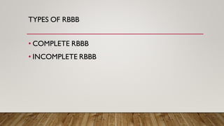
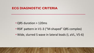
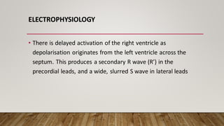
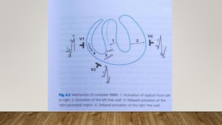
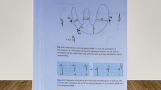
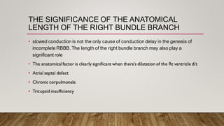
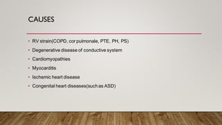
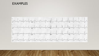
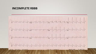
• RBBB
DR.VISHNUMOLAKALAADINARAYANA
JR1 GENERAL MEDICINE
RBBB
• Types of RBBB
• ECG Diagnostic Criteria Causes
• Electrophysiology
• Causes
• Examples
TYPES OF RBBB
• COMPLETE RBBB
• INCOMPLETE RBBB
ECG DIAGNOSTIC CRITERIA
• QRS duration > 120ms
• RSR’ pattern in V1-3 (“M-shaped” QRS complex)
• Wide, slurred S wave in lateral leads (I, aVL, V5-6)
ELECTROPHYSIOLOGY
• In normal cardiac conduction, impulses travel equally down
the left and right bundles, with the septum activated from
left to right ,followed by para septal activation followed by
free wall activation seperately.
• In RBBB, the left ventricle is activated normally, thus the
early part of the QRS complex correlating to septal
depolarisation is unchanged
ELECTROPHYSIOLOGY
• There is delayed activation of the right ventricle as
depolarisation originates from the left ventricle across the
septum. This produces a secondary R wave (R’) in the
precordial leads, and a wide, slurred S wave in lateral leads
SEQUENCE OF CONDUCTION IN RBBB:
• Septal depolarisationis thus unaffected, producing a normal
early QRS complex
• Left ventricular activation via the left bundle occurs normally
• Activation of the RV originates across the septum from LV
musculature
MECHANISM OF RSR PATTERN IN RT CHEST LEADS
• There is an initial small r wave due to left septal depolarisation
• This is followed by an S, or more likely a s, wave which is due
mainly to depolarisation of the left free wall , S wave is
attenuated and may even disappear completely.
• There is a terminal bizarre and slurred R wave (R') due to late
and anomalous right septal and right free wall depolarisation
• There is a small initial q wave, due to left septal
depolarization
• There is a relatively tall R wave, due mainly to
depolarization of the left free wall
• There is a terminal bizarre and slurred S wave due to late
and anomalous right septal and right free wall activation
MECHANISM OF SLURRED SWAVE IN LT CHEST
LEADS
THE ST SEGMENT AND T WAVE IN RIGHT BUNDLE
BRANCH BLOCK
• uncomplicated RBBB, the T wave and ST segment will reflect secondary changes.
• The T wave is opposite in direction to the terminal QRS deflection.
• Thus, if the terminal deflection is an R', as occurs, for example, in lead V1, the T wave will be inverted
• The associated ST segment will be slightly convex-upwards and, at times, minimally depressed.
• The terminal deflection is an S wave, as occurs, for example,
in lead V6, the T wave will be upright
• The associated ST segment will be slightly concave-upwards,
and, at times, minimally elevated.
• Any deviation from these manifestations usually represents a
primary change that is a primary myocardial abnormality in
addition to the RBBB.
THE ST SEGMENT AND T WAVE IN RIGHT BUNDLE
BRANCH BLOCK
INCOMPLETE RBBB
• There’s a diminition S wave in lead v2.
• A small R’ deflection will appear as the as the delay increases due to block.
• The R’ deflection becomes increasingly taller as the delay increases since larger
part of Rt free wall activation occurs after Lt free wall activation.
• While the ORS complex reflects a slight increase in duration, it is not increased
beyond 110 ms. The R' deflection itself is not widened beyond 40 ms.
THE SIGNIFICANCE OF THE ANATOMICAL
LENGTH OF THE RIGHT BUNDLE BRANCH
• slowed conduction is not the only cause of conduction delay in the genesis of
incomplete RBBB. The length of the right bundle branch may also play a
significant role
• The anatomical factor is clearly significant when there’s dilatation of the Rt ventricle d/t
• Atrial septal defect
• Chronic corpulmonale
• Tricupsid insufficiency
CAUSES
• RV strain(COPD, cor pulmonale, PTE, PH, PS)
• Degenerative disease of conductive system
• Cardiomyopathies
• Myocarditis
• Ischemic heart disease
• Congenital heart diseases(suchas ASD)
EXAMPLES
INCOMPLETE RBBB

right bundle branch block ,rbbb,right heart failure

  • 1.
  • 2.
    RBBB • Types ofRBBB • ECG Diagnostic Criteria Causes • Electrophysiology • Causes • Examples
  • 3.
    TYPES OF RBBB •COMPLETE RBBB • INCOMPLETE RBBB
  • 4.
    ECG DIAGNOSTIC CRITERIA •QRS duration > 120ms • RSR’ pattern in V1-3 (“M-shaped” QRS complex) • Wide, slurred S wave in lateral leads (I, aVL, V5-6)
  • 5.
    ELECTROPHYSIOLOGY • In normalcardiac conduction, impulses travel equally down the left and right bundles, with the septum activated from left to right ,followed by para septal activation followed by free wall activation seperately. • In RBBB, the left ventricle is activated normally, thus the early part of the QRS complex correlating to septal depolarisation is unchanged
  • 6.
    ELECTROPHYSIOLOGY • There isdelayed activation of the right ventricle as depolarisation originates from the left ventricle across the septum. This produces a secondary R wave (R’) in the precordial leads, and a wide, slurred S wave in lateral leads
  • 7.
    SEQUENCE OF CONDUCTIONIN RBBB: • Septal depolarisationis thus unaffected, producing a normal early QRS complex • Left ventricular activation via the left bundle occurs normally • Activation of the RV originates across the septum from LV musculature
  • 8.
    MECHANISM OF RSRPATTERN IN RT CHEST LEADS • There is an initial small r wave due to left septal depolarisation • This is followed by an S, or more likely a s, wave which is due mainly to depolarisation of the left free wall , S wave is attenuated and may even disappear completely. • There is a terminal bizarre and slurred R wave (R') due to late and anomalous right septal and right free wall depolarisation
  • 10.
    • There isa small initial q wave, due to left septal depolarization • There is a relatively tall R wave, due mainly to depolarization of the left free wall • There is a terminal bizarre and slurred S wave due to late and anomalous right septal and right free wall activation MECHANISM OF SLURRED SWAVE IN LT CHEST LEADS
  • 11.
    THE ST SEGMENTAND T WAVE IN RIGHT BUNDLE BRANCH BLOCK • uncomplicated RBBB, the T wave and ST segment will reflect secondary changes. • The T wave is opposite in direction to the terminal QRS deflection. • Thus, if the terminal deflection is an R', as occurs, for example, in lead V1, the T wave will be inverted • The associated ST segment will be slightly convex-upwards and, at times, minimally depressed.
  • 13.
    • The terminaldeflection is an S wave, as occurs, for example, in lead V6, the T wave will be upright • The associated ST segment will be slightly concave-upwards, and, at times, minimally elevated. • Any deviation from these manifestations usually represents a primary change that is a primary myocardial abnormality in addition to the RBBB. THE ST SEGMENT AND T WAVE IN RIGHT BUNDLE BRANCH BLOCK
  • 14.
    INCOMPLETE RBBB • There’sa diminition S wave in lead v2. • A small R’ deflection will appear as the as the delay increases due to block. • The R’ deflection becomes increasingly taller as the delay increases since larger part of Rt free wall activation occurs after Lt free wall activation. • While the ORS complex reflects a slight increase in duration, it is not increased beyond 110 ms. The R' deflection itself is not widened beyond 40 ms.
  • 16.
    THE SIGNIFICANCE OFTHE ANATOMICAL LENGTH OF THE RIGHT BUNDLE BRANCH • slowed conduction is not the only cause of conduction delay in the genesis of incomplete RBBB. The length of the right bundle branch may also play a significant role • The anatomical factor is clearly significant when there’s dilatation of the Rt ventricle d/t • Atrial septal defect • Chronic corpulmonale • Tricupsid insufficiency
  • 17.
    CAUSES • RV strain(COPD,cor pulmonale, PTE, PH, PS) • Degenerative disease of conductive system • Cardiomyopathies • Myocarditis • Ischemic heart disease • Congenital heart diseases(suchas ASD)
  • 18.
  • 19.