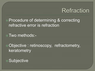
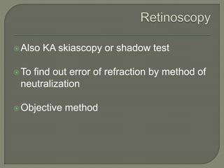
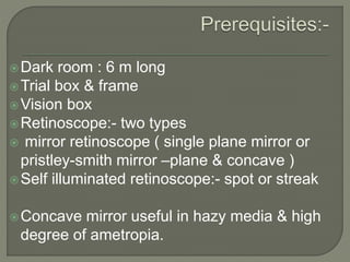
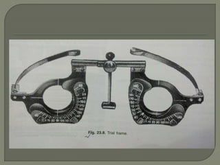
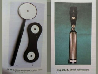
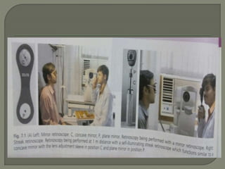
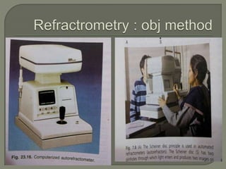
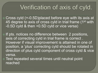
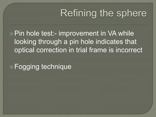
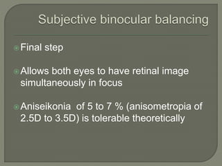
Dr. Atul Kumar Anand
Senior Resident
AIIMS Patna
Procedure of determining & correcting
refractive error is refraction
Two methods:-
Objective : retinoscopy, refractometry,
keratometry
Subjective
Also KA skiascopy or shadow test
To find out error of refraction by method of
neutralization
Objective method
Dark room : 6 m long
Trial box & frame
Vision box
Retinoscope:- two types
 mirror retinoscope ( single plane mirror or
pristley-smith mirror –plane & concave )
Self illuminated retinoscope:- spot or streak
Concave mirror useful in hazy media & high
degree of ametropia.
2 types:-
Dry retinoscopy ( without cyclopegics)
Wet retinoscopy
Done at 1 m distance usually
With plane mirror at 1 m, depending on
movement of red reflex, results are
No movement of red reflex indicate myopia
of 1D
With movement of red reflex indicate
myopia < 1D or emmetropia or hyper
Against movement of red reflex indicate
myopia > 1D
Movement of red reflex neutralized by
addition of increasing convex ( + )
spherical lens ( when red reflex move with
movement of retinoscope ) or concave (-)
spherical lens (when refex move against )
Deduction for distance 1D for 1 m or 1.5 D
for 2/3 m
Deduction for cycloplegic when used ( 1D for
atropine, 0.5D for homatropine or 0.75 D for
cyclopetolate )
Refractive error = retinoscopic finding –
distance - cycloplegic
For prescription of most suitable lens
Done after objective refraction ( retino or
AR )
When cycloplegic used, done after 3-4
days ( for homide or pentol), 3 wk for
topine :- PMT
Done by trial & error method (after retino
or AR) with trial box & frame, separately for
each eye
Most suitable lens prescribed
Strongest convex lens for hypermetropia
Weakest concave lens for myopia
Most suitable lens after subj. verification is
refined before final prescription
1st refine cylinder then sphere usually
Refining the cylinder: - by jackson’s cross
cylinder or astigmatic fan
Jackson’s cross cylinder: - combination of 2
cylinders of equal strength but oppst. Sign
placed with their axes at rt. Angle to each
other
Used to verify both strength & axis of cyld.
 Commonly used cross
cylinders are of +-
0.25D &
+-0.5D
To check power of cyld. , cross cyld. Of +-
0.25D is placed with its axis parallel to axis of
cylinder in trial frame 1st with same sign then
with oppst. Sign.
In 1st position, cylindrical correction is
enhanced by 0.25D & in 2nd diminished by
same amount.
When visual improvement does not occur in
either position, power of cyld. in trial frame
correct
 Cross cyld (+-0.5D)placed before eye with its axis at
45 degree to axis of cross cyld in trial frame (1st with
-0.5D cyld & then +0.5D cyld or vice versa)
 If pts. notices no difference between 2 positions,
axis of correcting cyld in trial frame is correct .
However if visual improvement is attained in one of
position, a ‘plus’ correcting cyld should be rotated in
direction of plus cyld component of cross cyld & vice
versa.
 Test repeated several times until neutral point
reached
Pin hole test:- improvement in VA while
looking through a pin hole indicates that
optical correction in trial frame is incorrect
Fogging technique
Final step
Allows both eyes to have retinal image
simultaneously in focus
Aniseikonia of 5 to 7 % (anisometropia of
2.5D to 3.5D) is tolerable theoretically
Done after 40 yr age
Better to under correct the presbyopia
Retinoscopy.pptx

Retinoscopy.pptx

  • 1.
    Dr. Atul KumarAnand Senior Resident AIIMS Patna
  • 2.
    Procedure of determining& correcting refractive error is refraction Two methods:- Objective : retinoscopy, refractometry, keratometry Subjective
  • 3.
    Also KA skiascopyor shadow test To find out error of refraction by method of neutralization Objective method
  • 4.
    Dark room :6 m long Trial box & frame Vision box Retinoscope:- two types  mirror retinoscope ( single plane mirror or pristley-smith mirror –plane & concave ) Self illuminated retinoscope:- spot or streak Concave mirror useful in hazy media & high degree of ametropia.
  • 7.
    2 types:- Dry retinoscopy( without cyclopegics) Wet retinoscopy Done at 1 m distance usually
  • 10.
    With plane mirrorat 1 m, depending on movement of red reflex, results are No movement of red reflex indicate myopia of 1D With movement of red reflex indicate myopia < 1D or emmetropia or hyper Against movement of red reflex indicate myopia > 1D
  • 12.
    Movement of redreflex neutralized by addition of increasing convex ( + ) spherical lens ( when red reflex move with movement of retinoscope ) or concave (-) spherical lens (when refex move against )
  • 14.
    Deduction for distance1D for 1 m or 1.5 D for 2/3 m Deduction for cycloplegic when used ( 1D for atropine, 0.5D for homatropine or 0.75 D for cyclopetolate ) Refractive error = retinoscopic finding – distance - cycloplegic
  • 17.
    For prescription ofmost suitable lens Done after objective refraction ( retino or AR ) When cycloplegic used, done after 3-4 days ( for homide or pentol), 3 wk for topine :- PMT
  • 18.
    Done by trial& error method (after retino or AR) with trial box & frame, separately for each eye Most suitable lens prescribed Strongest convex lens for hypermetropia Weakest concave lens for myopia
  • 19.
    Most suitable lensafter subj. verification is refined before final prescription 1st refine cylinder then sphere usually Refining the cylinder: - by jackson’s cross cylinder or astigmatic fan Jackson’s cross cylinder: - combination of 2 cylinders of equal strength but oppst. Sign placed with their axes at rt. Angle to each other Used to verify both strength & axis of cyld.
  • 20.
     Commonly usedcross cylinders are of +- 0.25D & +-0.5D
  • 21.
    To check powerof cyld. , cross cyld. Of +- 0.25D is placed with its axis parallel to axis of cylinder in trial frame 1st with same sign then with oppst. Sign. In 1st position, cylindrical correction is enhanced by 0.25D & in 2nd diminished by same amount. When visual improvement does not occur in either position, power of cyld. in trial frame correct
  • 22.
     Cross cyld(+-0.5D)placed before eye with its axis at 45 degree to axis of cross cyld in trial frame (1st with -0.5D cyld & then +0.5D cyld or vice versa)  If pts. notices no difference between 2 positions, axis of correcting cyld in trial frame is correct . However if visual improvement is attained in one of position, a ‘plus’ correcting cyld should be rotated in direction of plus cyld component of cross cyld & vice versa.  Test repeated several times until neutral point reached
  • 23.
    Pin hole test:-improvement in VA while looking through a pin hole indicates that optical correction in trial frame is incorrect Fogging technique
  • 24.
    Final step Allows botheyes to have retinal image simultaneously in focus Aniseikonia of 5 to 7 % (anisometropia of 2.5D to 3.5D) is tolerable theoretically
  • 25.
    Done after 40yr age Better to under correct the presbyopia