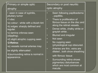
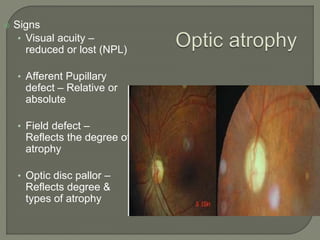
Dr. Atul Kumar Anand
Senior Resident
AIIMS Patna
Optic nerve shrinkage from any process
that produce degeneration of axons in the
ant.visual (Retinogeniculate) pathway
It is a term usually applied to the
condition of the optic disc when the optic
nerve is degenerated.
The Kestenbaum count is the number of
capillaries observed on the optic disc.
The normal count is approximately 10.
In optic atrophy, the number of these
capillaries reduces to less than 6; in a
hyperemic disc, the count is more than 12
1-Syphilis – Tabes dorsalis,
2-Pressure – pituitary tumour, glioma of
optic nerve
3-Tuberculous meningitis.
4-Glaucomatous optic atrophy
5-Circulatory optic atrophy – due to
occlusion of central retinal artery, arterio-
sclerosis.
6-Consecutive optic atrophy – due to
destruction of ganglion cells in retina as in
retinitis pigmentosa, choroiditis.
consecutive optic nerve atrophy : seen in
retinitis pigmentosa, choroiditis.
Disc: Yellow, waxy appearance, edge are
well defined.
Retina: vessels, show marked attenuation,
particularly in case of retinitis pigmentosa.
Surrounding retina shows evidence of retinitis
pigmentosa, choroditis (pigmentary
disturbance).
7-Traumatic optic atrophy: Avulsion of optic
nerve. fracture of skull, heamorrhage into
optic nerve sheath.
8-Toxic optic atrophy : exogenous toxins,
tobacco, alcohol,
9-Metabolic diabetes mellitus
10- Demyelinating disease – disseminated
sclerosis ,
11-Hereditary – Leber's disease.
PRIMARY-
SECONDARY –
 Post- papilloedemic optic atrophy
 Post-Neuritic optic atrophy
 Glaucomatous optic atrophy
 Consecutive optic atrophy
 Optic nerve fibers
degenerate in an orderly
manner and are replaced by
columns of glial cells without
alteration in the architecture
of the optic nerve head
 Pale disc
 Chalky white(full moon
against a dark red sky)
 Clear margin of disc/sharply
demarcated
 Normal cup
 Well seen lamina cribrosa
 Normal retinal vessels
 Optic nerve fibers exhibit marked degeneration, with
excessive proliferation of glial tissue
 The architecture is lost, resulting in indistinct margins.
The disc is grey or dirty grey , looks pale with a
greenish tinge
 The margins are poorly defined,
 The lamina cribrosa is obscured due to proliferating
fibroglial tissue.
 Hyaline bodies (corpora amylacea) or drusen may be
observed.
 Peripapillary sheathing of arteries as well as tortuous
veins may be observed.
Primary or simple optic
atrophy
Secondary or post neuritic
optic atrophy
• -seen in case of syphilis,
pituitary tumor
•1-disc
•a) colour : white with a bluish tint
•b) edges: sharply defined and
regular.
•c) lamina cribrosa seen
(stippling).
•d) slight atrophic cupping seen
•2-Retina:
•a) vessels normal arteries may
be slightly attenuated .
•b)surrounding retina is normal in
appearance.
• Seen following papilloedema
, papillitis.
• There is proliferation of
fibrous tissue on the disc and
along the retinal vessels.
• Dense white, chalky white or
grayish white .
• Blurred and irregular
• Not seen
• No cupping seen
(physiological cup obscured.
• Arteries are thin, veins are
dilated , both show sheathing
with fibrous tissue
• Surrounding retina shows
pigmentary disturbances
which are most common at
macula.
Symptoms
• That of causative condition
• Usually there is progressive diminution of vision .
• Visual loss – Depends on the degree of atrophy
• Partial atrophy – Partial visual loss
• Total atrophy – Total visual loss
Normal disc
OPTIC ATROPHY
Central Field Loss
 Signs
• Visual acuity –
reduced or lost (NPL)
• Afferent Pupillary
defect – Relative or
absolute
• Field defect –
Reflects the degree of
atrophy
• Optic disc pallor –
Reflects degree &
types of atrophy
When atrophy is complete the pupil is dilated
and fixed.
Treatment:
1-Directed at the cause –anti-syphilitic
treatment penicillin in high doses , removal of
pituitary tumour.
2-vasodilators: Tb. nicotinic acid.
3-Injection Vitamins B1, B12 in high doses.
OPTIC ATROPHY.pptx

OPTIC ATROPHY.pptx

  • 1.
    Dr. Atul KumarAnand Senior Resident AIIMS Patna
  • 2.
    Optic nerve shrinkagefrom any process that produce degeneration of axons in the ant.visual (Retinogeniculate) pathway It is a term usually applied to the condition of the optic disc when the optic nerve is degenerated.
  • 3.
    The Kestenbaum countis the number of capillaries observed on the optic disc. The normal count is approximately 10. In optic atrophy, the number of these capillaries reduces to less than 6; in a hyperemic disc, the count is more than 12
  • 5.
    1-Syphilis – Tabesdorsalis, 2-Pressure – pituitary tumour, glioma of optic nerve 3-Tuberculous meningitis. 4-Glaucomatous optic atrophy 5-Circulatory optic atrophy – due to occlusion of central retinal artery, arterio- sclerosis. 6-Consecutive optic atrophy – due to destruction of ganglion cells in retina as in retinitis pigmentosa, choroiditis.
  • 6.
    consecutive optic nerveatrophy : seen in retinitis pigmentosa, choroiditis. Disc: Yellow, waxy appearance, edge are well defined. Retina: vessels, show marked attenuation, particularly in case of retinitis pigmentosa. Surrounding retina shows evidence of retinitis pigmentosa, choroditis (pigmentary disturbance).
  • 7.
    7-Traumatic optic atrophy:Avulsion of optic nerve. fracture of skull, heamorrhage into optic nerve sheath. 8-Toxic optic atrophy : exogenous toxins, tobacco, alcohol, 9-Metabolic diabetes mellitus 10- Demyelinating disease – disseminated sclerosis , 11-Hereditary – Leber's disease.
  • 8.
    PRIMARY- SECONDARY –  Post-papilloedemic optic atrophy  Post-Neuritic optic atrophy  Glaucomatous optic atrophy  Consecutive optic atrophy
  • 9.
     Optic nervefibers degenerate in an orderly manner and are replaced by columns of glial cells without alteration in the architecture of the optic nerve head  Pale disc  Chalky white(full moon against a dark red sky)  Clear margin of disc/sharply demarcated  Normal cup  Well seen lamina cribrosa  Normal retinal vessels
  • 10.
     Optic nervefibers exhibit marked degeneration, with excessive proliferation of glial tissue  The architecture is lost, resulting in indistinct margins. The disc is grey or dirty grey , looks pale with a greenish tinge  The margins are poorly defined,  The lamina cribrosa is obscured due to proliferating fibroglial tissue.  Hyaline bodies (corpora amylacea) or drusen may be observed.  Peripapillary sheathing of arteries as well as tortuous veins may be observed.
  • 12.
    Primary or simpleoptic atrophy Secondary or post neuritic optic atrophy • -seen in case of syphilis, pituitary tumor •1-disc •a) colour : white with a bluish tint •b) edges: sharply defined and regular. •c) lamina cribrosa seen (stippling). •d) slight atrophic cupping seen •2-Retina: •a) vessels normal arteries may be slightly attenuated . •b)surrounding retina is normal in appearance. • Seen following papilloedema , papillitis. • There is proliferation of fibrous tissue on the disc and along the retinal vessels. • Dense white, chalky white or grayish white . • Blurred and irregular • Not seen • No cupping seen (physiological cup obscured. • Arteries are thin, veins are dilated , both show sheathing with fibrous tissue • Surrounding retina shows pigmentary disturbances which are most common at macula.
  • 13.
    Primary or simpleoptic atrophy Secondary or post neuritic optic atrophy • -seen in case of syphilis, pituitary tumor •1-disc •a) colour : white with a bluish tint •b) edges: sharply defined and regular. •c) lamina cribrosa seen (stippling). •d) slight atrophic cupping seen •2-Retina: •a) vessels normal arteries may be slightly attenuated . •b)surrounding retina is normal in appearance. • Seen following papilloedema , papillitis. • There is proliferation of fibrous tissue on the disc and along the retinal vessels. • Dense white, chalky white or grayish white . • Blurred and irregular • Not seen • No cupping seen (physiological cup obscured. • Arteries are thin, veins are dilated , both show sheathing with fibrous tissue • Surrounding retina shows pigmentary disturbances which are most common at macula.
  • 14.
    Symptoms • That ofcausative condition • Usually there is progressive diminution of vision . • Visual loss – Depends on the degree of atrophy • Partial atrophy – Partial visual loss • Total atrophy – Total visual loss
  • 15.
  • 16.
     Signs • Visualacuity – reduced or lost (NPL) • Afferent Pupillary defect – Relative or absolute • Field defect – Reflects the degree of atrophy • Optic disc pallor – Reflects degree & types of atrophy
  • 17.
    When atrophy iscomplete the pupil is dilated and fixed. Treatment: 1-Directed at the cause –anti-syphilitic treatment penicillin in high doses , removal of pituitary tumour. 2-vasodilators: Tb. nicotinic acid. 3-Injection Vitamins B1, B12 in high doses.